
Diphtheria
Reading File
Now I'll search the medical library for diphtheria content in parallel.
Finding Sources
Searching PubMed
diphtheria
Good - multiple textbook sections found. Now I'll read the most comprehensive sources in parallel.
Reading File
Reading File
Reading File
Reading File
Now let me read more of the Rosen's EM section which had the most content on pathophysiology and management:
Reading File
Finding Sources
Excellent - Harrison's 2025 has a dedicated section. Let me read it along with more of the Rosen's management section:
Reading File
Reading File
I now have rich, comprehensive content from multiple authoritative textbooks. Let me also fetch the key image showing diphtheria membrane/immunization graph:
Loading Image
I now have comprehensive, authoritative content from multiple major textbooks. Here is the full, well-organized answer:
Diphtheria
Definition and Organism
Diphtheria is an acute toxin-mediated infectious disease caused by Corynebacterium diphtheriae, a gram-positive, unencapsulated, nonmotile, non-spore-forming bacillus. Its name comes from two Greek roots: korynee (club, for its club-shaped morphology) and diphtheria (leather, for its characteristic membrane). Under the microscope, organisms form clusters of parallel "Chinese character" or palisade arrangements.
Less commonly, diphtheria-like disease is caused by C. ulcerans and C. pseudotuberculosis, both of which can carry the same toxin gene.
- Harrison's Principles of Internal Medicine, 22nd ed., p. 1265
- Rosen's Emergency Medicine, p. 2583
Pathogenesis and Toxin Mechanism
Toxigenic strains of C. diphtheriae are lysogenized by corynebacteriophage beta, which carries the structural gene tox encoding diphtheria toxin (DT). Under iron-limiting conditions (as found during infection), toxin expression is upregulated.
The toxin is a two-fragment (A-B) exotoxin:
- Fragment B binds to host cell surface receptors and facilitates entry
- Fragment A is the active enzyme - it catalyzes the ADP-ribosylation of Elongation Factor-2 (EF-2), irreversibly inactivating it and halting cellular protein synthesis
The local effects produce the pseudomembrane - composed of necrotic epithelial cells, leukocytes, erythrocytes, fibrin, and bacteria. This membrane is tough, grayish-white, and bleeds when removed. Systemic toxin absorption causes injury to the heart, nervous system, and kidneys.
- Rosen's Emergency Medicine, p. 2583
- Harrison's Principles of Internal Medicine, 22nd ed., p. 1265
Epidemiology
- Humans are the only reservoir
- Transmission: respiratory droplets, direct contact with secretions or skin lesions, rarely fomites
- R0: 1.7-4.3; untreated cases infectious for ~18.5 days
- Incubation: mean 1.4 days (range 1-10 days)
- In the pre-vaccine era, US incidence exceeded 100/100,000; 206,000 cases and 15,520 deaths in 1921 alone
- Vaccination has reduced this to near-zero in developed nations; only 2 cases in the US between 2004-2017
- Sporadic cases in inadequately immunized adolescents/adults; recent outbreaks in Yemen (2022-23) and Nigeria
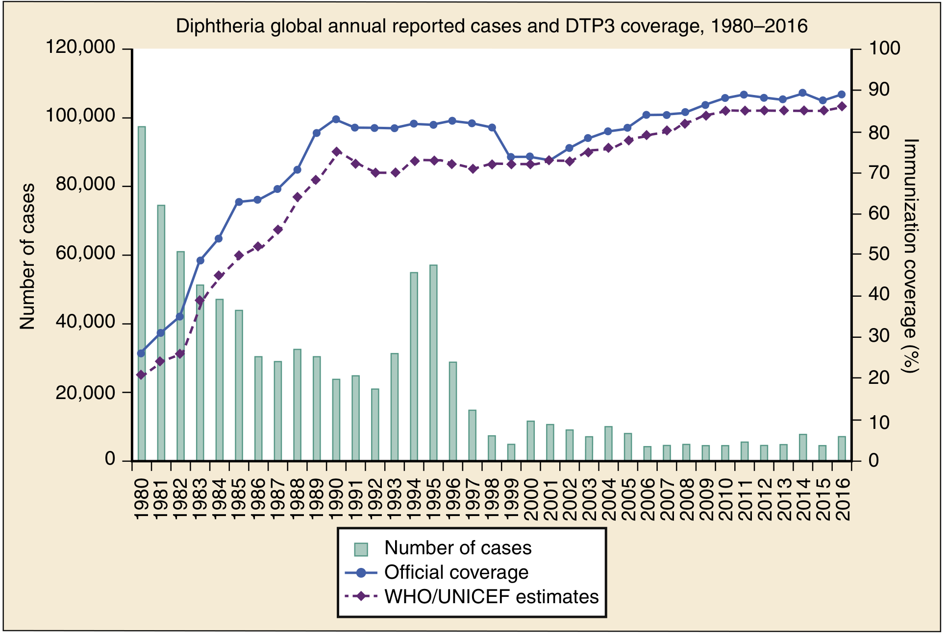
Important note on adult immunity: Even in countries with high childhood vaccination rates, >50% of adults over 40 lack protective antibody levels. Because childhood immunization eliminates circulating toxigenic strains (which previously served as natural boosters), adult immunity wanes.
Types of Diphtheria
| Type | Location | Key Features |
|---|---|---|
| Faucial (pharyngeal/tonsillar) | Tonsils, soft palate | Most common; greatest systemic toxicity |
| Nasal | Nasal passages | Serosanguineous discharge; milder systemic effects |
| Laryngeal (tracheobronchial) | Larynx/trachea | Barking cough, stridor, hoarseness; high risk of airway obstruction |
| Cutaneous | Skin | Chronic ulcers; less toxin; more common in tropics; most nontoxigenic |
Clinical Features
Incubation: 2-4 days (range 1-8 days)
Early symptoms (indistinguishable from other URTIs):
- Low-grade fever, sore throat
- Malaise, weakness, dysphagia, headache
- Voice changes (hoarseness)
Pharyngeal diphtheria:
- The hallmark is the pseudomembrane - initially white, becoming gray, tough, and adherent; bleeds on removal
- Limited to tonsils = milder disease; extends across the entire pharynx = severe disease
- "Bull neck": marked cervical lymphadenopathy with neck tissue edema due to cellulitis - characteristic of malignant diphtheria
- Malignant form: high fever, severe muscle weakness, vomiting, diarrhea, restlessness, delirium
Laryngeal diphtheria:
- Classic "barking" cough, inspiratory stridor, hoarseness
- Marked risk of acute airway obstruction and death
Complications
1. Myocarditis (Cardiac Toxicity)
- Appears 1-2 weeks after illness onset (earlier in severe cases)
- ECG changes (ST-T wave changes, AV block, dysrhythmias) in up to two-thirds of patients
- Clinical myocarditis in 10-25% of cases
- Dilated or hypertrophic cardiomyopathy on echo
- Serum troponin correlates with severity
- Can cause sudden death from arrhythmia or heart failure
2. Neuropathy (Neurologic Toxicity)
The nervous system involvement follows a predictable biphasic pattern:
Early (days 5-12):
- Palatal paralysis - most common cranial nerve involvement; nasal voice, regurgitation, dysphagia
- Other cranial nerves: trigeminal, facial, vagus, hypoglossal
Weeks 2-3:
- Ciliary body paralysis - loss of accommodation, blurred vision, but pupillary light reaction preserved (opposite of Argyll Robertson pupil)
- Rarely, extraocular muscle weakness
Weeks 5-8 (delayed):
- Sensorimotor polyneuropathy - may resemble Guillain-Barre syndrome (ascending paralysis, elevated CSF protein, acellular CSF)
- About 5% of respiratory cases develop polyneuritis; 75% of severe cases have some form of neuropathy
- Recovery is usually complete but may take months
The toxin reaches Schwann cells via the bloodstream within 24-48 hours but its metabolic effects (demyelination in proximal spinal nerves, dorsal root ganglia, spinal roots) unfold over weeks.
- Adams and Victor's Principles of Neurology, 12th ed.
- Rosen's Emergency Medicine, p. 2583
Diagnosis
Clinical suspicion drives initial management - do not wait for culture confirmation.
Suspect case: Pharyngitis + absent/low-grade fever + gray adherent pseudomembrane that bleeds when manipulated
Probable case: Above + stridor, bull neck, or toxic circulatory collapse
Laboratory workup:
- Notify the laboratory specifically that C. diphtheriae is suspected (routine cultures will miss it)
- Throat/nasopharyngeal swab before antibiotics; plate on tellurite selective medium (Loeffler's or potassium tellurite)
- Toxin detection: Elek test (immunodiffusion), or PCR for the tox gene (more sensitive)
- Mass spectrometry detection methods are emerging
- Up to 30% of diphtheria patients also test positive for Group A streptococcus - this does NOT exclude diphtheria
- Leukocytosis, mild thrombocytopenia, proteinuria are common but nonspecific
- ECG, troponin, echocardiogram for cardiac monitoring
Management
Three goals: protect the airway, limit toxin effects, stop further toxin production
1. Airway Management
- Respiratory isolation immediately
- Early intubation for laryngeal involvement
- Manage fluid resuscitation carefully - myocardial toxicity may limit fluid tolerance
2. Diphtheria Antitoxin (DAT)
- Most critical intervention - neutralizes only unbound circulating toxin; toxin already bound to tissues cannot be neutralized
- Must be given on clinical suspicion without waiting for culture confirmation - delay increases mortality
- Dose: 10,000-100,000 units depending on severity and membrane extent
- Test for equine hypersensitivity before administration (DAT is horse-derived)
- Administer IV for severe/late cases, IM for mild cases
3. Antibiotics
- Penicillin (benzylpenicillin) or erythromycin are first-line
- Purpose: halt toxin production, eliminate the organism, reduce transmission
- Antibiotics alone are insufficient - antitoxin is required
- Treatment continues until two consecutive negative cultures are obtained
4. Supportive Care
- Cardiac monitoring (continuous ECG)
- Respiratory support, including mechanical ventilation if needed
- Nasogastric feeding if palatal paralysis prevents swallowing
5. Contacts and Public Health
- All close contacts: nasopharyngeal cultures + prophylactic antibiotics (erythromycin or penicillin)
- Unimmunized contacts: diphtheria toxoid
- Report to public health authorities
Differential Diagnosis
| Condition | Distinguishing Feature |
|---|---|
| Streptococcal pharyngitis | No membrane, positive rapid strep test |
| Infectious mononucleosis | Splenomegaly, atypical lymphocytes, positive monospot |
| Peritonsillar abscess | Unilateral, uvular deviation, fluctuance |
| Epiglottitis | No membrane, drooling, tripod position |
| Viral croup | Younger age, preceding URTI, no membrane |
| Ludwig's angina | Floor of mouth induration, no membrane |
| Candida (thrush) | White plaques, scrapes off easily, immunocompromised host |
Vaccination
- DTaP (diphtheria, tetanus, pertussis) for children: primary series at 2, 4, 6 months; boosters at 15-18 months and 4-6 years
- Tdap/Td boosters every 10 years in adults
- Vaccine prevents toxin-mediated disease but does not prevent carriage
-
95% efficacy with complete primary series
Key Points (High-Yield Summary)
- Causative organism: C. diphtheriae; gram-positive club-shaped bacillus; toxin encoded by bacteriophage beta
- Toxin mechanism: ADP-ribosylation of EF-2 → inhibits protein synthesis
- Hallmark: Gray, adherent, bleeding pseudomembrane
- Systemic complications: Myocarditis (1-2 weeks), palatal paralysis (5-12 days), ciliary paralysis with accommodation loss but intact pupillary light reflex (2-3 weeks), delayed polyneuropathy (5-8 weeks)
- Treatment: Antitoxin (give immediately on clinical grounds) + penicillin/erythromycin
- Critical point: Antitoxin neutralizes only free (unbound) toxin - early administration is essential
Sources: Rosen's Emergency Medicine, Tintinalli's Emergency Medicine, Harrison's Principles of Internal Medicine (22nd ed., 2025), Adams & Victor's Principles of Neurology, Scott-Brown's Otorhinolaryngology, Robbins & Cotran Pathologic Basis of Disease
This is a shared conversation. Sign in to Orris to start your own chat.