
Prokaryotic reflection image.
A detailed scientific diagram of a prokaryotic cell with labeled structures including cell wall, cell membrane, cytoplasm, nucleoid region with DNA, ribosomes, flagella, pili, capsule, and plasmids. Clean educational illustration style with clear labels and arrows.
404 Not Found. {'message': '{\n "error": {\n "code": 404,\n "message": "Publisher model `projects/engaged-beaker-474810-u2/locations/global/publishers/google/models/gemini-3-pro-image-preview` was not found or your project does not have access to it. Ensure you are using a valid model name and that the model is available in the specified region. For more information, see: https://docs.cloud.google.com/gemini-enterprise-agent-platform/resources/locations.",\n "status": "NOT_FOUND"\n }\n}\n', 'status': 'Not Found'}
We encountered an error while processing your request. Please try again or reach out to us for help.
Prokaryotic replication
Prokaryotic DNA Replication
Overview
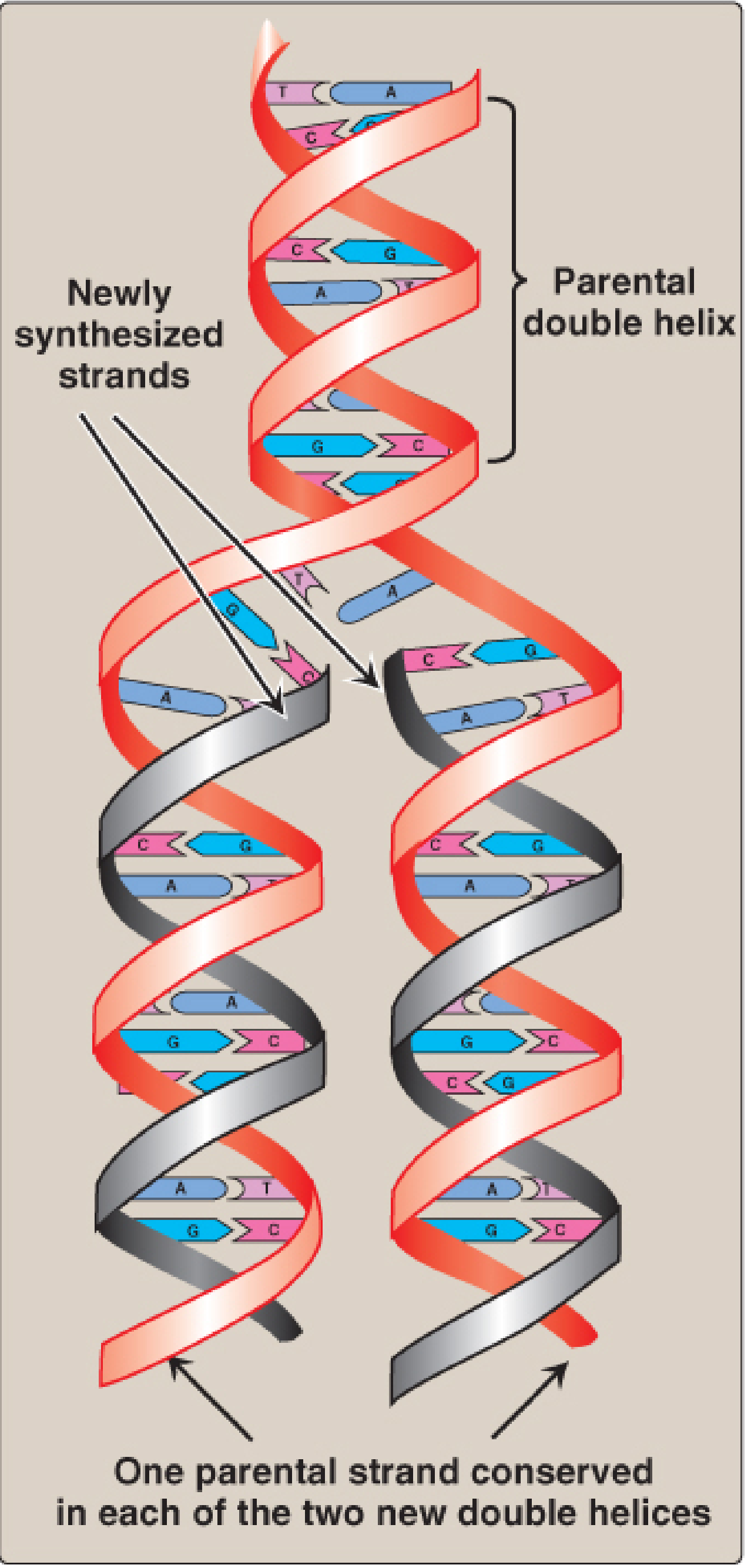
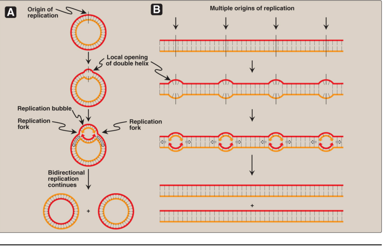
Step 1 - Initiation at the Origin (oriC)
- DnaA protein binds to DnaA boxes within oriC, causing the AT-rich DNA unwinding element to melt open, creating short regions of ssDNA.
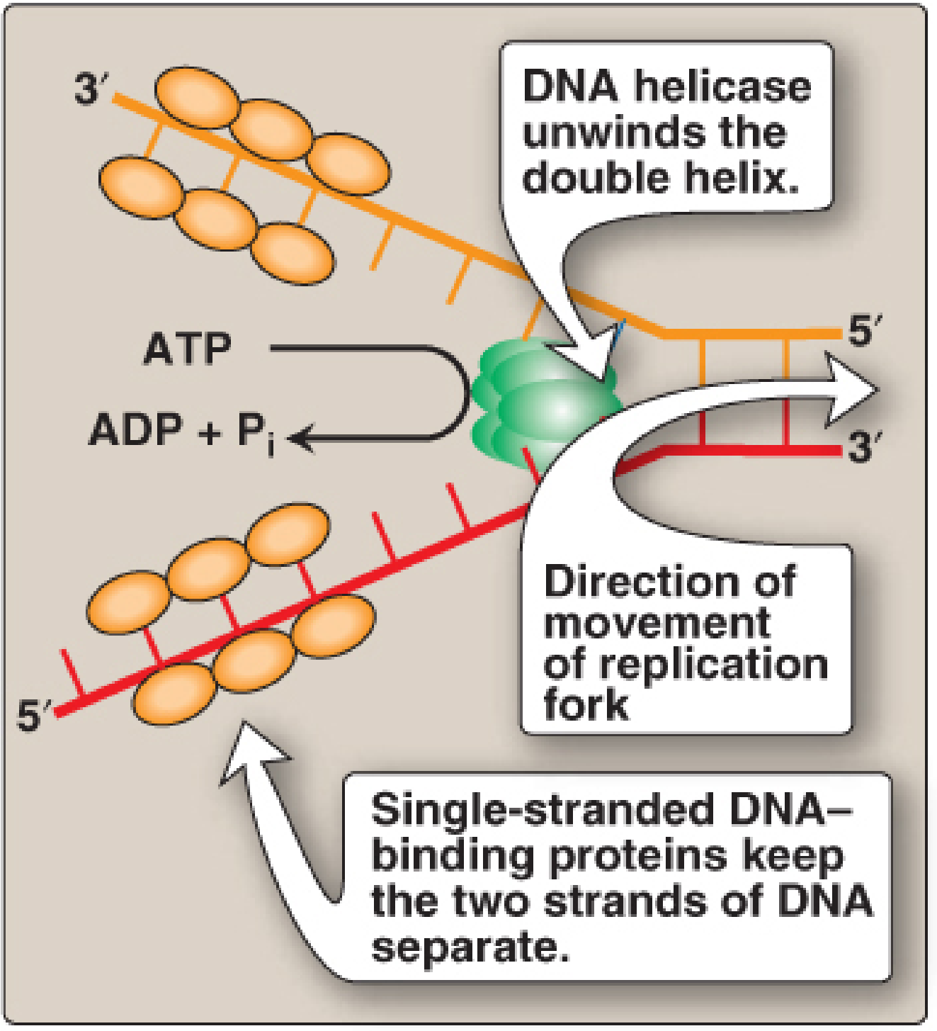
Step 2 - Replication Fork Formation
| Protein | Function |
|---|---|
| DnaB helicase | Unwinds the double helix using ATP hydrolysis |
| SSB proteins (single-stranded DNA-binding) | Bind cooperatively to ssDNA to keep strands separated and protect from nucleases |
| Topoisomerases | Relieve torsional stress (positive supercoils) that builds ahead of the fork |
- Type I topoisomerases: nick one strand, rotate, and reseal - no ATP needed
- Type II topoisomerases (DNA gyrase): cut both strands, pass a segment through, and reseal - uses ATP; introduces negative supercoils to neutralize positive ones
Clinical note: Fluoroquinolones (e.g., ciprofloxacin) inhibit bacterial DNA gyrase - a type II topoisomerase unique to bacteria. This makes it an excellent antibiotic target.
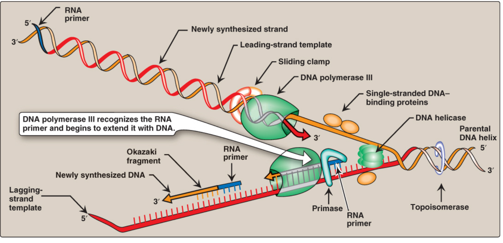
Step 3 - Priming
- Primase (part of the primosome complex) synthesizes a short RNA primer in the 5'→3' direction
- The primosome = prepriming complex + primase
- RNA primers are made for the leading strand (once) and for each Okazaki fragment on the lagging strand
Step 4 - Elongation by DNA Pol III
- Extends from the 3'-OH of the RNA primer
- Synthesizes in the 5'→3' direction only
- Is highly processive - stays on the template without dissociating, due to its β-subunit sliding clamp (a ring that encircles the template strand)
- Uses dNTPs (dATP, dTTP, dCTP, dGTP); releases PPi which is hydrolyzed to 2 Pi (making the reaction irreversible)
Step 5 - Primer Removal and Gap Filling (DNA Pol I)
- 5'→3' exonuclease: removes the RNA primer ahead of it
- 5'→3' polymerase: simultaneously fills the gap with DNA
Step 6 - Ligation
Summary Table of Key Enzymes
| Enzyme | Role |
|---|---|
| DnaA | Initiates opening at oriC |
| DnaB (helicase) | Unwinds double helix, ATP-dependent |
| SSB proteins | Stabilize ssDNA at replication fork |
| DNA gyrase (Topo II) | Relieves positive supercoils ahead of fork |
| Primase | Synthesizes RNA primers |
| DNA Pol III | Main chain elongation (5'→3'), high fidelity, processive |
| DNA Pol I | Removes RNA primers; fills gaps (nick translation) |
| DNA ligase | Seals nicks between Okazaki fragments |