
Fluidmosaic model and factiors affecting the fluidity of cell membrane
Reading File
The fluid mosaic model, proposed by Singer and Nicolson in 1972, describes the cell membrane as a dynamic, two-dimensional fluid structure in which proteins float within or on a phospholipid bilayer. Factors affecting membrane fluidity include temperature, cholesterol content, fatty acid saturation, and phospholipid chain length.
Searching sources...
Finding Sources
Finding Sources
Reading File
Reading File
Finding Sources
Reading File
Reading File
Reading File
Reading File
Reading File
Now let me fetch the key diagram images to include in the answer.
Loading Image
Loading Image
Loading Image
I now have comprehensive information from multiple authoritative textbooks (Harper's Biochemistry, Histology: A Text and Atlas, Basic Medical Biochemistry, and Medical Physiology) along with diagrams. Here is the complete answer:
Fluid Mosaic Model & Factors Affecting Membrane Fluidity
1. The Fluid Mosaic Model
The fluid mosaic model was first proposed by S.J. Singer and G.L. Nicolson in 1972. It describes the plasma membrane as a dynamic, two-dimensional fluid structure in which a mosaic of lipid and protein molecules can, for the most part, move laterally in the plane of the membrane.
The term "fluid mosaic" captures two key ideas:
- Fluid - lipid and most protein molecules diffuse laterally within the bilayer
- Mosaic - proteins of varying shapes and functions are embedded throughout the bilayer, like tiles in a mosaic
Key Structural Components
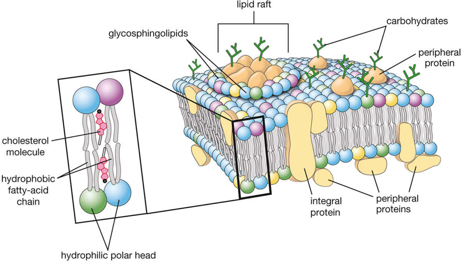
Modified fluid-mosaic model showing lipid bilayer with integral proteins, peripheral proteins, lipid rafts, and carbohydrate chains (Histology: A Text and Atlas, p. 134)
A. Lipid Bilayer - The Foundation
The backbone of the membrane is a phospholipid bilayer. All major lipids are amphipathic (containing both hydrophobic and hydrophilic regions):
- Hydrophilic polar head groups face the aqueous environment (outer and inner surfaces)
- Hydrophobic fatty acid tails face each other, forming the membrane's interior
The principal phospholipids are:
- Phosphatidylcholine (lecithin) - most abundant; concentrated in the outer leaflet
- Phosphatidylethanolamine - concentrated in the inner leaflet
- Phosphatidylserine - concentrated in the inner leaflet
- Sphingomyelin - outer leaflet
- Phosphatidylinositol - inner leaflet (also functions in signal transduction)
The lipid composition is asymmetric - the two leaflets have different compositions. Choline-containing phospholipids (phosphatidylcholine and sphingomyelin) predominate in the outer leaflet; aminophospholipids (phosphatidylserine and phosphatidylethanolamine) concentrate in the inner leaflet. - Harper's Illustrated Biochemistry, 32nd Ed
B. Membrane Proteins
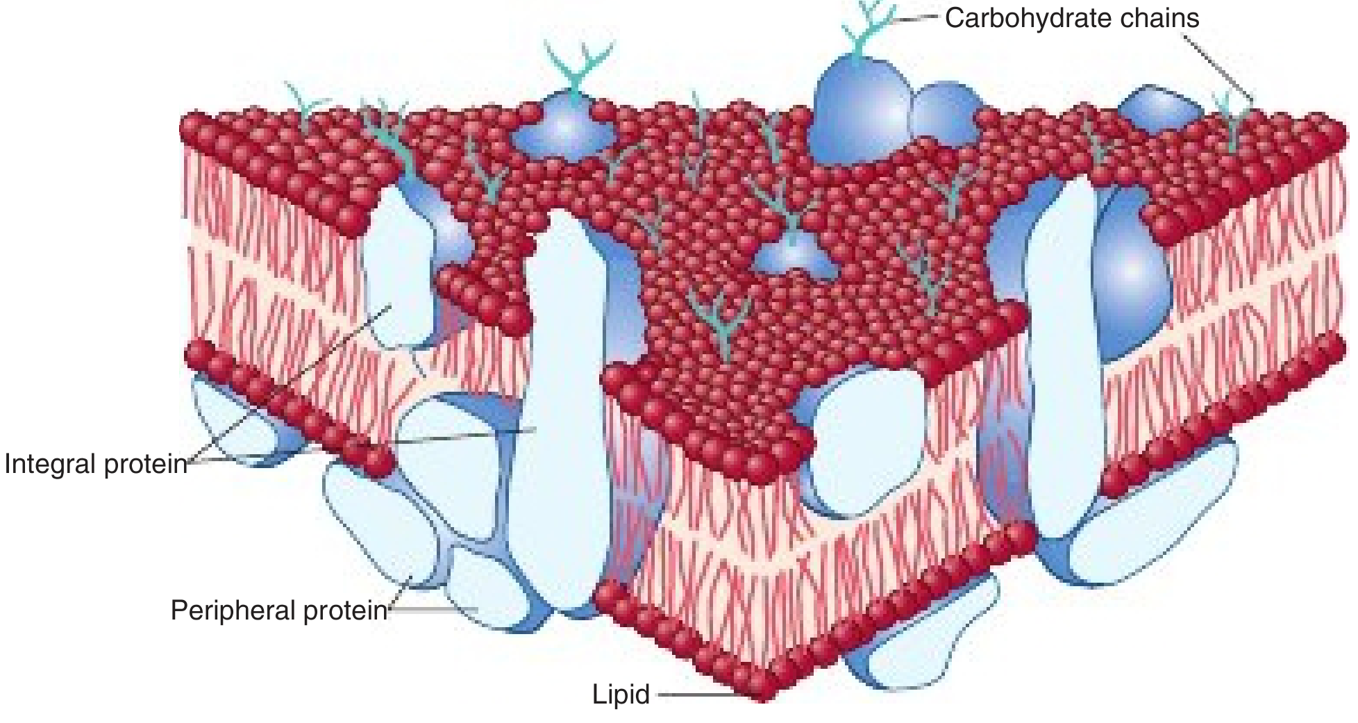
Fluid mosaic model: integral proteins firmly embedded in the lipid bilayer, peripheral proteins loosely associated with the surface (Harper's Illustrated Biochemistry, 32nd Ed)
Proteins constitute approximately half of the total membrane mass and are classified into two types:
| Type | Location | Interaction | Removal |
|---|---|---|---|
| Integral (intrinsic) | Embedded within or spanning the bilayer | Hydrophobic interactions with lipid acyl chains | Require detergents or organic solvents |
| Peripheral (extrinsic) | Associated with inner or outer surface | Electrostatic bonds to lipids or integral proteins | Mild conditions (high salt, pH change) |
Integral proteins that span the entire bilayer are called transmembrane proteins. They serve as channels, transporters, receptors, and structural proteins. Their membrane-spanning domains are typically alpha-helices rich in hydrophobic amino acids.
C. Glycocalyx
Carbohydrate chains (oligosaccharides) are covalently attached to proteins (forming glycoproteins) and lipids (forming glycolipids) exclusively on the external surface of the membrane - none project from the inner face. This external carbohydrate layer is called the glycocalyx and functions in:
- Cell recognition
- Cell-cell interactions
- Protection
D. Lipid Rafts
Lipid rafts are specialized microdomains within the membrane enriched in glycosphingolipids, cholesterol, and specific signaling proteins. Due to the high cholesterol content and longer, highly saturated fatty acid chains, lipid rafts are thicker and exhibit less fluidity than the surrounding plasma membrane. They appear to serve as platforms for signal transduction. - Histology: A Text and Atlas, p. 134
E. Types of Lipid Movement in the Membrane
| Movement | Direction | Rate |
|---|---|---|
| Lateral diffusion | Within same leaflet | Very fast (hallmark of fluid membrane) |
| Rotational diffusion | Spinning in place | Very fast |
| Flip-flop (transverse diffusion) | Between leaflets | Extremely slow (half-life: weeks in synthetic bilayers; requires flippases in vivo) |
2. Factors Affecting Membrane Fluidity
Fluidity refers to how easily lipids and proteins move laterally. It is determined by several key factors:
A. Temperature
- At high temperatures, thermal energy exceeds interaction energy between lipids → lateral diffusion is rapid → membrane is in the sol (fluid) state
- At low temperatures, interaction energies dominate → phospholipids cannot escape neighbors → membrane is in the gel (rigid) state
- The temperature at which the membrane transitions from gel to sol is called the transition temperature (Tm)
"At high temperatures, the thermal energy of any given lipid molecule is greater than the interaction energy that would tend to hold adjacent lipid molecules together. Under these conditions, lateral diffusion can proceed rapidly." - Medical Physiology (Boron & Boulpaep)
B. Cholesterol Content
Cholesterol has a biphasic (dual) effect on fluidity - arguably the most interesting factor:
- At physiological temperature: Cholesterol reduces fluidity by inserting its rigid steroid ring structure between phospholipids, restricting the movement of fatty acid chains (immobilizing them)
- At low temperature: Cholesterol prevents the membrane from freezing (prevents gel state formation) by disrupting the tight packing of fatty acid chains
The result is that cholesterol acts as a "fluidity buffer" - maintaining membrane fluidity within a physiologically useful range across varying temperatures.
"Cholesterol molecules are interspersed less evenly throughout the lipid bilayer; cholesterol affects the packing of the fatty acid chains, with a major effect on membrane fluidity." - Junqueira's Basic Histology, 17th Ed
C. Fatty Acid Saturation
This is a major determinant:
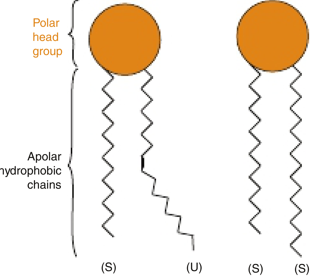
Saturated (S) tails pack tightly and reduce fluidity; unsaturated (U) tails have kinks that prevent tight packing and increase fluidity (Harper's Illustrated Biochemistry, 32nd Ed)
- Saturated fatty acids form straight, rigid tails → pack tightly together → reduce fluidity → higher transition temperature
- Example: Dioctadecanoic phosphatidylcholine (two 18-carbon saturated chains) has Tm = 55.5°C
- Unsaturated fatty acids (especially cis double bonds) form "kinked" tails → cannot pack closely → increase fluidity → lower transition temperature
- Introducing one double bond into an 18-carbon chain dramatically lowers the Tm
- More double bonds = more kinks = more fluid membrane
- Trans fatty acids (found in processed foods) behave more like saturated fats and reduce fluidity, which is why trans fats are biologically problematic
"As the number of double bonds within the lipid side chains increase, the number of kinks in the tails increases. As a consequence, the membrane lipids become less tightly packed and the membrane more fluid." - Harper's Illustrated Biochemistry, 32nd Ed
D. Fatty Acid Chain Length
- Shorter chains have fewer van der Waals interactions with neighbors → increase fluidity → lower Tm
- Example: Reducing chain length from 18 to 14 carbons drops Tm from 55.5°C to 23°C
- Longer chains pack more tightly → decrease fluidity
- Chain length also determines bilayer thickness (C16 chains → 2.47 nm wide bilayer; C14 chains → 2.3 nm)
E. Sphingolipid Content
Sphingolipids (like sphingomyelin) tend to pack more tightly due to their fully saturated chains and the ability to form hydrogen bonds via their amide groups. High sphingolipid content reduces fluidity, which is why lipid rafts (enriched in sphingolipids + cholesterol) are less fluid than the surrounding membrane.
F. Proteins
- The presence of large transmembrane proteins can locally restrict lipid movement
- Some proteins are anchored to the cytoskeleton and are essentially immobile, creating fixed zones within the otherwise fluid membrane
G. Presence of Other Molecules
- Ethanol intercalates between membrane lipids, increasing fluidity - this is the molecular basis of the short-term CNS effects of alcohol (changes ion channel and neurotransmitter receptor function). - Basic Medical Biochemistry, 6th Ed
Summary Table: Factors Affecting Membrane Fluidity
| Factor | Effect on Fluidity |
|---|---|
| High temperature | Increases |
| Low temperature | Decreases |
| Unsaturated fatty acids (cis) | Increases |
| Saturated fatty acids | Decreases |
| Short fatty acid chains | Increases |
| Long fatty acid chains | Decreases |
| Cholesterol (at body temp) | Decreases (buffers extremes) |
| High sphingolipid content | Decreases |
| Ethanol | Increases |
| Trans fatty acids | Decreases |
Sources:
- Harper's Illustrated Biochemistry, 32nd Ed (Chapter 40)
- Histology: A Text and Atlas with Correlated Cell and Molecular Biology (Chapter 2)
- Junqueira's Basic Histology, 17th Ed
- Basic Medical Biochemistry: A Clinical Approach, 6th Ed (Chapter 10)
- Medical Physiology (Boron & Boulpaep)
Phagocytosis in short
Phagocytosis is the process by which cells engulf large particles (>0.5 µm) such as bacteria, dead cells, and debris by forming a large vesicle called a phagosome. Key phagocytes include neutrophils and macrophages.
Searching sources...
Finding Sources
Reading File
Reading File
Loading Image
Loading Image
Phagocytosis
Phagocytosis is the process by which specialized cells (phagocytes - mainly neutrophils and macrophages) engulf and destroy large particles (>0.5 µm) such as microbes, dead cells, and foreign debris.
Cells Involved
- Neutrophils - first responders
- Macrophages - professional phagocytes; more sustained killing
- Dendritic cells, monocytes (to a lesser extent)
Steps of Phagocytosis
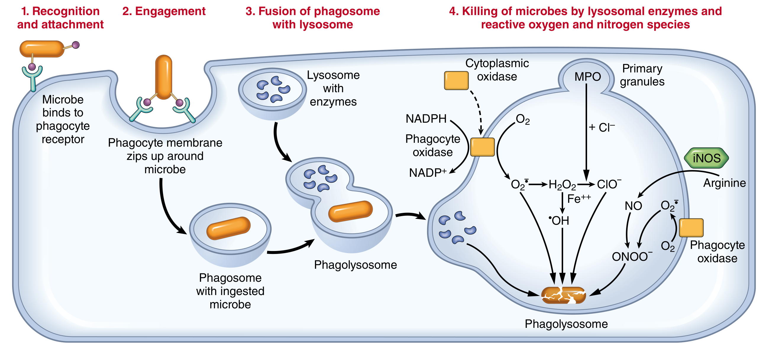
Phagocytosis and intracellular destruction of microbes - Robbins & Cotran Pathologic Basis of Disease
Step 1 - Recognition and Attachment
Phagocytes recognize microbes via surface receptors:
| Receptor | Ligand |
|---|---|
| Fc receptors | Fc region of IgG antibodies coating the microbe |
| Complement receptors (CR) | C3b deposited on microbial surface |
| Mannose receptors | Terminal mannose/fucose on microbial glycoproteins (not on host cells) |
| Scavenger receptors | Modified LDL, polyanionic molecules on bacteria |
| Toll-like receptors (TLRs) | PAMPs (LPS, peptidoglycan, lipoteichoic acid) |
Opsonization greatly enhances phagocytosis. Opsonins (IgG, C3b, mannose-binding lectin) coat the microbe like a "flag," making it easier to bind and engulf.
Step 2 - Engulfment
- Cytoplasmic extensions called pseudopods flow around the bound particle
- Pseudopods fuse at the tips, enclosing the particle
- The plasma membrane pinches off, forming an intracellular vesicle called a phagosome
- This requires actin cytoskeleton remodeling (depolymerization and repolymerization of actin filaments)
Step 3 - Phagolysosome Formation
- The phagosome fuses with lysosomes (containing hydrolytic enzymes)
- This creates a phagolysosome
- Both specific granules and azurophilic (primary) granules fuse with the phagosome - this is called degranulation
Step 4 - Killing and Digestion
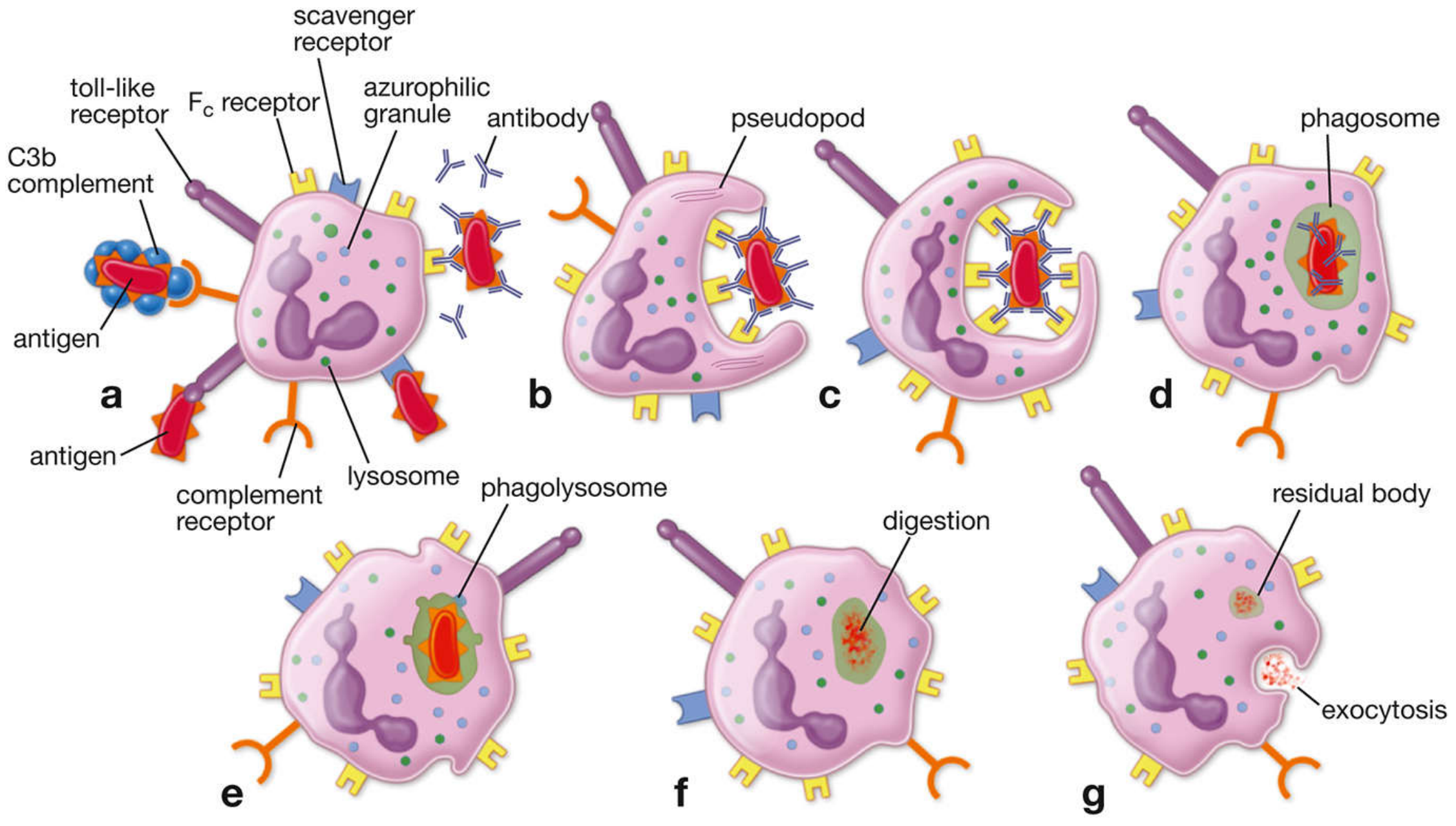
Neutrophil phagocytosis from recognition to digestion and exocytosis - Histology: A Text and Atlas
Killing occurs by two major mechanisms:
A. Oxygen-Dependent (Respiratory Burst)
- NADPH oxidase converts O₂ → superoxide (O₂•⁻)
- O₂•⁻ → H₂O₂ (hydrogen peroxide)
- Myeloperoxidase (MPO) converts H₂O₂ + Cl⁻ → HOCl (hypochlorite - the most potent microbicidal agent)
- Via Fenton reaction: H₂O₂ + Fe²⁺ → •OH (hydroxyl radical)
- iNOS produces NO, which combines with O₂•⁻ → ONOO⁻ (peroxynitrite) - reactive nitrogen species
B. Oxygen-Independent
- Lysosomal enzymes: acid hydrolases, proteases, lysozyme, defensins, lactoferrin
- These degrade the microbial components at low pH within the phagolysosome
Step 5 - Disposal
- Digested material is either exocytosed into the extracellular space
- Or stored as residual bodies within the phagocyte
Summary in One Line
Phagocytosis = Recognition → Engulfment (phagosome) → Fusion with lysosome (phagolysosome) → Killing (ROS + lysosomal enzymes) → Exocytosis
Sources: Robbins & Cotran Pathologic Basis of Disease; Histology: A Text and Atlas with Correlated Cell and Molecular Biology
This is a shared conversation. Sign in to Orris to start your own chat.