
lung abscess
"lung abscess" AND treatment
lung abscess chest x-ray cavity with air-fluid level
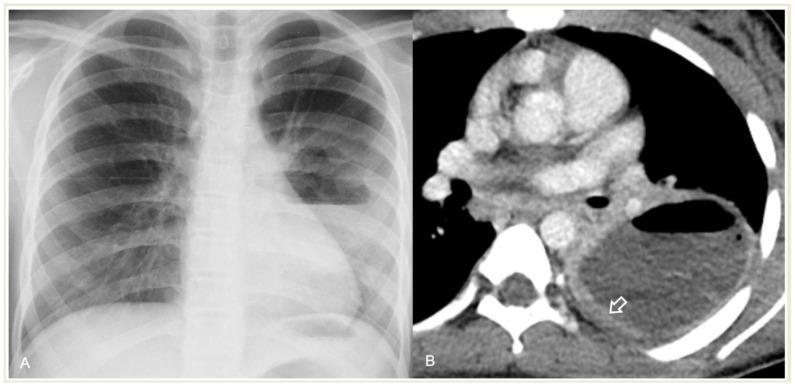
This dual-panel image displays diagnostic imaging of a 12-year-old female with a lung abscess complicating community-acquired pneumonia (CAP). Panel (A) is a frontal chest radiograph (X-ray) showing a large, well-circumscribed cavitary lesion in the right lung field. The lesion is characterized by a prominent horizontal air-fluid level, where the upper portion is radiolucent (air) and the lower portion is opaque (fluid/pus). Panel (B) is a transverse axial contrast-enhanced CT (CECT) scan of the thorax at the level of the hila. It demonstrates a large, hypodense fluid-filled cavity within the right lung parenchyma. Key features include a thick, irregular, and strongly enhancing peripheral wall (indicated by an arrow), which is a classic diagnostic sign of an abscess. The CT further confirms the presence of an air-fluid level. These findings are essential for differentiating a lung abscess from other complications like necrotizing pneumonia or loculated empyema, which typically lack such a well-defined enhancing wall.
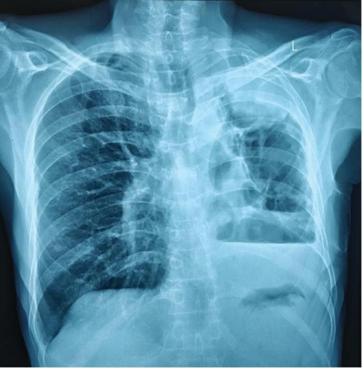
This diagnostic image is a posteroanterior (PA) chest X-ray demonstrating a severely diseased and contracted left lung, indicative of a 'destroyed lung' phenotype. The left hemithorax shows significant volume loss, characterized by an ipsilateral mediastinal shift (tracheal and cardiac deviation to the left) and narrowing of the intercostal spaces. Multiple large, thin-walled cavitary lesions are visible in the upper and middle zones of the left lung, with a prominent air-fluid level suggestive of a large abscess or infected cavity. A dense opacity is noted in the left apex. The left costophrenic angle is obliterated by a hazy density consistent with a moderate pleural effusion or pleural thickening. In contrast, the right lung field appears hyperinflated, likely due to compensatory hypertrophy. This radiographic presentation is clinically relevant for chronic pulmonary infections such as necrotizing pneumonia (e.g., Klebsiella) or sequelae of pulmonary tuberculosis, illustrating the long-term anatomical distortions caused by extensive parenchymal destruction.
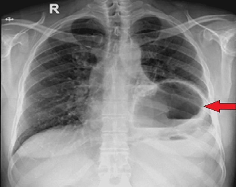
This diagnostic image is an anteroposterior (AP) chest X-ray demonstrating a significant abnormality in the left hemithorax. The primary finding is a large, well-defined cystic structure or gas-filled viscus containing a prominent horizontal air-fluid level, highlighted by a red arrow. This structure occupies a substantial portion of the left lower and mid-thorax, resulting in the compression of the adjacent left lung parenchyma and potential obscuration of the left hemidiaphragm silhouette. A slight rightward mediastinal shift may be present due to the mass effect of the lesion. While the right lung appears relatively clear, the radiographic appearance in the left hemithorax is highly suggestive of a large diaphragmatic hernia (such as a hiatal hernia) with gastric herniation and subsequent gastric outlet obstruction, or potentially a large lung abscess or infected bulla. The clinical context provided confirms this as a diaphragmatic hernia with herniation of the stomach into the thoracic cavity.
Lung Abscess
Definition
Etiology and Pathogenesis
- Altered consciousness (alcoholism, drug overdose, seizures, anesthesia, coma)
- Bulbar/neuromuscular dysfunction, prior stroke
- Esophageal dysmotility, strictures, tumors, or reflux
- Poor dentition/periodontal disease - lung abscess is rare in edentulous patients in the absence of an obstructing lesion (Harrison's, p. 842-843; Robbins Basic Pathology, p. 1127)
- Bronchial obstruction (tumor, foreign body) causing impaired drainage and distal necrosis
- Necrotizing pneumonia (S. aureus, Klebsiella pneumoniae, Pseudomonas, S. pyogenes)
- Septic emboli (right-sided endocarditis, Lemierre's syndrome from Fusobacterium necrophorum)
- Hematogenous spread (staphylococcal bacteremia → often multiple abscesses)
- Immunocompromise (HIV, transplant) with unusual organisms - Nocardia, Aspergillus, Mucorales, Cryptococcus, Rhodococcus equi, Pneumocystis (Sabiston, p. 2166; Harrison's Table 132-1)
Clinical Manifestations
- Initially resembles pneumonia: fever, cough, purulent sputum, chest pain, dyspnea
- Anaerobic abscesses often have an indolent course over weeks: night sweats, weight loss, fatigue, anemia
- Foul-smelling/foul-tasting ("putrid") sputum in anaerobic disease
- S. aureus and gram-negative abscesses tend to be more fulminant with high fevers
- Exam: fever, poor dentition/gingival disease, amphoric or cavernous breath sounds, digital clubbing, absent gag reflex (Harrison's, p. 908-910)
Diagnosis
- Imaging is central: chest radiograph or CT shows a cavity, often with an air-fluid level and a thick, irregular, enhancing wall on contrast CT - this enhancing wall helps distinguish abscess from empyema or necrotizing pneumonia.
- Sputum Gram stain/culture; blood cultures if systemic toxicity. Anaerobic cultures require special technique and are often not pursued given limited impact on management.
- Differential diagnosis: lung infarction, cavitary malignancy, sequestration, organizing pneumonia, granulomatosis with polyangiitis, infected bullae/cysts, and septic emboli (Harrison's, p. 913-915).
Treatment
- Antibiotics are first-line for most cases:
- Historically clindamycin or penicillin; now typically a beta-lactam/beta-lactamase inhibitor (e.g., ampicillin-sulbactam) or a carbapenem, which cover anaerobes and streptococci well.
- Duration is guided by clinical/radiographic resolution - typically continued until fever, sputum production, and radiographic fluid collections resolve or stabilize, often 2-3 weeks or longer, sometimes months for large or chronic cavities.
- For secondary abscesses, tailor coverage to the specific pathogen (e.g., anti-pseudomonal agents, antifungals) (Fishman's Pulmonary Diseases, p. 1165).
- Postural drainage / chest physiotherapy may assist clearance, though evidence is limited.
- Drainage procedures for abscesses that fail to respond to antibiotics (persistent sepsis, large cavity, inability to drain via the airway):
- Percutaneous transthoracic catheter drainage
- Endobronchial/endoscopic drainage
- A 2025 Bayesian network meta-analysis (PMID: 41585276) compared drainage methods for lung abscess and may offer updated comparative-effectiveness data on percutaneous vs. endobronchial approaches - worth reviewing if considering procedural drainage, as this is more recent than the textbook coverage.
- Surgery (lobectomy/segmentectomy) is reserved for abscesses refractory to medical therapy, massive hemoptysis, suspected malignancy, or when a bronchopleural fistula/empyema complicates the picture (Sabiston/Schwartz's, general surgery texts).