Case study Topic pneumonia Q1. What is the most likely diagnosis? Q2.what are the differential diagnosis? Q3 . What are the treatment and management?

Asking for Preferences
Reading File
Finding Sources
Finding Sources
Reading File
Reading File
Finding Sources
Reading File
Finding Sources
Searching Images

community acquired pneumonia chest X-ray consolidation

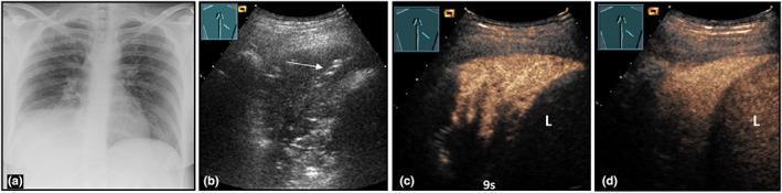
This composite figure illustrates the multimodal diagnostic imaging of community-acquired pneumonia in a 36-year-old female. (a) A posteroanterior chest X-ray reveals a dense, right-sided lower lobe consolidation. (b) B-mode lung ultrasound (LUS) demonstrates a corresponding hypoechoic peripheral consolidation featuring hyperechoic linear structures consistent with air bronchograms (indicated by an arrow). (c) Contrast-enhanced ultrasound (CEUS) at 9 seconds shows early pulmonary arterial perfusion within the lesion. (d) At 2 minutes, the lesion exhibits marked, homogeneous enhancement, reaching an intensity similar to the adjacent liver (L), which serves as a vascular reference. The progression from early arterial perfusion to late homogeneous enhancement is a hallmark of uncomplicated lobar pneumonia. This series demonstrates how CEUS adds functional perfusion data to the structural findings of B-mode LUS, assisting in the differentiation between pneumonic consolidation and other pulmonary pathologies like abscesses or infarcts.

This composite figure illustrates the multimodal diagnostic imaging of community-acquired pneumonia in a 36-year-old female. (a) A posteroanterior chest X-ray reveals a dense, right-sided lower lobe consolidation. (b) B-mode lung ultrasound (LUS) demonstrates a corresponding hypoechoic peripheral consolidation featuring hyperechoic linear structures consistent with air bronchograms (indicated by an arrow). (c) Contrast-enhanced ultrasound (CEUS) at 9 seconds shows early pulmonary arterial perfusion within the lesion. (d) At 2 minutes, the lesion exhibits marked, homogeneous enhancement, reaching an intensity similar to the adjacent liver (L), which serves as a vascular reference. The progression from early arterial perfusion to late homogeneous enhancement is a hallmark of uncomplicated lobar pneumonia. This series demonstrates how CEUS adds functional perfusion data to the structural findings of B-mode LUS, assisting in the differentiation between pneumonic consolidation and other pulmonary pathologies like abscesses or infarcts.

Multi-modal diagnostic imaging of community-acquired pneumonia in a 37-year-old female. Panel (a) is a posterior-anterior chest X-ray showing right-sided basal consolidation. Panel (b) is a B-mode Lung Ultrasound (LUS) demonstrating an inhomogeneous, hypoechoic consolidation with visible air bronchograms (arrows). Panels (c), (d), and (e) present a Contrast-Enhanced Ultrasound (CEUS) time-series. At 3 seconds (c), the consolidation shows an inhomogeneous pulmonary arterial enhancement pattern with a significant non-perfused area (NPA), representing a primary absence of pulmonary arterial perfusion. At 9 seconds (d), the consolidation displays delayed secondary bronchial arterial perfusion within the previously non-perfused tissue. At 3 minutes (late phase, e), the area remains demarcated as a hypoechoic region (arrows). The sequence illustrates the hemodynamic differentiation between pulmonary and bronchial arterial blood supply in pneumonic lung tissue, highlighting the utility of CEUS in evaluating complex perfusion disturbances in pulmonary pathology.

Multi-modal diagnostic imaging of community-acquired pneumonia in a 37-year-old female. Panel (a) is a posterior-anterior chest X-ray showing right-sided basal consolidation. Panel (b) is a B-mode Lung Ultrasound (LUS) demonstrating an inhomogeneous, hypoechoic consolidation with visible air bronchograms (arrows). Panels (c), (d), and (e) present a Contrast-Enhanced Ultrasound (CEUS) time-series. At 3 seconds (c), the consolidation shows an inhomogeneous pulmonary arterial enhancement pattern with a significant non-perfused area (NPA), representing a primary absence of pulmonary arterial perfusion. At 9 seconds (d), the consolidation displays delayed secondary bronchial arterial perfusion within the previously non-perfused tissue. At 3 minutes (late phase, e), the area remains demarcated as a hypoechoic region (arrows). The sequence illustrates the hemodynamic differentiation between pulmonary and bronchial arterial blood supply in pneumonic lung tissue, highlighting the utility of CEUS in evaluating complex perfusion disturbances in pulmonary pathology.

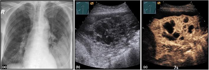
This composite figure illustrates a case of community-acquired abscessed pneumonia using three imaging modalities. Panel (a) is a posterior-anterior chest X-ray showing a low-field consolidation in the left lung. Panel (b) presents a B-mode Lung Ultrasound (LUS) of the same region, demonstrating a complex, hypoechoic consolidation. Within this consolidation, central anechoic areas are visible, representing confluent regions of necrosis or early abscess formation. Panel (c) displays a Contrast-Enhanced Ultrasound (CEUS) image at 7 seconds (pulmonary arterial phase). It reveals an inhomogeneous enhancement pattern throughout the consolidated lung tissue. Notably, there are multiple well-defined anechoic zones characterized by a total absence of enhancement, which confirms the presence of avascular abscess cavities. This multi-modal approach highlights the utility of CEUS in differentiating simple pneumonia from complicated, necrotizing, or abscessed pneumonia by visualizing microvascular perfusion and identifying non-perfused necrotic tissue.

This composite figure illustrates a case of community-acquired abscessed pneumonia using three imaging modalities. Panel (a) is a posterior-anterior chest X-ray showing a low-field consolidation in the left lung. Panel (b) presents a B-mode Lung Ultrasound (LUS) of the same region, demonstrating a complex, hypoechoic consolidation. Within this consolidation, central anechoic areas are visible, representing confluent regions of necrosis or early abscess formation. Panel (c) displays a Contrast-Enhanced Ultrasound (CEUS) image at 7 seconds (pulmonary arterial phase). It reveals an inhomogeneous enhancement pattern throughout the consolidated lung tissue. Notably, there are multiple well-defined anechoic zones characterized by a total absence of enhancement, which confirms the presence of avascular abscess cavities. This multi-modal approach highlights the utility of CEUS in differentiating simple pneumonia from complicated, necrotizing, or abscessed pneumonia by visualizing microvascular perfusion and identifying non-perfused necrotic tissue.

Reading File
Finding Sources
Reading File
Reading File
Reading File
Reading File
I now have comprehensive information. Let me compile the full answer.

Pneumonia - Case Study Answer

Note: No specific case details were provided, so this uses a classic Community-Acquired Pneumonia (CAP) presentation - a standard clinical scenario used in medical education. The framework applies to any pneumonia case.

Illustrative Case Scenario

Patient: 55-year-old male, smoker, with a 3-day history of:
  • Sudden-onset fever (39.2°C) and rigors
  • Productive cough with rusty/purulent sputum
  • Right-sided pleuritic chest pain
  • Dyspnea
  • On examination: dullness to percussion, bronchial breath sounds, and egophony over the right lower lobe
  • CXR: right lower lobe consolidation
  • WBC: 18,000/mm³ with neutrophilia
  • SpO2: 94% on room air

Q1. Most Likely Diagnosis

Community-Acquired Pneumonia (CAP) - Lobar (Bacterial) Pneumonia

Most likely causative organism: Streptococcus pneumoniae (pneumococcal pneumonia)
Diagnostic basis:
FeatureClassic Bacterial (Pneumococcal) Pneumonia
OnsetAbrupt, acute
FeverHigh-grade (>38.5°C) with rigors
CoughProductive, rusty/blood-tinged sputum
Chest painPleuritic (peripheral, localized)
WBCElevated (>15,000/mm³), neutrophilia
CXRLobar consolidation, alveolar infiltrates
Physical examDullness to percussion, bronchial breath sounds, egophony
According to the Textbook of Family Medicine 9e: "A rapid onset of cough and shortness of breath with a high fever can indicate classic bacterial lobar pneumonia such as that produced by a pneumococcus. Physical findings after consolidation occurs include decreased breath sounds, dullness to percussion, and egophony on the affected side."
Chest imaging - what you would see:
CXR showing right lower lobe consolidation with air bronchograms in community-acquired pneumonia
Right-sided basal consolidation with air bronchograms on chest X-ray and lung ultrasound in a case of community-acquired pneumonia
Key diagnostic steps:
  1. Clinical assessment - history + physical examination
  2. Chest X-ray - lobar or segmental consolidation (may lag 1-2 days behind clinical course)
  3. Blood tests - CBC (elevated WBC + neutrophilia), CRP, LFT, renal function
  4. Sputum Gram stain and culture (low yield but useful when positive)
  5. Blood cultures - mandatory before antibiotics for hospitalized patients
  6. Urine antigen tests - for Legionella and S. pneumoniae
  7. Cold agglutinin test - positive in Mycoplasma pneumoniae

Q2. Differential Diagnoses

The following conditions can mimic pneumonia and must be considered:

Primary Pulmonary Differentials

DiagnosisDistinguishing Features
Atypical Pneumonia (Mycoplasma, Chlamydia, Legionella)Gradual onset, low-grade fever, dry/nonproductive cough, normal or mildly elevated WBC, bilateral patchy interstitial pattern on CXR
Pulmonary TuberculosisChronic onset (>2 weeks), night sweats, weight loss, upper lobe infiltrates, cavitation, AFB smear +ve
Lung AbscessFoul-smelling purulent sputum, cavitating lesion on CXR, history of aspiration/alcoholism
Aspiration Pneumonitis/PneumoniaHistory of impaired consciousness, dysphagia, right lower or upper lobe infiltrates
Viral Pneumonia (Influenza, COVID-19)Bilateral patchy/interstitial infiltrates, myalgia, headache, preceding URTI

Cardiovascular / Other Important Differentials

DiagnosisDistinguishing Features
Pulmonary EmbolismSudden pleuritic chest pain + dyspnea, raised D-dimer, risk factors (DVT, immobility), CTPA confirms
Acute Heart Failure (Pulmonary Edema)Bilateral basal crepitations, raised JVP, cardiomegaly, bilateral infiltrates ("bat-wing" pattern), BNP elevated
COPD ExacerbationBackground COPD, hyperinflation, no consolidation
Lung Cancer with Post-obstructive PneumoniaRecurrent pneumonia same site, weight loss, haemoptysis, hilar mass
Cryptogenic Organizing Pneumonia (COP)Migratory infiltrates, subacute onset, poor response to antibiotics
Hypersensitivity PneumonitisOccupational/environmental antigen exposure, bilateral micronodular pattern
As noted in the Textbook of Family Medicine 9e: "Pulmonary thromboembolism (arguably the most common cause of 'atypical pneumonia'), congestive heart failure...hypersensitivity pneumonitis...cryptogenic organizing pneumonia...idiopathic acute eosinophilic pneumonia...foreign body aspiration, connective tissue diseases..." all enter the differential.

Q3. Treatment and Management

Step 1 - Assess Severity (CURB-65 Score)

Use the CURB-65 tool to decide site of care (Harrison's Principles of Internal Medicine 22E):
ParameterScore
C - Confusion (new disorientation)1
U - Urea >7 mmol/L (BUN >20 mg/dL)1
R - Respiratory rate ≥30/min1
B - Blood pressure: systolic ≤90 or diastolic ≤60 mmHg1
65 - Age ≥65 years1
Interpretation:
  • Score 0 - Outpatient treatment (30-day mortality ~1.5%)
  • Score 1-2 - Consider hospitalization
  • Score ≥3 - Hospital admission; consider ICU (mortality ~22%)
ICU admission required if:
  • Septic shock needing vasopressors
  • Respiratory failure needing mechanical ventilation
  • ≥3 of the minor criteria: RR ≥30, PaO2/FiO2 ≤250, multilobar infiltrates, confusion, BUN ≥20 mg/dL, WBC <4000, platelets <100,000, hypothermia, hypotension needing fluids

Step 2 - Empirical Antibiotic Therapy

Based on ATS/IDSA guidelines:

A. Outpatient Treatment (No Comorbidities)

  • Amoxicillin 1g PO three times daily, OR
  • Doxycycline 100mg PO twice daily, OR
  • Azithromycin 500mg day 1, then 250mg daily (if macrolide resistance <25%)

B. Outpatient Treatment (With Comorbidities - diabetes, cardiac, liver, renal disease, immunosuppression, or recent antibiotic use)

  • Respiratory fluoroquinolone - Levofloxacin 750mg OD or Moxifloxacin 400mg OD, OR
  • Beta-lactam + macrolide combination - Amoxicillin-clavulanate + Azithromycin

C. Inpatient (Non-ICU)

  • Beta-lactam (Cefotaxime or Ceftriaxone) + Azithromycin, OR
  • Respiratory fluoroquinolone monotherapy (Levofloxacin or Moxifloxacin)

D. Inpatient ICU (Severe CAP)

  • Beta-lactam (Cefotaxime/Ceftriaxone) + Azithromycin, OR
  • Beta-lactam + respiratory fluoroquinolone
  • If MRSA suspected: Add Vancomycin or Linezolid
  • If Pseudomonas suspected (bronchiectasis, recent hospitalization): Piperacillin-tazobactam or Cefepime + fluoroquinolone
Duration of treatment:
  • Minimum 5 days and at least 48-72 hours beyond last fever and clinical instability (Textbook of Family Medicine 9e)
  • Most uncomplicated CAP: 5-7 days

Step 3 - Supportive Management

MeasureDetails
Oxygen therapyTarget SpO2 ≥94%; high-flow O2 or NIV if needed
IV fluidsFor dehydration or sepsis
Antipyretics/AnalgesicsParacetamol for fever and pleuritic pain
PhysiotherapyDeep breathing exercises, chest physiotherapy, postural drainage
DVT prophylaxisFor hospitalized patients (LMWH + compression stockings)
Nutritional supportEarly enteral nutrition if intubated
Early switch to oralWhen clinically stable, switch IV to oral antibiotics to reduce hospital stay

Step 4 - Prevention

  • Influenza vaccine annually - all persons >6 months, especially >50 years, chronic disease patients, and healthcare workers
  • Pneumococcal vaccine (PCV13/PPSV23) - one-time for adults >65 years; re-vaccinate after 10 years if first vaccine was given before age 65
  • Smoking cessation
  • Good hand hygiene
(Textbook of Family Medicine 9e: "Approximately half of all cases of adult pneumonia can be prevented by annual administration of influenza vaccine plus a one-time pneumococcal vaccine.")

Quick Summary Table

AspectKey Points
Most likely DxLobar bacterial CAP - Streptococcus pneumoniae
DifferentialsAtypical pneumonia, PE, heart failure, TB, viral pneumonia, lung abscess, COP
Severity toolCURB-65 (score ≥2 = admit; ≥3 = ICU)
Outpatient RxAmoxicillin or Doxycycline
Inpatient RxBeta-lactam + Macrolide or Respiratory Fluoroquinolone
ICU RxBeta-lactam + Macrolide ± Vancomycin (if MRSA risk)
DurationMin 5 days, 48-72h afebrile
PreventionInfluenza + Pneumococcal vaccines
Sources: Harrison's Principles of Internal Medicine 22E, Textbook of Family Medicine 9e, Fishman's Pulmonary Diseases and Disorders

Case study Topic pneumonia Q1. What is the most likely diagnosis? Q2.what are the differential diagnosis? Q3 . What are the treatment? In short notes

Asking for Preferences
Using a standard CAP case. Here is the concise short-note format answer:

Pneumonia - Case Study (Short Notes)

Standard Case: 52-year-old male, 4-day history of sudden fever (39°C), rigors, productive cough with rusty sputum, right-sided pleuritic chest pain, dyspnea. O/E: dullness to percussion + bronchial breathing right lower lobe. CXR: right lower lobe consolidation. WBC 18,000/mm³ (neutrophilia). SpO2 94%.

Q1. Most Likely Diagnosis

Community-Acquired Pneumonia (CAP) - Right Lower Lobe Lobar Pneumonia Causative organism: Streptococcus pneumoniae (most common)
Basis:
FeatureFinding
OnsetAcute/sudden
FeverHigh-grade + rigors
SputumRusty/purulent (classic for pneumococcus)
Chest painPleuritic (peripheral)
ExaminationDullness, bronchial breath sounds, egophony
CXRLobar consolidation
WBCElevated with neutrophilia

Q2. Differential Diagnoses

ConditionKey Distinguishing Feature
Atypical pneumonia (Mycoplasma, Chlamydia, Legionella)Gradual onset, dry cough, bilateral patchy infiltrates, normal WBC
Pulmonary TuberculosisChronic course, night sweats, upper lobe cavitation, AFB +ve
Pulmonary EmbolismSudden dyspnea, raised D-dimer, normal/wedge-shaped CXR, risk factors (DVT, immobility)
Acute Pulmonary EdemaBilateral basal crackles, raised JVP, cardiomegaly, bat-wing CXR, elevated BNP
Lung AbscessFoul sputum, cavitation on CXR, predisposing aspiration history
Viral Pneumonia (Influenza, COVID-19)Bilateral interstitial infiltrates, myalgia, preceding URTI
Lung Cancer + Post-obstructive PneumoniaRecurrent same-site pneumonia, haemoptysis, weight loss, hilar mass
Cryptogenic Organizing Pneumonia (COP)Migratory infiltrates, no response to antibiotics, requires steroids

Q3. Treatment (Short Notes)

A. Assess Severity - CURB-65

Confusion + Urea >7 mmol/L + RR ≥30 + BP <90/60 + Age ≥65
  • Score 0 → Outpatient
  • Score 1-2 → Hospitalize
  • Score ≥3 → Consider ICU

B. Antibiotics (Empirical - ATS/IDSA Guidelines)

Outpatient (mild, no comorbidities):
  • Amoxicillin 1g TDS, OR
  • Doxycycline 100mg BD, OR
  • Azithromycin 500mg OD
Outpatient (with comorbidities / recent antibiotics):
  • Respiratory fluoroquinolone (Levofloxacin 750mg OD or Moxifloxacin 400mg OD), OR
  • Amoxicillin-clavulanate + Azithromycin
Inpatient (non-ICU):
  • Ceftriaxone + Azithromycin, OR
  • Levofloxacin/Moxifloxacin monotherapy
ICU (severe CAP):
  • Ceftriaxone + Azithromycin
  • Add Vancomycin if MRSA suspected
  • Add Piperacillin-tazobactam if Pseudomonas suspected
Duration: Minimum 5 days, at least 48-72 hours after last fever

C. Supportive Management

  • O2 therapy - Target SpO2 ≥94%
  • IV fluids - For dehydration/sepsis
  • Antipyretics - Paracetamol 1g TDS
  • Analgesia - For pleuritic pain (NSAIDs or paracetamol)
  • Chest physiotherapy - Deep breathing, postural drainage
  • DVT prophylaxis - LMWH + compression stockings (hospitalized patients)
  • Early switch - IV → oral antibiotics once afebrile and tolerating orally

D. Prevention

  • Annual Influenza vaccine
  • Pneumococcal vaccine (PCV13/PPSV23) - age >65 or chronic illness
  • Smoking cessation

Sources: Harrison's Principles of Internal Medicine 22E; Textbook of Family Medicine 9e; ATS/IDSA CAP Guidelines
This is a shared conversation. Sign in to Orris to start your own chat.