
Dermatomes
Reading File
This is a broad/visual topic. Let me search headings and paragraphs in parallel.
Finding Sources
Finding Sources
The THIEME Atlas of Anatomy has the most dedicated dermatome section. Let me read it along with the rheumatology sensory content and look for images.
Reading File
Reading File
Now let me read the dermatome map section and fetch the key images.
Reading File
Loading Image
Now let me fetch the main dermatome map image and the lower limb cutaneous innervation image in parallel.
Loading Image
Loading Image
Loading Image
Excellent - I have the full dermatome map (anterior + posterior views) and the lower limb diagram. Now I have everything needed to give a complete, well-illustrated answer.
Dermatomes
A dermatome is the area of skin supplied by the sensory fibers of a single posterior (dorsal) spinal nerve root. The term comes from the embryonic dermatome - the part of each somite that migrates to form the skin, carrying its segmental innervation along with it.
Core Anatomy
Sensory (afferent) fibers travel from the skin through peripheral nerves, converge in a plexus, and ultimately enter the spinal cord through a single dorsal root. The dorsal root ganglion houses the cell bodies. Because C1 is purely motor, it has no corresponding dermatome - sensory coverage of the body begins at C2.
On the trunk, the segmental arrangement is simple and roughly horizontal (T2-T12 form bands across the chest and abdomen). In the limbs, embryonic migration and plexus formation cause the dermatomes to spiral and interdigitate, making limb dermatomal maps less straightforward.
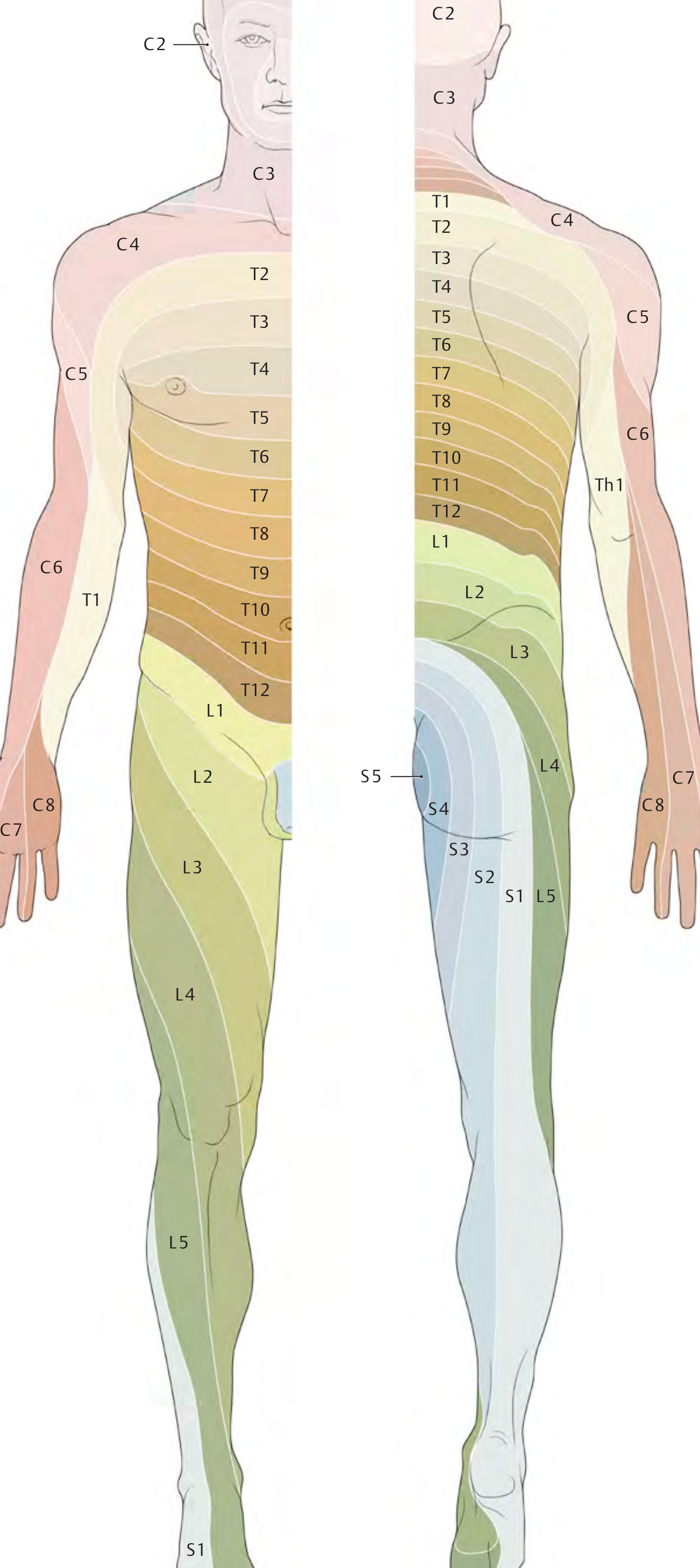
Radicular (segmental) dermatome map - anterior view (left) and posterior view (right). Note the horizontal banding of thoracic segments on the trunk vs. the spiraling pattern in the limbs. - THIEME Atlas of General Anatomy & Musculoskeletal System, p. 107
Key Clinical Dermatomes at a Glance
| Region | Landmark Dermatome(s) |
|---|---|
| Top of head / scalp | C2 |
| Neck | C3 |
| Shoulder / clavicle | C4 |
| Lateral arm | C5 |
| Thumb / lateral forearm | C6 |
| Middle finger / dorsal hand | C7 |
| Little finger / medial forearm | C8 |
| Medial arm / axilla | T1 |
| Nipple line | T4 |
| Umbilicus | T10 |
| Inguinal ligament | L1 |
| Anterior thigh | L2-L3 |
| Anterior knee / medial shin | L4 |
| Lateral calf / dorsum of foot / great toe | L5 |
| Lateral foot / sole / posterior calf | S1 |
| Perineum / saddle area | S2-S4 |
Dermatome vs. Peripheral Nerve Territory
This is a critical clinical distinction:
- Radicular lesion (e.g., herniated disc, shingles/herpes zoster) - sensory loss follows the dermatome
- Peripheral nerve lesion (e.g., trauma, entrapment) - sensory loss follows the autonomous territory of that peripheral nerve, which differs significantly from the dermatome
Because adjacent dermatomes broadly overlap, the clinically detectable area of sensory loss from a single root lesion is always smaller than the full anatomical dermatome. The autonomous area of a cutaneous nerve is the region it alone supplies, with no overlap from adjacent segments.
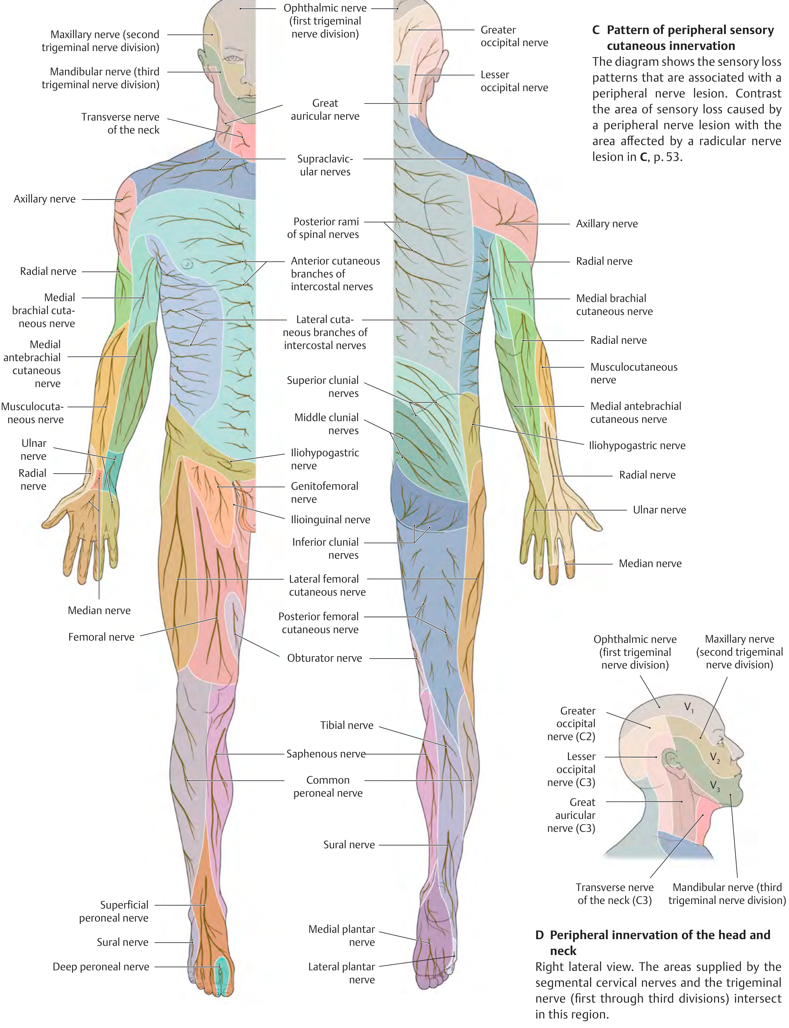
Peripheral nerve territories (named nerves) vs. radicular dermatomes. Compare with the segmental map above to understand why the clinical deficit differs by lesion level. - THIEME Atlas, p. 108
Lower Limb in Detail
The lower limb dermatomes are especially tested clinically for lumbar disc pathology:
- L4 - anterior thigh, anterior knee, pretibial region of the shin, medial foot
- L5 - buttock, lateral thigh, anterolateral calf, dorsal foot, great toe
- S1 - buttock, posterior thigh, posterior calf, lateral foot
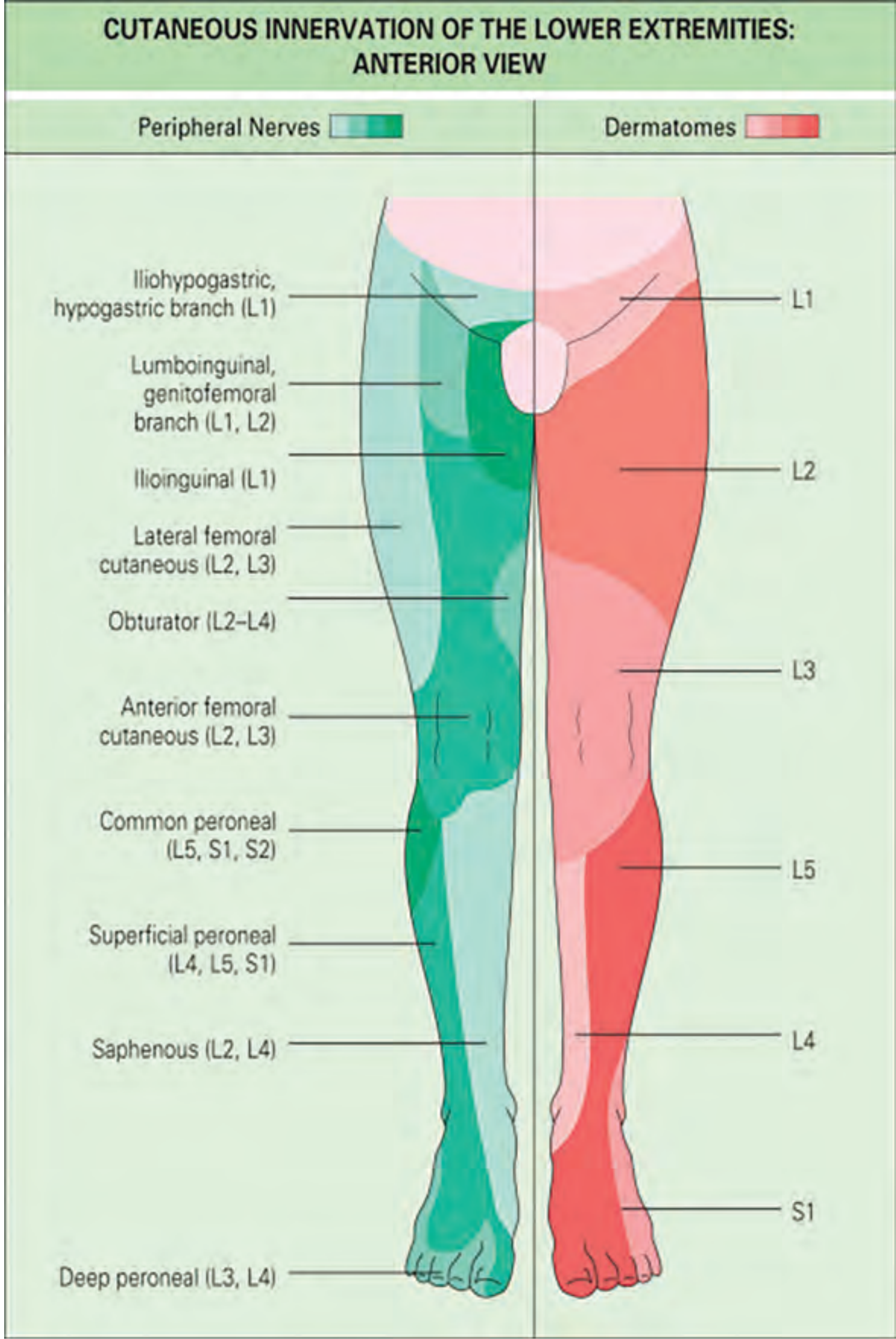
Left side = peripheral nerve territories; right side = dermatomal territories (L1-S1). Note how a single peripheral nerve (e.g. saphenous = L2-L4) spans multiple dermatomes. - Rheumatology 2-Volume Set (Elsevier 2022), p. Fig. 78.2
Plexus Formation and Why Limb Dermatomes Spiral
During embryonic development, somites split into sclerotome, myotome, and dermatome components. As limb buds develop, dermatome primordia migrate and carry their spinal cord segment's axons with them. This is why:
- A single peripheral nerve in the limb contains fibers from multiple spinal roots
- A single dermatome is served by multiple peripheral nerves
- The dermatome spiral in the limbs follows the embryonic migration path
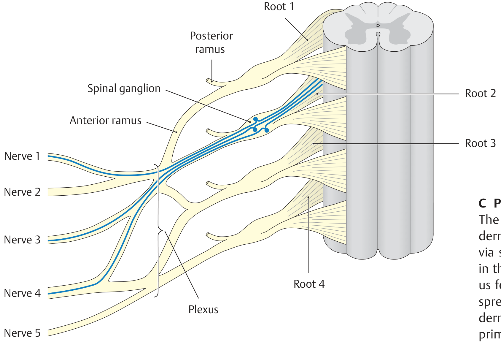
One spinal root (blue fibers) sends afferents via multiple peripheral nerves to a single dermatome. Damage to one nerve = partial loss; damage to the root = full dermatomal deficit. - THIEME Atlas, p. 105
Head: Trigeminal Override
The face and anterior scalp are NOT supplied by cervical roots but by cranial nerve V (trigeminal nerve):
- V1 (ophthalmic) - forehead, cornea, nose bridge
- V2 (maxillary) - cheeks, upper lip, lateral nose
- V3 (mandibular) - jaw, lower lip, chin, temporomandibular area
A central lesion of the trigeminal sensory nucleus produces an "onion-skin" pattern of concentric sensory loss around the mouth (Solder lines), whereas a peripheral V branch lesion produces a divisional pattern.
Clinical Applications
| Scenario | Affected Level |
|---|---|
| Disc herniation causing numbness in great toe | L5 root |
| Loss of Achilles reflex + lateral foot numbness | S1 root |
| Weakness of foot dorsiflexion ("foot drop") | L4-L5 |
| Herpes zoster in a band across the chest | Single thoracic dermatome |
| Saddle anesthesia (perineum) | S2-S4 - cauda equina emergency |
| Umbilical pain referred from appendix (early) | T10 dermatome |
Sources:
- THIEME Atlas of General Anatomy and Musculoskeletal System, pp. 105-108
- Rheumatology, 2-Volume Set (Elsevier 2022), p. sensory examination section
- Langman's Medical Embryology - somite differentiation section
This is a shared conversation. Sign in to Orris to start your own chat.