
Asymptomatic proteinuria workup and management
"proteinuria" AND "evaluation" AND "management"
evaluation of isolated asymptomatic proteinuria flowchart algorithm
Summary : This flowchart provides a comprehensive decision-making algorithm for the evaluation and management of solid focal liver lesions detected on ultrasound or non-diagnostic imaging, including initial workup, imaging choices, diagnosis, and subsequent management pathways for various lesion types and patient scenarios. flowchart: # Nodes : • Start (rectangle): "Focal Solid Liver Lesion on Ultrasound or Nondiagnostic Imaging" • Decision (parallelogram): "Evaluate history, physical, laboratory workup" • Decision (diamond): "If cirrhosis present, refer to AASLD guidance on hepatocellular carcinoma" • Branch 1 (rectangle): "Patient <40 years of age with unremarkable PMH, exam, labs" • Branch 2 (rectangle): "All other patients" • Branch 3 (rectangle): "History of malignancy" • Imaging (rectangle): "MRI with hepatobiliary imaging" • Imaging (rectangle): "Triple phase contrast enhanced CT OR Dynamic MRI with hepatobiliary contrast OR Dynamic MRI with extracellular contrast" • Guidance (rectangle): "See appropriate guidance for follow-up based on known type of prior malignancy or multidisciplinary discussion for further management recommendations" • Diagnosis (rectangle): "Diagnosis made" • No Diagnosis (rectangle): "Diagnosis not made" • Multidisciplinary (rectangle): "Multidisciplinary discussion" • Surveillance (rectangle, yellow): "Surveillance imaging, usually 3-6 months" • Lesion types (rectangles): "Adenoma*", "Focal Nodular Hyperplasia", "Hemangioma", "Hepatic hemangioendothelioma", "Fibrolamellar Hepatocellular Carcinoma", "Angiosarcoma" • Management (rectangles): "Stop OCPs or hormone impregnated IUDs, weight loss if appropriate", "No follow up needed", "Consider resection. If unresectable, transarterial embolization", "Asymptomatic", "Symptomatic (i.e. pain)", "Resect or alternative therapy (RF/AMWA/EBRT/TAE)", "Resect or transplant, even if extrahepatic disease is present. Alternative: ablation, SBRT", "Resect or transplant. Alternative: radiotherapy, chemotherapy, immunotherapy", "Resect; consider adjuvant chemo/XRT" • Evaluation (rectangle): "Evaluate MRI features to determine subtype & consider biopsy if concern for beta catenin mutation" • Gender branches (rectangles): "Men", "Women" • Biopsy/Resection (rectangles): "NO biopsy needed. Resect regardless of size OR treat definitively with alternative method of treatment if not resectable", "Greater than 5 cm", "Stop hormonal therapy", "Observe 6–12 mos for regression", "Remains >5 cm", "Resect", "Shrink to <5 cm", "Monitor with contrast enhanced MRI every 6 mos", "Less than 5 cm", "If concern for beta catenin", "Biopsy", "If positive resect", "If negative", "Surveillance imaging every 6 mos, for 2 yrs, then annual", "If growth >20% resect", "Multiple adenomas", "Unilobar", "Resect", "Multilobar", "Focus on treatment of dominant lesion", "In pregnancy", "Ultrasound q 6-12 weeks", "No growth <6.5 cm, US, continue US follow-up", "If growth >6.5 cm or exophytic", "Resect if early in pregnancy OR Embolize if >26 weeks OR Consider resection if third trimester" • Footnotes (rectangles): "*See text for special circumstances, e.g. adenomas, rupture, malignant transformation", "*Note that most FNH lesions are incidental & asymptomatic; see text for further discussion" # Connectors : • Arrows connect nodes in a top-down, left-to-right branching structure. • Initial decision splits into three branches: age <40, all other patients, history of malignancy. • Each branch leads to specific imaging recommendations. • Imaging leads to diagnosis or no diagnosis; if no diagnosis, multidisciplinary discussion and surveillance imaging. • Diagnosis leads to lesion-specific management pathways (adenoma, FNH, hemangioma, etc.). • Adenoma management splits by gender, lesion size, and pregnancy status, with further branches for biopsy, resection, observation, and surveillance. • FNH and hemangioma management split by symptoms (asymptomatic/symptomatic). • Malignant lesions (hemangioendothelioma, fibrolamellar HCC, angiosarcoma) direct to resection, transplant, or adjuvant therapies. # Layout : • Flowchart is organized in a vertical, hierarchical structure with three main branches at the top. • Each branch subdivides into further diagnostic and management pathways. • Color coding: blue for standard nodes, yellow for surveillance imaging. • Decision diamonds and rectangles are used for choices and actions. • Footnotes and special circumstances are indicated with asterisks and referenced to the text. # Analysis : • The flowchart provides a systematic approach for evaluating solid focal liver lesions, integrating patient age, history, and risk factors to guide imaging and management. • It distinguishes between benign and malignant lesions, with tailored recommendations for each, including observation, resection, and advanced therapies. • Surveillance intervals and criteria for intervention are clearly defined, especially for adenomas and lesions in pregnancy. • The algorithm emphasizes multidisciplinary discussion for indeterminate cases and incorporates gender-specific management for adenomas. • Overall, the flowchart enables clinicians to follow a stepwise, evidence-based pathway for liver lesion evaluation and treatment.
Summary : This flowchart presents an algorithm for the management of duplex system ureteroceles (DSU) in infants after the first 3–6 months of life, outlining diagnostic and treatment pathways based on symptoms, presence of hydronephrosis/obstruction, vesicoureteric reflux (VUR), and renal function. flowchart: # Nodes : • Start (rectangle): DSU • Decision (rectangle): Asymptomatic, No severe HUN or obstruction • Decision (rectangle): Symptomatic or severe HUN or obstruction • Decision (rectangle): No VUR (from Asymptomatic) • Decision (rectangle): VUR (from Asymptomatic) • Decision (rectangle): VUR (from Symptomatic) • Decision (rectangle): No VUR (from Symptomatic) • Decision (rectangle): Low grade (from VUR, Asymptomatic) • Decision (rectangle): High grade or multiple infections (from VUR, Asymptomatic) • Decision (rectangle): Good function (from No VUR, Symptomatic) • Decision (rectangle): No/poor function (from No VUR, Symptomatic) • Action (rectangle, green): Observation • Action (rectangle, green): Bladder surgery or endoscopic management • Action (rectangle, green): Ectopic: upper to lower tract anastomosis • Action (rectangle, green): Intravesical endoscopic decompression • Action (rectangle, green): Ectopic UPPN # Connectors : • DSU splits into two main branches: Asymptomatic/No severe HUN or obstruction and Symptomatic/severe HUN or obstruction. • Asymptomatic branch splits into No VUR and VUR. • No VUR leads to Observation. • VUR splits into Low grade (leading to Observation) and High grade or multiple infections (leading to Bladder surgery or endoscopic management). • Symptomatic/severe HUN or obstruction splits into VUR and No VUR. • VUR leads to Ectopic: upper to lower tract anastomosis. • No VUR splits into Good function (leading to Intravesical endoscopic decompression) and No/poor function (leading to Ectopic UPPN). # Layout : • The flowchart is organized in a top-down manner, starting with DSU at the top, splitting into two main diagnostic branches, and further subdividing based on clinical findings and test results. • Treatment and follow-up actions are shown at the bottom in green rectangles. • Color coding: Diagnosis (light blue), Treatment (green), Follow-up (dark blue). # Analysis : • The algorithm prioritizes observation for asymptomatic cases without VUR or with low-grade VUR. • Surgical or endoscopic intervention is recommended for high-grade VUR, multiple infections, or symptomatic cases with obstruction. • The presence and grade of VUR, as well as renal function, are key determinants in management decisions. • The flowchart provides a clear, stepwise approach to guide clinicians in selecting appropriate management strategies for infants with DSU.
Summary : This flowchart presents an algorithm for the management of pregnant patients with atrial ectopy, outlining decision pathways based on symptom severity and recommended interventions. flowchart: # Nodes : • "Pregnant patient with atrial ectopy" (rectangle, top node) • "Premature atrial contractions" (rectangle, second row) • "Asymptomatic or tolerable symptoms" (rectangle, third row, left branch) • "Intolerable symptoms" (rectangle, third row, right branch) • "Reassurance with no intervention (COR 1)" (rounded rectangle, green, fourth row, left branch) • "Beta-blocker treatment, preferably metoprolol or propranolol (COR 1)" (rounded rectangle, green, fourth row, right branch) # Connectors : • Downward arrows connect each node to the next step. • After "Premature atrial contractions", the flow splits into two branches: left for "Asymptomatic or tolerable symptoms", right for "Intolerable symptoms". • Each symptom node leads to its respective intervention node. # Layout : • Vertical arrangement with a split into two parallel branches at the symptom assessment stage. • The final row contains two green recommendation nodes side by side. # Analysis : • The algorithm prioritizes symptom assessment in pregnant patients with atrial ectopy. • Asymptomatic or tolerable cases are managed conservatively with reassurance and no intervention. • Intolerable symptoms prompt pharmacological intervention, specifically recommending beta-blockers (metoprolol or propranolol). • Both recommendations are classified as COR 1, indicating strong support for these management strategies. • The flowchart provides a clear, stepwise approach for clinicians, emphasizing minimal intervention unless symptoms are significant.
Asymptomatic Proteinuria: Workup and Management
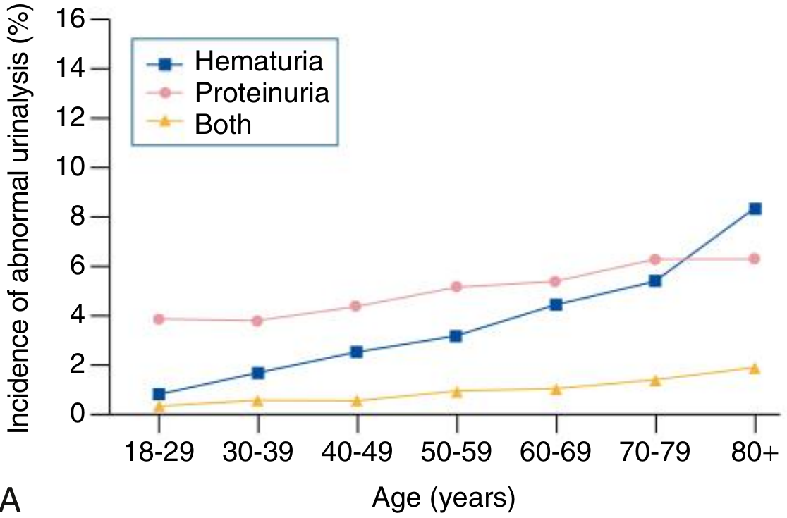
1. Prevalence and Significance
2. Normal Physiology
- Normal urine protein: <150 mg/day (mostly Tamm-Horsfall glycoprotein)
- GFR produces ~180 L of filtrate/day; the glomerular filtration barrier (endothelium + GBM + podocytes with slit diaphragms) restricts albumin (67 kDa, negatively charged) by size and charge selectivity
- Only ~0.01% of filtered protein appears in final urine - Brenner and Rector's The Kidney, p. 1002
3. Classification of Proteinuria
| Type | Mechanism | Key Feature |
|---|---|---|
| Functional (transient) | Hemodynamic - fever, exercise, heart failure, hyperadrenergic states | Resolves with cause; benign |
| Orthostatic | Positional - absent in recumbent urine | Only in upright position; <1 g/day |
| Tubular | Impaired reabsorption of filtered proteins (α₁-, β₂-microglobulin) | Low-grade (<2 g/day); suggests tubulointerstitial disease |
| Glomerular - fixed non-nephrotic | Glomerular barrier damage | 0.5-3.5 g/day; requires workup |
| Nephrotic-range | Severe glomerular injury | >3.5 g/day; associated with edema, hypoalbuminemia |
4. Workup - Step-by-Step Algorithm
Step 1: Confirm and Quantify Proteinuria
| Test | Threshold |
|---|---|
| Dipstick | ≥1+ if specific gravity ≤1.015; ≥2+ if specific gravity >1.015 |
| Urine protein:creatinine ratio (UPCr) | >0.2 in adults; >0.25 in children >2 yr |
| 24-hr urine protein | 30-300 mg/day = microalbuminuria; >300 mg = albuminuria; >3.5 g = nephrotic range |
| Albumin:creatinine ratio (ACR) | >30 mg/g warrants diagnostic evaluation |
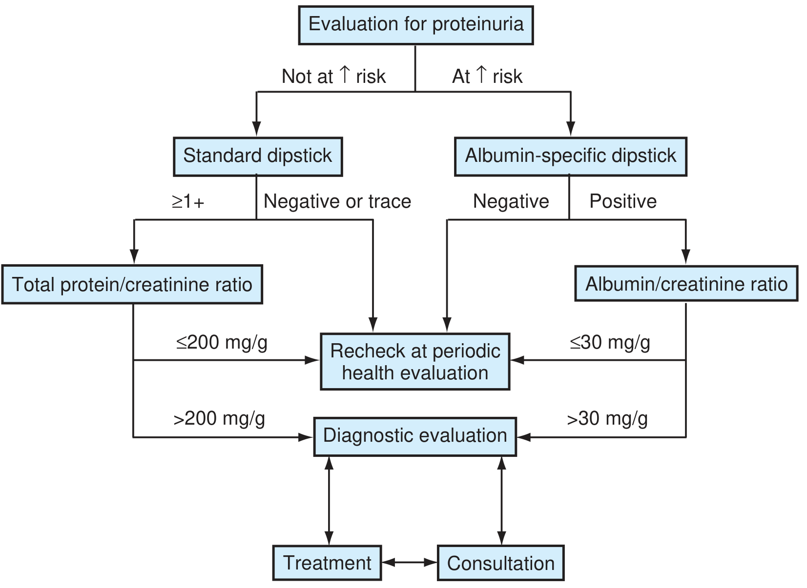
Step 2: Measure GFR and Test for Orthostatic Proteinuria
- Measure serum creatinine / eGFR
- Test for orthostatic proteinuria - collect first-morning urine before ambulation; if UPCr is normal in this sample, orthostatic proteinuria is confirmed
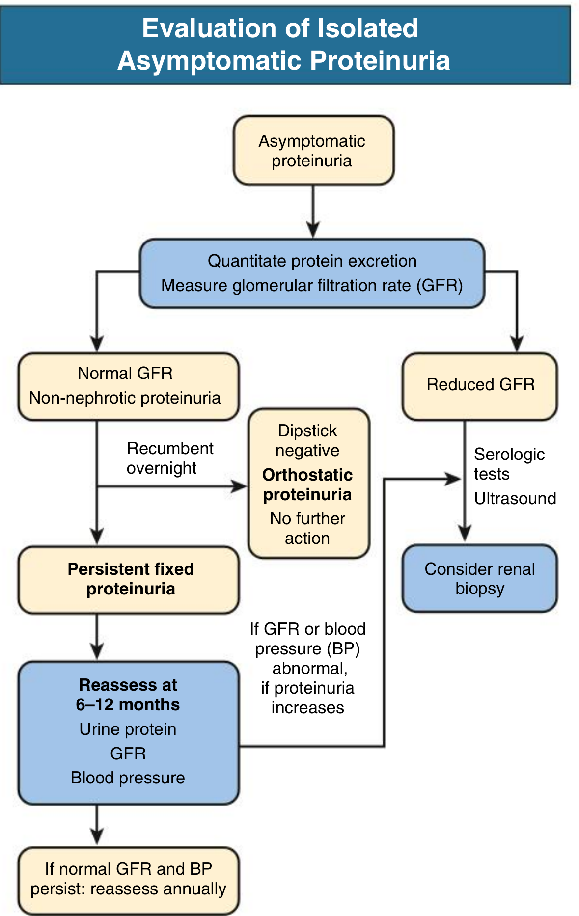
Step 3: Detailed Evaluation Based on GFR and Quantity
A. Normal GFR + Non-nephrotic Proteinuria + First-Morning Urine Negative → Orthostatic Proteinuria
- No further workup needed; excellent prognosis
- Biopsy not indicated (usually normal morphology or minimal change)
B. Normal GFR + Non-nephrotic Proteinuria + Persistent (Fixed) → Reassess at 6-12 months
- Measure: urine protein, GFR, blood pressure
- If all remain stable: annual reassessment
- If GFR/BP becomes abnormal or proteinuria increases: renal biopsy consideration
C. Reduced GFR → Expanded Workup
- Serologic panel: ANA, ANCA, anti-dsDNA, anti-GBM, complement (C3/C4), SPEP/UPEP, cryoglobulins, hepatitis B/C, HIV
- Renal ultrasound: exclude structural/anatomic causes, assess kidney size and echogenicity
- Consider renal biopsy: strongly indicated if GFR is reduced, especially with proteinuria >1 g/day
D. Nephrotic-Range Proteinuria (>3.5 g/day)
- Full nephrotic workup; serology; almost always requires biopsy
Step 4: Urine Microscopy
- Dysmorphic RBCs or RBC casts → glomerular origin; suspect GN even at low proteinuria levels
- Combined hematuria + proteinuria carries a much greater risk for significant glomerular injury, hypertension, and progressive kidney dysfunction than either finding alone - Comprehensive Clinical Nephrology
- Biopsy is often performed even at 0.5-1 g/day if combined with persistent microhematuria + casts and/or declining GFR
Step 5: Common Underlying Causes to Consider
| Category | Diseases |
|---|---|
| Primary glomerular | IgA nephropathy, FSGS, membranous nephropathy, minimal change disease, MPGN |
| Secondary glomerular | Diabetic nephropathy, lupus nephritis, amyloidosis, Alport syndrome |
| Tubulointerstitial | Reflux nephropathy, obstructive uropathy, interstitial nephritis, PKD |
| Systemic vasculitis | ANCA-associated GN (MPA, GPA) - ranges from asymptomatic proteinuria to RPGN |
| Overflow | Multiple myeloma (Bence-Jones protein), hemolysis (hemoglobinuria) |
5. Management
Orthostatic Proteinuria
- No treatment needed; reassure patient
- Transient or fixed; prognosis is uniformly excellent
Functional (Transient) Proteinuria
- Treat the underlying trigger (fever, exercise, heart failure)
- Recheck urine after resolution; no further workup if resolved
Fixed Non-Nephrotic Proteinuria with Normal GFR
- Conservative approach: regular monitoring
- Blood pressure control - target <130/80 mmHg
- ACE inhibitor or ARB: first-line antiproteinuric therapy; reduces intraglomerular pressure and proteinuria by ~30%. Consider initiating if proteinuria is >0.5-1 g/day even in the absence of hypertension
- Some nephrologists perform biopsy if nonnephrotic proteinuria persists >1 g/day after ACE-I/ARB initiation - Comprehensive Clinical Nephrology
Proteinuria with Reduced GFR / Nephrotic Range
- Nephrology referral
- Biopsy to establish diagnosis and guide immunosuppression
- Disease-specific therapy (e.g., immunosuppression for LN, MN; SGLT2 inhibitors for diabetic nephropathy)
- Antiproteinuric measures: ACE-I/ARB, dietary protein restriction (0.8 g/kg/day), sodium restriction
Children and Adolescents - Special Considerations
- Orthostatic proteinuria accounts for up to 60% of all asymptomatic proteinuria in children - Brenner & Rector's Kidney
- Isolated non-orthostatic proteinuria lasting >1 year may represent significant renal pathology
- Follow-up at least annually: BP, renal function, serum albumin, urinalysis, UPCr
- Subnephrotic proteinuria (UPCr <2.0 mg/mg) + normal GFR + minimal biopsy sclerosis → ACE-I/ARB; more severe disease may require immunosuppression
6. Follow-Up Schedule Summary
| Scenario | Follow-Up |
|---|---|
| Orthostatic / transient | No further workup; recheck periodically |
| Fixed non-nephrotic, normal GFR, stable | Reassess every 6-12 months (urine protein, GFR, BP); if stable, annually |
| Proteinuria increasing or BP/GFR abnormal | Expedite nephrology referral; consider biopsy |
| Nephrotic range / reduced GFR | Urgent nephrology referral + biopsy |
| Combined proteinuria + hematuria | Lower threshold for biopsy even at <1 g/day |
- Comprehensive Clinical Nephrology, 7th Edition, Ch. 16 - Urinary Abnormalities
- Brenner and Rector's The Kidney, 2-Volume Set - Chapters 13, 39, 40
- Textbook of Family Medicine, 9th Edition, Ch. 40