
Standard deviation psm
Standard deviation (SD) is a measure of how spread out values are around the mean.
Quick formula: SD = sqrt[ Σ(xi - x̄)² / (n-1) ] for a sample
In PSM (Propensity Score Matching), SD is used mainly in the standardized mean difference (SMD), which checks covariate balance between matched groups.
Searching sources...
Searching the Web
standard deviation propensity score matching PSM statistics
Searching the Web
standardized mean difference PSM balance checking formula
Reading Web Page
https://pmc.ncbi.nlm.nih.gov/articles/PMC6351359
Carpal tunnel syndrome with images
Reading File
Finding Sources
Finding Sources
Good sources found. I'll read the most comprehensive ones - Campbell's Operative Orthopaedics (for surgical detail) and Goldman-Cecil Medicine (for clinical overview), plus Gray's Anatomy for Students (for anatomy with images), and Firestein & Kelley's Rheumatology (for clinical detail).
Reading File
Reading File
Reading File
Reading File
Good content so far. Let me read more of Campbell's (treatment/surgical section) and Rheumatology, and also get the image URLs for fetching.
Reading File
Reading File
Now let me read the surgical treatment section from Campbell's (the main block) and fetch the key images.
Reading File
Loading Image
Excellent anatomy image. Now let me get the Campbell's surgical treatment content and the wrist splint image from Rheumatology, plus look for images showing Phalen/Tinel tests and thenar atrophy.
Reading File
Finding Sources
Finding Sources
Now let me get the thenar atrophy image, the wrist splint image, and the treatment section from Campbell's.
Reading File
Reading File
Now let me fetch the thenar atrophy image and look for additional surgical/clinical images.
Loading Image
Finding Sources
I now have comprehensive information and two excellent images. Let me also look for the nerve distribution image and the wrist splint image from Rheumatology.
Finding Sources
Reading File
Loading Image
I now have three excellent images and comprehensive content. Let me compile the full response.
Carpal Tunnel Syndrome (CTS)
CTS is the most common upper extremity compression neuropathy, caused by compression of the median nerve within the carpal tunnel. It affects up to 10% of the population and is twice as common in women. Peak incidence is between ages 30-60 years.
1. Anatomy of the Carpal Tunnel
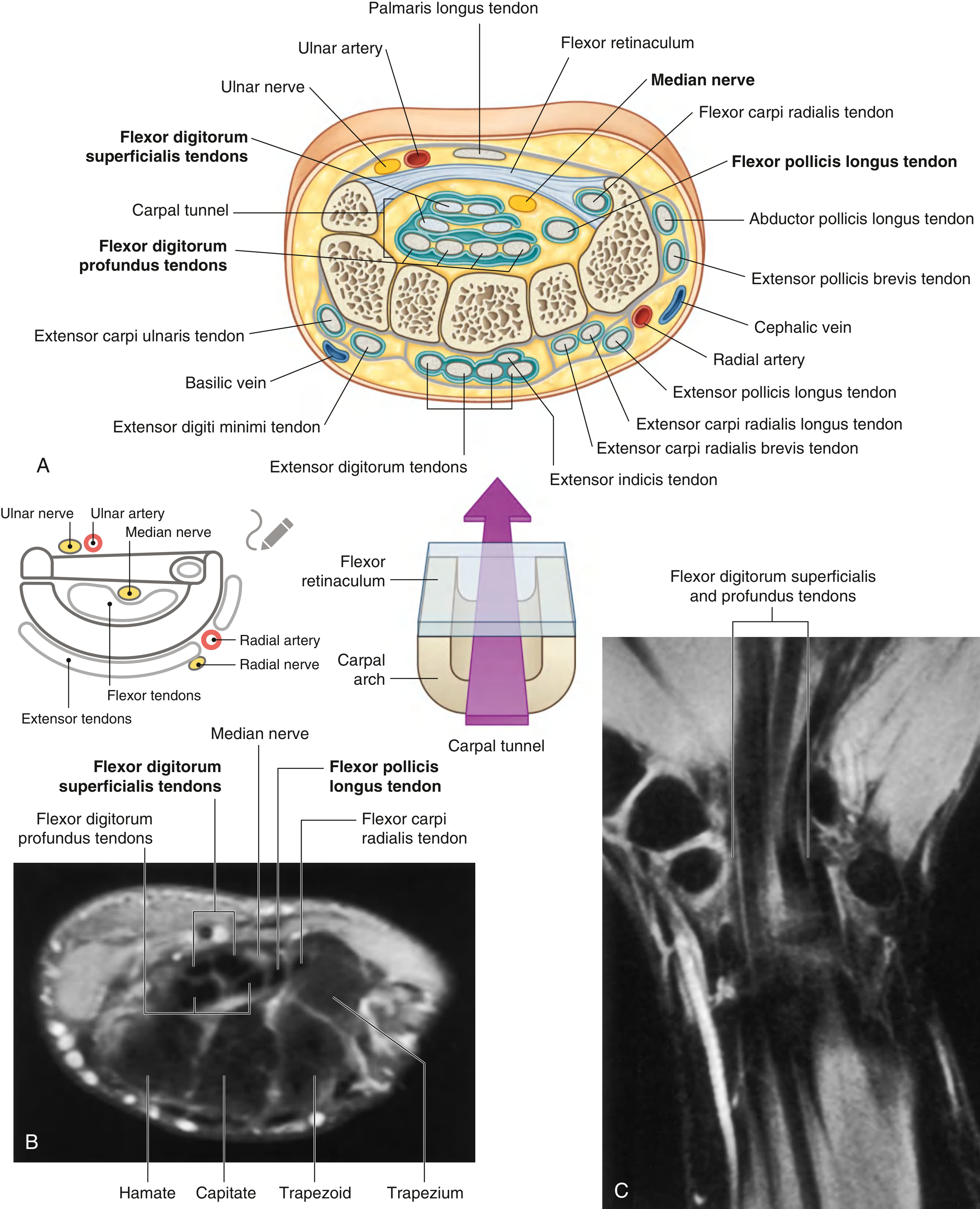
The carpal tunnel is a fibro-osseous canal at the wrist:
- Floor and walls (dorsal/lateral): Arching carpal bones (hamate, pisiform medially; scaphoid tubercle and trapezial ridge laterally)
- Roof (palmar aspect): Flexor retinaculum = deep forearm fascia proximally + transverse carpal ligament (TCL) over the wrist + aponeurosis between thenar/hypothenar muscles distally
- Contents: The median nerve (most palmar structure) + 9 flexor tendons (FDS ×4, FDP ×4, FPL ×1)
2. Pathophysiology
Elevated carpal tunnel pressure (>20-30 mmHg) impedes epineurial blood flow, producing nerve edema and anoxic capillary damage to the median nerve. Causes include:
| Category | Examples |
|---|---|
| Patient factors | Female sex, obesity, smoking, physical inactivity |
| Decreased tunnel size | Bony abnormalities, acromegaly, wrist flexion/extension |
| Inflammatory | Rheumatoid arthritis, gout, nonspecific tenosynovitis, infection |
| Fluid balance changes | Pregnancy, menopause, hypothyroidism, renal failure, hemodialysis |
| Autoimmune | Amyloidosis, lupus, scleroderma |
| External forces | Vibration, repetitive forceful wrist/finger flexion-extension, direct pressure, trauma |
The cause is idiopathic in most patients.
3. Clinical Features
Classic symptoms:
- Nocturnal paresthesia in the thumb, index, and middle fingers (often wakes the patient)
- Pain and "pins and needles" in the median nerve distribution
- Symptoms relieved by shaking or elevating the hand
- Forearm and elbow aching (poorly localized)
- Weakness or clumsiness of the hand
Late signs (advanced disease):
- Objective sensory loss in median nerve distribution
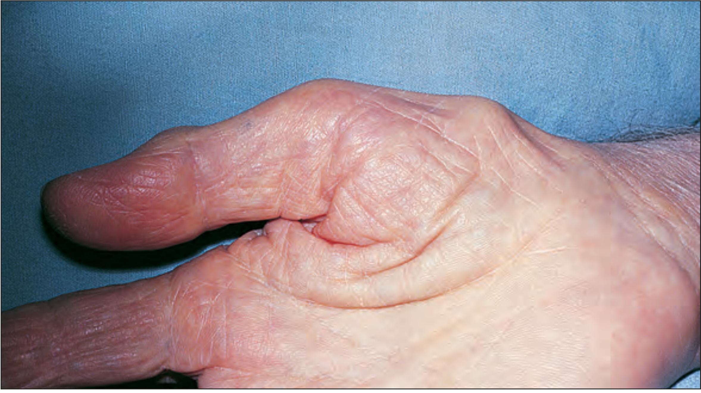
- Thenar muscle atrophy and weakness (abductor pollicis brevis most affected)
4. Diagnosis
Clinical Diagnostic Tests
| Test | Method | Notes |
|---|---|---|
| Phalen test | Sustained extreme wrist flexion for 1 minute; positive if paresthesias reproduced | Most sensitive test |
| Tinel sign | Percussion over carpal tunnel (with wrist extended); positive if radiating paresthesia | Most specific, least sensitive; percussion with wrist in extension is key |
| Durkan (carpal compression) test | Direct thumb compression over median nerve for 30 seconds | 90% specific, 87% sensitive - outperforms both Phalen and Tinel |
CTS-6 Diagnostic Score
| Finding | Points |
|---|---|
| Nocturnal symptoms | 4 |
| Thenar atrophy or weakness | 5 |
| Positive Phalen test | 5 |
| Positive Tinel sign | 4 |
| Numbness in median distribution | 3.5 |
| Loss of 2-point discrimination (>5 mm) | 4.5 |
Score ≥12 = positive (sensitivity 95%, specificity 91% - comparable to EMG/NCS but without cost or discomfort)
Electrodiagnostic Studies (Nerve Conduction Studies / EMG)
- Abnormal values: Distal motor latency >4.5 ms; sensory latency >3.5 ms
- NCS: ~90% sensitive, 60% specific
- EMG findings: increased insertional activity, positive sharp waves, fibrillations, decreased motor recruitment
- Note: studies can be normal even with symptomatic CTS; bilateral studies recommended
Differential Diagnosis
Cervical disc disease/radiculopathy, thoracic outlet syndrome, diabetic neuropathy, pronator teres syndrome, cubital tunnel syndrome
5. Treatment
Conservative (Mild CTS, No Thenar Atrophy)
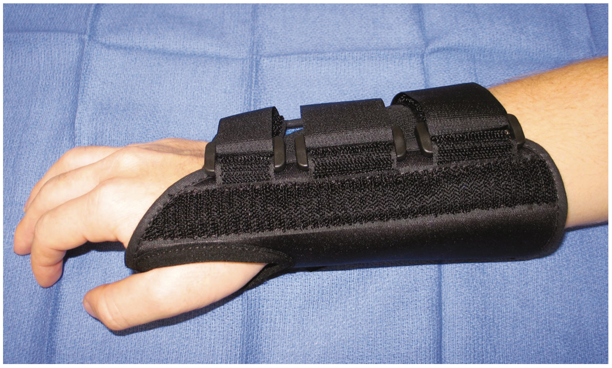
- Wrist splinting in neutral position (not >10° extension), especially at night - reduces provocative wrist positioning
- NSAIDs for pain control
- Corticosteroid injection into the carpal tunnel - effective short-term; long-term efficacy is mixed; risk of nerve injury if injected incorrectly. Best used when condition is temporary (e.g., pregnancy) or surgery must be deferred
- Vitamin B6 (100-200 mg/day) - some benefit reported, not confirmed in RCTs
Surgical (Intermediate/Advanced CTS, or Failed Conservative)
Absolute indications for early surgery: objective sensory loss or thenar atrophy (late findings)
Carpal tunnel release (CTR) - division of the transverse carpal ligament:
- Open CTR: Incision ≤3 cm along palm skin creases; gold standard
- Endoscopic CTR: Single or dual portal technique; faster return to work, less post-operative pain at the incision site, but technically more demanding
Outcomes:
- Significant improvements in most patients at 12 months (except pinch strength)
- Diabetic patients have similar short-term outcomes but worse long-term results (10 years)
- Severe electrophysiologic disease predicts prolonged/incomplete recovery at 1 year
- Internal neurolysis and epineurotomy add no benefit over release alone
6. Special Populations
- Pregnancy: CTS is common; usually resolves after delivery. Corticosteroid injection preferred over surgery
- Children: Rare; associated with congenital bone abnormalities, hypothyroidism, lysosomal storage disease, myopathic contractures
- Dialysis patients: Amyloid deposition in tunnel is a recognized cause
Sources: Campbell's Operative Orthopaedics 15th Ed 2026 | Firestein & Kelley's Textbook of Rheumatology | Goldman-Cecil Medicine | Gray's Anatomy for Students | Rheumatology, 2-Volume Set (Elsevier)
This is a shared conversation. Sign in to Orris to start your own chat.