
Azelaic acid use
"azelaic acid" AND dermatology
azelaic acid acne rosacea skin treatment
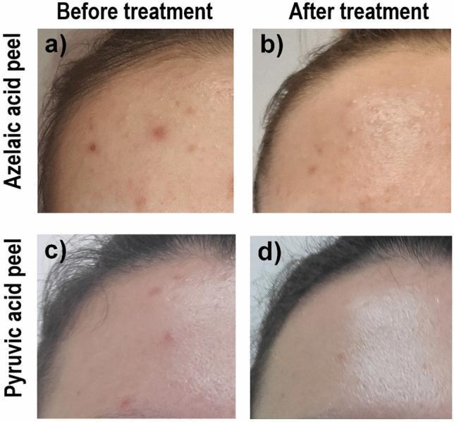
This clinical photograph comparison demonstrates the efficacy of chemical peels in treating acne vulgaris on the forehead. The image is divided into four quadrants (a-d) illustrating results from two different treatment groups: Azelaic Acid (AA) and Pyruvic Acid (PA). In the 'before' images (a and c), the forehead skin exhibits active acne lesions, including erythematous inflammatory papules, blackheads, and uneven texture associated with seborrhea. In the corresponding 'after' images (b and d), there is a visible reduction in lesion count and severity. Specifically, image (b) shows decreased erythema and flattening of papules following AA treatment. Image (d) displays significantly smoother skin texture and a reduction in inflammatory markers after PA treatment, with the skin appearing more reflective, suggesting improved surface regularity and potentially altered oiliness. This visual comparison serves as an educational tool for dermatological assessment using the Scale of Hellegren-Vincent Severity Symptoms (SHVSS) and highlights the role of organic acids in managing mild to moderate late acne symptoms in young adults.
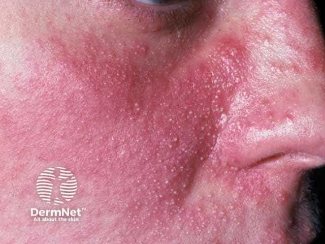
This is a high-resolution clinical dermatology photograph of the lateral left midface (cheek, nasal bridge and perioral region) captured in a frontal-oblique view to document steroid-induced papulopustular rosacea (SR). modality: in vivo skin photography; technique: standard color photography under diffuse, clinical lighting. The image shows diffuse facial erythema with numerous small papules and pustules scattered across the central face, involving cheeks and nasal ala, with mild edema and fine scaling. The papules are 1–3 mm, some pustular, follicular-based, with perifollicular erythema indicating inflammatory papulopustular eruption. There is no obvious comedones; telangiectasias may be subtle. The distribution pattern is centralized rather than peripheral; appearance may reflect steroid-exacerbated rosacea or rosacea-like dermatitis following topical corticosteroid use. Noting steroid history is critical to differentiate from acne vulgaris. Management implications include cessation of offending steroid, initiation of rosacea-directed therapy (metronidazole, azelaic acid, ivermectin, doxycycline in appropriate doses) and patient education on steroid-sparing regimens. This image is valuable for educational comparisons with acneiform eruptions and rosacea phenotypes, supports differential diagnosis, and monitors treatment response in SR-patient cohorts. Clinical utility includes documenting response to therapy, guiding patient counseling, and enabling comparative studies of steroid-induced rosacea across patient cohorts and dermatology education programs for clinical learning purposes.
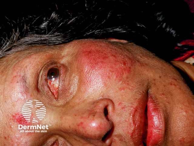
Clinical photography of the face, frontal close-up, high-resolution. Anatomical region: central facial skin including cheeks, nasal dorsum, perioral area, and periocular tissues. Primary subject: papulopustular rosacea with ocular involvement. Appearance: diffuse erythema of the central face with a constellation of inflammatory papules and pustules scattered across the malar region and nose; mild telangiectasia along the cheeks; lips and surrounding skin show no ulceration. Periocular redness and lid-margin irritation are present, compatible with ocular rosacea; eyelids appear mildly edematous without obvious keratitis in this image. Skin texture shows slight roughness and residual flaking. The overall pattern is chronic inflammatory facial dermatitis centered on the midface, rather than acne conglobata or lupus. Diagnostic significance: classic papulopustular rosacea with probable ocular involvement; warrants ophthalmology referral to assess ocular surface, blepharitis, and potential corneal involvement; informs systemic versus topical therapy decisions. Differential considerations: acne vulgaris (less likely distribution), steroid-induced rosacea, seborrheic dermatitis, lupus erythematosus mimicking rosacea, contact dermatitis. Clinical relevance: important for educational purposes, differential diagnosis of facial erythema, guiding treatment planning (topical metronidazole/azelaic acid, doxycycline in adequate dosing, eyelid hygiene) and patient counseling on triggers and chronicity. This image illustrates rosacea heterogeneity and emphasizes the need for multidisciplinary management. Guides comprehensive patient care.
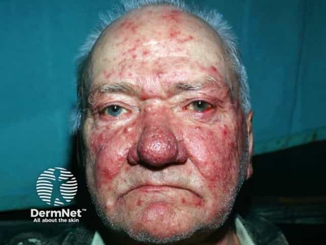
Comprehensive Description: This clinical photograph shows an elderly male with an advanced rosacea phenotype, dominated by severe papulopustular eruption and rhinophyma. Modality is digital clinical photography in visible light, frontal/anterior view of the face. Anatomical localization centers on centrofacial skin, with marked nasal bulbar hypertrophy and thickened, irregular nasal skin; accompanying erythema and telangiectasia extend to the cheeks, forehead, and perioral regions. Visual features include diffuse intense erythema, coalescing papules and pustules, nodules, and edema of the nasal pyramid; the nose exhibits notable rhinophymatous remodeling with bulbous contour and sebaceous prominence. There is no evident ocular involvement in this image. The pattern is pathognomonic for rosacea with rhinophyma change: centrofacial distribution, vascular ectasia, inflammatory acneiform lesions, and smooth-to-nodular nasal thickening. Differential considerations include acne rosacea versus acne vulgaris in older adults, seborrheic dermatitis, granulomatous diseases such as sarcoidosis or lupus pernio, and mucocutaneous infections; however the combination of nasal hypertrophy and facial erythema strongly supports rosacea with rhinophyma. Clinical significance: this stage informs treatment planning, including anti-inflammatory therapies (topical metronidazole, azelaic acid; oral doxycycline), and consideration of surgical rhinophyma reduction (laser therapy, dermabrasion, scalpel/clipping). Use cases: dermatology education, case repositories, teledermatology review, and research on rosacea progression for clinician education.
Azelaic Acid - Clinical Uses
What Is It?
- Dermatology 2-Volume Set 5e
- Fitzpatrick's Dermatology, Vol. 1 & 2
Indications (What It's Used For)
| Condition | Notes |
|---|---|
| Acne vulgaris | Inflammatory and comedonal acne; first-line alternative |
| Rosacea (papulopustular) | 15% gel is well-studied; non-inferior to adapalene |
| Melasma | Reduces abnormal melanin production |
| Post-inflammatory hyperpigmentation (PIH) | Selective effect on hyperactive melanocytes |
Mechanisms of Action
-
Antimicrobial - Active against Cutibacterium (Propionibacterium) acnes and Staphylococcus epidermidis, reducing bacterial load in acne and rosacea.
-
Anticomedogenic / Comedolytic - Normalizes/reverses abnormal follicular keratinization, helping to clear and prevent comedones.
-
Anti-inflammatory - Inhibits free radical oxygen production by neutrophils, reducing oxidative tissue injury at sites of inflammation.
-
Skin-lightening (depigmenting):
- Competitive inhibitor of tyrosinase (the key enzyme in melanin synthesis)
- Reversible inhibitor of cytochrome P450 reductase and 5α-reductase
- Selective cytotoxic/antiproliferative effect on hyperactive and abnormal melanocytes, with little effect on normal melanocytes
Formulations and Dosing
| Formulation | Concentration | Status |
|---|---|---|
| Cream | 20% | Prescription |
| Gel | 15% | Prescription |
| Cream/foam | 10%-14% | Over-the-counter |
- Applied twice daily
- Takes 4-8 weeks for visible results in acne; up to 3 months for pigmentation
Side Effects
- Mild-to-moderate irritant contact dermatitis: dryness, peeling, burning, and erythema at the application site
- These effects typically subside within 2 to 4 weeks with continued use
- Compared with adapalene, azelaic acid 15% gel shows greater tolerability while maintaining similar efficacy
Use in Pregnancy
- Fitzpatrick's Dermatology; confirmed by a 2024 review on acne management in pregnancy (PMID: 38453786)
Position in Acne Treatment Algorithm
- Comedonal acne: Second-line agent (after topical retinoids)
- Papular/pustular acne: Alternate agent in combination regimens
- Melasma: Listed as a first-line topical therapy alongside hydroquinone
Clinical Comparisons
- In a randomized controlled trial, azelaic acid 15% gel was non-inferior to adapalene 0.1% gel in inflammatory acne with better tolerability
- A 2024 review (PMID: 39464747) confirms its role in acne, rosacea, melasma, and PIH, summarizing its multi-target mechanism
- 2024 AAD acne guidelines (PMID: 38300170) endorse azelaic acid as a valid topical alternative
Example Clinical Images
Summary
Acne - Rosacea - Melasma - Post-inflammatory Hyperpigmentation (PIH)