
Give me a picture of myotonic dystrophy patient relevant to ddx
myotonic dystrophy patient clinical features photograph facial appearance ddx
Myotonic Dystrophy (DM1) - Clinical Images for DDx
Image 1 - DDx Comparison: FSH Dystrophy (A) vs. Myotonic Dystrophy (B)
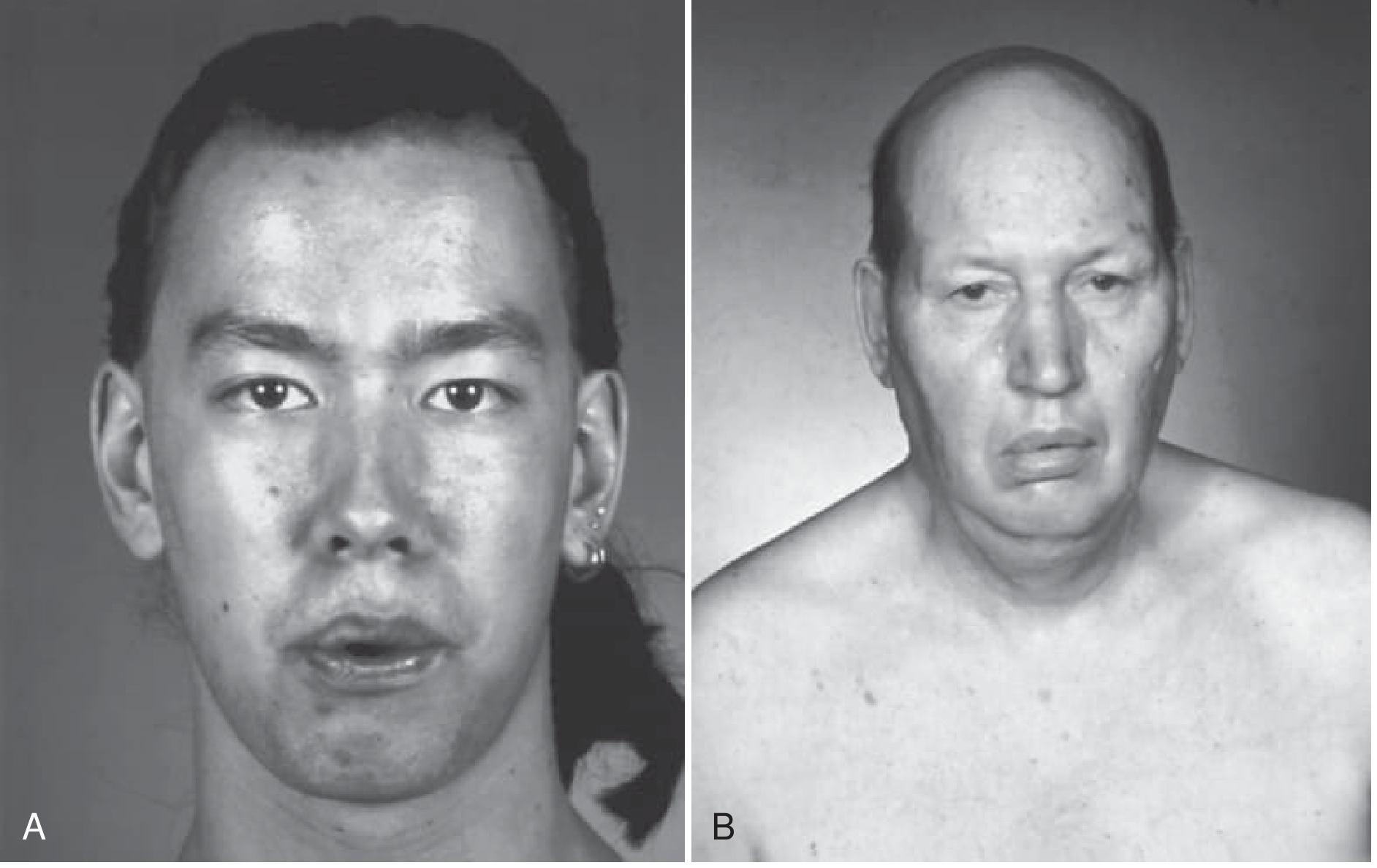
- Panel A (left): Patient with facioscapulohumeral (FSH) dystrophy - unable to purse the lips when attempting to whistle; note the preserved temporalis bulk
- Panel B (right): Patient with myotonic dystrophy type 1 - note the classic triad:
- Frontal balding
- Temporalis + masseter wasting (hollowed temples)
- Ptosis + facial weakness (long, expressionless face)
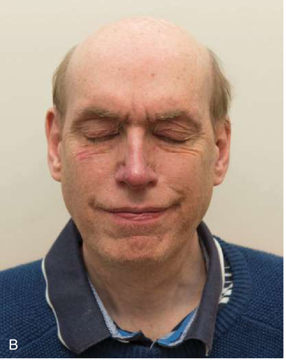
Images 2 & 3 - DM1 Type 1 (58-year-old man) - Pathognomonic Features
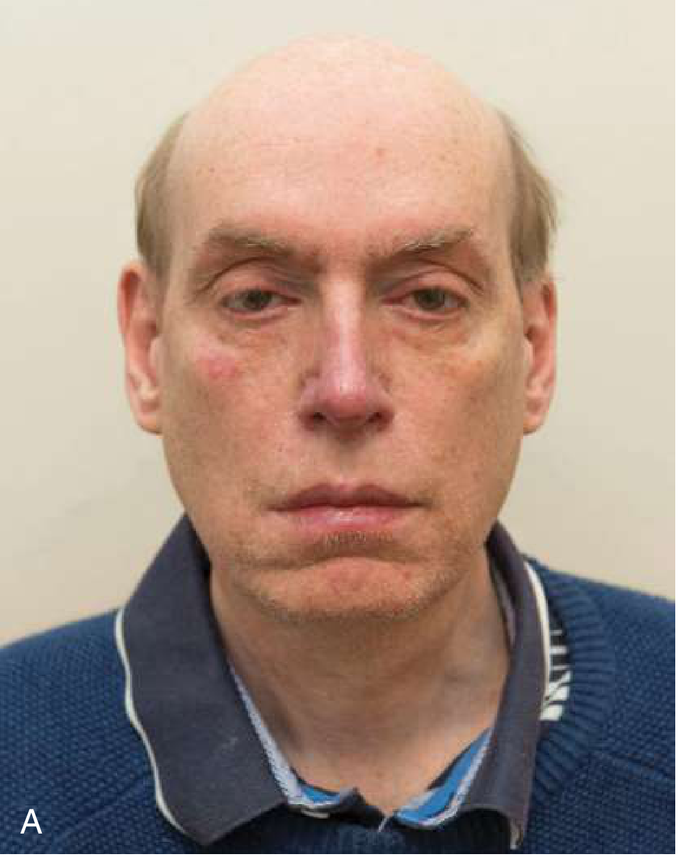
- Panel A: Frontal balding, bilateral ptosis, temporal muscle atrophy - "hatchet face" appearance
- Panel B: Forced eye closure - note the inability to bury the eyelashes (orbicularis oculi weakness), a sign of orbicularis weakness distinct from facioscapulohumeral dystrophy where orbicularis is equally or more severely affected
DDx Key Distinguishing Features at a Glance
| Feature | Myotonic Dystrophy (DM1) | FSH Dystrophy | Oculopharyngeal Dystrophy | Mitochondrial Myopathy (CPEO) |
|---|---|---|---|---|
| Ptosis | Yes (bilateral, mild-mod) | Rare | Yes | Prominent |
| Facial weakness | Yes (bifacial, expressionless) | Yes (lip/orbicularis prominent, asymmetric) | Mild | Mild |
| Temporal wasting | Yes (hallmark) | Absent | Absent | Variable |
| Frontal balding | Yes (men) | No | No | No |
| Myotonia (grip/percussion) | Yes - pathognomonic | No | No | No |
| Distal > proximal weakness | Yes | No | No | No |
| Cataracts | Yes (posterior subcapsular) | No | No | Yes |
| Cardiac involvement | Yes (conduction defects) | Sometimes | Rare | Yes |
| Genetics | CTG repeat, chr 19 (DM1) | D4Z4 deletion, chr 4q35 | PABPN1, chr 14 | mtDNA/nuclear |
DDx of vomiting in a 36 Y asian immigrant female
vomiting differential diagnosis young female Asian immigrant clinical approach
DDx of Vomiting - 36-Year-Old Asian Immigrant Female
STEP 1: RULE OUT FIRST (Critical & Common)
| Condition | Why Top Priority | Key Clue |
|---|---|---|
| Pregnancy / Hyperemesis gravidarum | Prevalence 50-80% of pregnancies; HEG in 0.3-3% | Morning timing, amenorrhea, +uHCG |
| GI obstruction | Requires urgent surgical Rx | Bilious vomiting, distension, absent bowel sounds |
| Acute abdomen (appendicitis, perforated viscus, mesenteric ischemia) | Life-threatening | Severe abdominal pain + vomiting |
| Raised ICP (CNS mass, hemorrhage) | Fatal if missed | Projectile, positional, headache, papilledema |
| DKA / metabolic crisis | Treatable emergency | Polyuria, polydipsia, hyperglycemia |
| Acute MI | Atypical presentation in women | Diaphoresis, chest/jaw/arm discomfort |
STEP 2: GASTROENTEROLOGICAL (Most Common Overall)
Acute vomiting WITHOUT significant abdominal pain
- Infectious gastroenteritis - most common single cause globally; viral (Norovirus, Rotavirus) >> bacterial
- Food poisoning (Staph aureus, Bacillus cereus, Salmonella) - immigrant status increases exposure to unfamiliar food preparation
- Hepatitis A/B/E - higher prevalence in Asia; hepatitis E especially in recent immigrants
- Medications / drug toxicity - OCPs, NSAIDs, antibiotics, herbal remedies (common in Asian cultures)
- Migraine headache - peak incidence in women aged 25-55
Acute vomiting WITH abdominal pain
- Peptic ulcer disease (PUD) / H. pylori - H. pylori seroprevalence in East/Southeast Asia is 50-80% vs ~35% in Western countries; strongly associated with nausea, epigastric pain, vomiting
- Acute cholecystitis / biliary colic - "Fat, fertile, forty, female" - Asian women are NOT exempt; common in Chinese, Japanese, Korean women
- Pancreatitis - gallstone-related predominates in Asian women
- Appendicitis - still in the age range
Chronic/Recurrent vomiting (>1 month)
- Gastroparesis - idiopathic or post-viral
- GERD / esophagitis - regurgitation may be misreported as vomiting
- Cyclic vomiting syndrome - episodic, stereotyped; associated with migraine
- Small bowel or gastric outlet obstruction - undigested food hours after eating = gastric obstruction
STEP 3: IMMIGRANT/ASIAN-SPECIFIC ELEVATED PRIORITIES
| Condition | Reason Elevated in This Patient |
|---|---|
| Helicobacter pylori gastritis/PUD | H. pylori prevalence ~60-80% in many Asian nations (China, Japan, Korea, Vietnam); immigrants carry this burden |
| Intestinal parasitosis (Ascaris, Giardia, Strongyloides, liver flukes) | Endemic in many parts of Southeast/South Asia; may present with nausea, vomiting, abdominal discomfort |
| Hepatitis E (HEV) | Endemic in South/Southeast Asia; acute hepatitis with nausea, jaundice, vomiting |
| Typhoid fever (Salmonella typhi) | Endemic in South Asia, SE Asia; vomiting, step-ladder fever, relative bradycardia |
| Tuberculosis (GI/abdominal TB) | Higher TB burden in Asian immigrants; abdominal TB causes vomiting, ascites, weight loss |
| Gastric cancer | Japan, Korea, China have some of the world's highest gastric cancer rates; while 36 is young, consider if alarm features present (weight loss, dysphagia, early satiety) |
| Lactose intolerance | ~80-90% of East Asians are lactose intolerant; dietary change after immigration to dairy-heavy Western diet can cause bloating, nausea, vomiting |
| Traditional herbal medicine toxicity | TCM, Ayurvedic, and other herbal preparations are commonly used; many are hepatotoxic or directly emetic |
| Acculturative stress / anxiety | Immigration-related psychosocial stress is a real precipitant of functional GI disorders |
STEP 4: SYSTEMIC / NON-GI CAUSES
| System | Conditions |
|---|---|
| Endocrine | Hypothyroidism, Addison's disease, hypercalcemia, hyperthyroidism (Graves - higher in Asian women) |
| Renal | Uremic vomiting (CKD, pyelonephritis) |
| Cardiac | Inferior MI (vagal), heart failure |
| Neurological | Vestibular neuritis, labyrinthitis, Ménière's disease, posterior fossa lesion |
| Psychiatric | Bulimia nervosa, anxiety, depression with somatization |
| Iatrogenic | NSAIDs, antibiotics, opioids, iron supplements, OCP-induced nausea |
STEP 5: DIAGNOSTIC APPROACH FRAMEWORK
VOMITING IN THIS PATIENT
│
├─ Pregnancy test (urine hCG) ──► FIRST, ALWAYS
│
├─ +Abdominal pain? ──► CBC, CMP, lipase, LFTs, RUQ US, CT abdomen
│
├─ Acute (<1 week)? ──► Consider infectious, medication, pregnancy
│
├─ Chronic (>1 month)? ──► EGD (H. pylori + biopsy), motility studies, CT
│
├─ Alarm features (weight loss, dysphagia, hematemesis)?
│ ──► Urgent EGD (exclude gastric cancer, ulcer)
│
└─ Immigrant-specific workup:
- H. pylori stool antigen / breath test
- Stool O&P (parasites)
- Hepatitis serologies (A, B, E)
- TB screening (IGRA/Mantoux)
- LFTs (herbal toxicity)
Mnemonic: "VINDICATE P" applied here
- H. pylori gastritis/PUD (by far most probable given Asian origin)
- Infectious gastroenteritis (viral/bacterial)
- Biliary colic / cholecystitis
- Medication/herbal toxicity
- Functional nausea/vomiting with psychosocial overlay (immigration stress)
now this asian/indian woman also has multiple sexual partners but there is no infectious or suspicious vaginal discharge or lower abdominal pain, now what should I further consider in this case of VOMITING?
What Multiple Sexual Partners Adds to the Vomiting DDx
1. PREGNANCY - Risk Now Substantially Higher
- Intrauterine pregnancy / Morning sickness - #1 still; now even more likely
- Ectopic pregnancy - CRITICAL addition. Prior STIs (even silent ones like Chlamydia) cause subclinical salpingitis → tubal scarring → ectopic implantation. The patient may have no pelvic pain yet if the ectopic is early and unruptured. Nausea/vomiting + amenorrhea + positive hCG + adnexal tenderness on exam = ectopic until proven otherwise. This is a surgical emergency if ruptured.
Risk factors for ectopic: prior PID, multiple partners, prior STI, smoking - this patient may have several of these silently.
2. ACUTE HIV / ACUTE RETROVIRAL SYNDROME (ARS) - Must Not Miss
"The most common symptoms seen in the acute retroviral syndrome are fever, lymphadenopathy, pharyngitis, and myalgias/arthralgias - all seen in >75% of cases. Other common symptoms include non-pruritic maculopapular rash (upper body), nausea, vomiting, and diarrhea. Up to 50% of patients experience severe headaches and may have signs of meningitis or encephalitis."
- Occurs 2-4 weeks after HIV exposure
- Standard HIV ELISA/Western blot may be negative during this window - need HIV RNA PCR (viral load) or p24 antigen (4th-generation HIV combo test)
- Mimics infectious mononucleosis ("the great imitator")
- No vaginal symptoms expected - this is a systemic viremia
- High viral load = high transmissibility = public health urgency
- EBV infectious mononucleosis (monospot, EBV PCR)
- CMV mononucleosis
- Toxoplasmosis
- Secondary syphilis
- Viral hepatitis (A, B, C)
- Drug reaction (DRESS)
3. VIRAL HEPATITIS - Sexually Transmitted Forms
| Hepatitis | Sexual Transmission | Nausea/Vomiting? | Relevant Here? |
|---|---|---|---|
| Hepatitis B (HBV) | Yes - highly, especially with multiple partners | Yes - prominent prodrome | HIGH - Asian immigrants already at elevated HBV risk; MSP amplifies further |
| Hepatitis C (HCV) | Yes - lower efficiency, risk with multiple partners | Yes | Moderate |
| Hepatitis A (HAV) | Yes - fecal-oral, also oro-anal | Yes - prominent | Moderate (also endemic in South Asia) |
4. SECONDARY SYPHILIS - The "Great Imitator"
- Secondary syphilis is classically systemic: rash (palms/soles), fever, malaise, nausea, lymphadenopathy, mucous patches
- No genital symptoms required - primary chancre may have healed silently weeks earlier
- Syphilitic hepatitis can occur in secondary syphilis - causing nausea, vomiting, elevated LFTs, RUQ discomfort
- Check RPR/VDRL + TPHA/FTA-ABS
5. DISSEMINATED GONOCOCCAL INFECTION (DGI) - Silent Locally, Systemic
- Disseminated N. gonorrhoeae can occur even without urogenital symptoms or discharge in ~25% of women (the primary infection in the endocervix is often asymptomatic)
- Systemic features: fever, nausea, polyarthralgia/tenosynovitis, petechial/pustular rash
- Can cause gonococcal perihepatitis (Fitz-Hugh-Curtis syndrome) - RUQ pain + nausea + vomiting without significant lower abdominal pain
- Also seen with Chlamydia causing Fitz-Hugh-Curtis
6. FITZ-HUGH-CURTIS SYNDROME (Perihepatitis)
- Caused by Chlamydia trachomatis or N. gonorrhoeae ascending to the liver capsule
- Presents as RUQ pain + nausea/vomiting mimicking cholecystitis or hepatitis
- May have no discharge, no cervicitis, no lower abdominal pain at presentation
- "Violin-string" adhesions between liver capsule and peritoneum (visible on laparoscopy)
- Liver enzymes may be mildly elevated
- Diagnose with cervical/vaginal NAAT (nucleic acid amplification test) for Chlamydia/GC
7. MEDICATION EFFECTS FROM CONTRACEPTION / POST-EXPOSURE TREATMENT
- Emergency contraception (Plan B / levonorgestrel) - nausea and vomiting are the #1 side effects (incidence 23-50%)
- Oral contraceptive pills - nausea common, especially in first weeks
- HIV PrEP (tenofovir/emtricitabine) - nausea is a known early side effect
- HIV PEP (post-exposure prophylaxis) - if the patient recently had a risk event and started PEP, nausea/vomiting is extremely common with PEP regimens
8. PSYCHOSOCIAL / BEHAVIOURAL OVERLAY
- Shame, stigma, fear of STI diagnosis in culturally conservative Asian contexts can manifest somatically as nausea/vomiting
- Eating disorders (higher prevalence in young women with high-risk sexual behaviour contexts)
- Substance use (alcohol, recreational drugs) in some sexual risk contexts
Updated Priority DDx Given Full Context
| Priority | Diagnosis | Why |
|---|---|---|
| #1 | Intrauterine pregnancy / HEG | Reproductive age, MSP, high probability |
| #2 | Ectopic pregnancy | Silent prior STI → tubal damage; can present without pain early |
| #3 | Acute HIV / ARS | MSP = major risk; nausea/vomiting is a cardinal ARS symptom; window period means tests may be negative |
| #4 | Hepatitis B (acute) | Asian immigrant + MSP = double risk |
| #5 | Fitz-Hugh-Curtis (Chlamydia/GC perihepatitis) | No discharge/pelvic pain required; presents as RUQ nausea/vomiting |
| #6 | Secondary syphilis | Silent primary, systemic secondary |
| #7 | Emergency contraception / OCP side effects | Common, overlooked |
| #8 | H. pylori PUD | Still in the differential from before |
Immediate Investigations to Add to Previous List
Previous workup + ADD:
MANDATORY:
□ urine/serum β-hCG → if positive: transvaginal ultrasound to exclude ectopic
□ 4th-gen HIV combo test (Ag/Ab) → if negative but high suspicion: HIV RNA PCR
□ HBsAg, HBcAb IgM, HCV Ab, HAV IgM
□ RPR (syphilis screen) + TPHA
□ Cervical/vaginal NAAT for Chlamydia + GC (even without symptoms)
□ LFTs (hepatitis, Fitz-Hugh-Curtis, syphilitic hepatitis)
CONSIDER:
□ Emergency contraception history (timing of last sexual encounter?)
□ RUQ ultrasound (if LFTs elevated or RUQ tenderness → Fitz-Hugh-Curtis vs cholecystitis)
□ Monospot + EBV/CMV serology (ARS mimics mono)
what if the same indian women with vomiting also states that she also have a headache but no changes in vision, only headache and vomiting, with normal regular periods, no abdominal pain, no discharge, no fever, no lymphadenopathy
Headache + Vomiting, No Fever, No Visual Changes, No Pelvic Symptoms, Normal Periods
THE CLINICAL LOGIC
- Projectile (without preceding nausea in classic ICP)
- Morning-predominant (worsens lying flat overnight, ICP peaks in early morning)
- Worse with Valsalva (coughing, straining, bending forward)
PRIORITY DDx: HEADACHE + VOMITING
1. IDIOPATHIC INTRACRANIAL HYPERTENSION (IIH) / Pseudotumor Cerebri - TOP DIAGNOSIS
"Primary idiopathic intracranial hypertension occurs in obese women of childbearing age. Incidence is 19-20 per 100,000 in obese women aged 15-55."
"The most prominent symptoms include headache (84%), transient visual obscurations (68%), pulsatile tinnitus (52%). Only 32% of patients report visual loss."
- Young woman (36 years) ✓
- Asian women are also susceptible, especially with weight gain ✓
- Headache + vomiting ✓
- No visual changes - still consistent (visual obscurations and papilledema can be absent early)
- A variant without papilledema exists - diagnosed by MRI neuroimaging criteria (empty sella, posterior globe flattening, perioptic subarachnoid space distension, transverse sinus stenosis)
| Trigger | Relevance |
|---|---|
| Tetracyclines / minocycline | Common in India for acne/malaria prophylaxis |
| Vitamin A excess / retinoids | Isotretinoin (acne treatment) |
| Fluoroquinolone antibiotics | Widely used |
| Lithium | Psychiatric medication |
| OCP / hormonal contraception | Relevant given her sexual history |
| Rapid weight gain | Especially pertinent |
2. MIGRAINE - Most Common Primary Headache + Vomiting
"In primary care, <10% of patients presenting with headache >3 months without neurologic symptoms have tension-type headache; >90% have migraine."
- Headache + nausea/vomiting (cardinal symptom pair)
- No visual changes required - only migraine with aura has visual prodrome
- Photophobia, phonophobia, worse with movement
- Young woman: peak migraine incidence is 25-55 years in females
- Hormonal trigger: menstrual migraine (around menstrual cycle even with normal periods)
- Indian/Asian women have similar migraine prevalence to the general population
- ≥5 attacks lasting 4-72h
- Unilateral, pulsating, moderate-severe, worsened by routine activity
- Nausea/vomiting OR photophobia + phonophobia
3. CEREBRAL VENOUS SINUS THROMBOSIS (CVST) - Must Not Miss
- Headache is the presenting feature in >90% of CVST
- Vomiting from raised ICP
- No fever, no focal deficit required early in the course
- OCP use is the #1 risk factor in young women - and this patient is likely using contraception
- Indian women have additional risk: antiphospholipid syndrome, inherited thrombophilias (Factor V Leiden less common in Asians but protein C/S deficiency more common)
- Thunderclap or progressive subacute headache worsening over days-weeks
Red flag per Tintinalli's: "Wakens from sleep, early morning headache, progressive frequency/severity" → Elevated ICP, CVST must be excluded
4. INTRACRANIAL MASS LESION (Brain Tumor / Abscess)
- Any space-occupying lesion raises ICP → headache + vomiting
- Morning headache worsened by Valsalva is the classic pattern
- No focal neuro deficits required early
- In Indian immigrants: CNS tuberculoma is a genuine concern (TB is endemic in South Asia; CNS TB presents as ring-enhancing lesion, chronic headache, vomiting)
- Primary brain tumors (glioma, meningioma), metastases
- Brain abscess (consider given MSP - risk of bacterial endocarditis if IV drug use; or dental/ENT source)
5. SUBARACHNOID HEMORRHAGE (SAH) - Thunderclap Variant
- Classic: sudden "worst headache of life" (thunderclap)
- BUT: sentinel headaches can precede full SAH by days-weeks, presenting as recurrent moderate headaches + vomiting
- No focal deficit, no meningism required in early/small bleeds
- No fever
- Must be excluded in any new headache + vomiting pattern in a young adult
- CT head is the first-line test; if negative and suspicion remains → LP (xanthochromia)
6. CARBON MONOXIDE POISONING
"DDx includes alcoholic intoxication, cerebrovascular accident, influenza, meningitis, migraine, myocardial infarction, food poisoning."
- Cooking with gas stoves in poorly ventilated homes
- Headache + vomiting without fever is the classic presentation
- Multiple household members affected simultaneously is the clue
- Ask: does the headache improve when leaving the house/environment?
7. TENSION-TYPE HEADACHE + FUNCTIONAL VOMITING
- Least alarming
- Bilateral, band-like, not worsened by activity, no vomiting typically
- However: immigration-related chronic psychosocial stress → tension headache + functional nausea/vomiting
- Diagnosis of exclusion - never assign without ruling out structural causes first
8. MEDICATION / SUBSTANCE EFFECT
- OCP - headache is a recognized side effect; combined OCP can trigger migraines (estrogen withdrawal during pill-free week)
- Emergency contraception - headache + vomiting after levonorgestrel
- NSAID overuse headache (rebound/analgesic overuse headache)
- Herbal remedies common in Indian communities
RED FLAG FEATURES TO ACTIVELY SEEK
| Red Flag | What It Suggests |
|---|---|
| Sudden onset ("thunderclap") | SAH |
| Wakes from sleep / early morning | Raised ICP, mass |
| Progressive worsening over weeks | Mass, CVST, IIH |
| Worse lying flat, better sitting up | Raised ICP |
| Worse with Valsalva/cough/straining | Raised ICP |
| Neck stiffness | Meningitis/SAH |
| New headache in patient on OCP | CVST |
| Pulsatile tinnitus | IIH |
Prioritized DDx for This Specific Patient
| Rank | Diagnosis | Why |
|---|---|---|
| #1 | Migraine without aura | Most common; young woman, headache + vomiting, no visual Δ |
| #2 | IIH (Pseudotumor cerebri) | Young woman, headache + vomiting, papilledema/visual Δ may be absent early; OCP/tetracycline triggers |
| #3 | CVST | Young woman, likely OCP use, Indian origin (thrombophilia risk), headache + raised ICP signs |
| #4 | CNS Tuberculoma / Abscess | Indian immigrant, space-occupying, chronic progressive headache + vomiting |
| #5 | SAH (sentinel bleed) | Must exclude; CT + LP if suspicion |
| #6 | CO poisoning | Environmental, no fever, headache + vomiting classic |
| #7 | OCP / medication-induced | History check |
| #8 | Tension headache + functional vomiting | Exclusion diagnosis; psychosocial stress |
Immediate Investigation Priority
STEP 1 - URGENT (exclude emergencies):
□ Non-contrast CT head → exclude SAH, mass, herniation
□ If CT negative + SAH still suspected → LP (xanthochromia)
□ MR venography if CVST suspected (young woman on OCP)
STEP 2 - TARGETED:
□ MRI brain with gadolinium → better for mass, tuberculoma, abscess, IIH signs
□ Fundoscopy (papilledema? → IIH; normal → doesn't exclude)
□ LP with opening pressure (if imaging safe) → IIH (>250 mmH₂O), meningitis
STEP 3 - SYSTEMIC:
□ CO-oximetry / carboxyhaemoglobin (if CO poisoning suspected)
□ OCP/medication history
□ Formal visual fields (IIH - visual acuity preserved until late)
□ urine hCG (periods can be present in early pregnancy)
I am preparing for MRCP (UK) PACES exam and for that exam I am asking you questions, in this exam we have short cases and long cases for which I have to prepare, we need to take History and examination as per the presenting complaint and cover all the possible list of DDx and then work our way through the most likely Diagnosis, please answer any firther question by keeping that in mind as well, now can you please explain the scenerio in which IIH will be the most likely diagnosis on Hx and Physical Examination (PE)
IIH as the Most Likely Diagnosis: MRCP PACES History & Examination Guide
THE MODEL PATIENT PRESENTATION
A 34-year-old obese Indian woman presents with a 3-month history of daily generalised headache and vomiting. She has been using minocycline for acne.
PART 1: HISTORY (Hx)
SOCRATES - The Headache
| Component | What IIH Classically Gives You |
|---|---|
| Site | Generalised, bifrontal, or retro-orbital ("behind the eyes") - not unilateral |
| Onset | Gradual, progressive over weeks to months |
| Character | Dull, pressure-like, or pulsating; occasionally throbbing |
| Radiation | May radiate to neck and upper back |
| Associations | Nausea, vomiting (from raised ICP); pulsatile tinnitus ("whooshing sound" in ears, synchronous with pulse); transient visual obscurations |
| Timing | Wakes patient from sleep in the early morning hours - classic; worse on waking, eases through the day |
| Exacerbating | Coughing, straining, Valsalva, bending forward, lying flat - all raise ICP further |
| Relieving | Sitting upright, analgesia partially |
| Severity | Moderate to severe; headache may have been present for months before diagnosis |
PACES tip: The examiner loves "wakes from sleep" + "worse on bending/coughing" + "retro-orbital pain" - this trio strongly points to raised ICP rather than migraine or tension headache.
Asking About the "IIH Symptom Cluster"
-
Visual symptoms:
- "Do you get brief episodes of blurred or greyed-out vision, especially when you stand up quickly or bend forward?" → Transient visual obscurations (TVOs) - almost pathognomonic for IIH
- "Do you see double?" → Diplopia (CN VI palsy - false localising sign)
- "Any permanent change in vision?" - Loss is late and the patient may not volunteer this
- Note: TVOs occur in 68% but the patient may not have reported them unless asked directly - always ask
-
Pulsatile tinnitus:
- "Do you hear a whooshing or pulsing sound in one or both ears?" - present in ~52%; synchronous with heartbeat; often louder on one side
-
Neck/back pain:
- Referred pain from raised CSF pressure; reported in >50%
-
Cognitive/mood symptoms:
- Brain fog, concentration difficulties in chronic IIH
Systematic Enquiry - Risk Factors to Actively Seek
| Risk Factor | How to Ask |
|---|---|
| Obesity / recent weight gain | "Have you gained weight recently?" - the single strongest risk factor |
| Medications | "Are you on any tablets, creams, or supplements?" - then specifically: |
| - Tetracyclines (minocycline, doxycycline) | Common in Indian women for acne/skin care |
| - Isotretinoin (Roaccutane) | Acne treatment - directly causes IIH |
| - Oral contraceptive pill / progesterone | Relevant in this patient with MSP |
| - Vitamin A / retinoids (excess) | Including high-dose supplements |
| - Steroids (and steroid withdrawal) | |
| - Fluoroquinolones, nalidixic acid | |
| - Trimethoprim-sulfamethoxazole | Common in immunocompromised |
| - Tamoxifen | Relevant in older women |
| - Growth hormone | |
| Pregnancy | Must always exclude |
| Sleep apnoea | "Do you snore? Wake unrefreshed?" - OSA is a secondary IIH cause |
| Endocrine disorders | Hypothyroidism, Addison's, hypoparathyroidism |
| Family history | Rare but genetic component exists |
Negative History (Equally Important in PACES)
- No fever → excludes meningitis, encephalitis, abscess
- No thunderclap onset → against SAH
- No focal neurological symptoms → against mass lesion, stroke
- No neck stiffness → against meningitis/SAH
- No preceding viral illness → against post-infectious causes
- No immunosuppression / HIV → against opportunistic CNS infection
- No known malignancy → against metastases
- Normal periods → pregnancy less likely (but test anyway)
PART 2: PHYSICAL EXAMINATION (PE)
General Inspection (First 10 Seconds)
"This is an obese young woman who appears well and alert with no focal neurological deficit at rest."
- BMI >30 is present in ~90% of IIH cases
- Patient looks well - not acutely unwell, no signs of systemic sepsis
- No rash (excludes meningococcaemia, secondary syphilis)
Vitals
- BP - may be normal or mildly raised (raised ICP → hypertension; Cushing response is a late/severe sign)
- Heart rate - normal (bradycardia + hypertension = Cushing triad = late, severe)
- Temperature - normal (fever → infectious/inflammatory cause)
Cranial Nerve Examination - The Key Findings
| CN | Expected Finding in IIH | Significance |
|---|---|---|
| CN II (Optic) | Acuity usually preserved (until late); colour vision normal | Important - distinguishes papilledema from optic neuritis |
| CN II - Fundoscopy | Bilateral papilledema - the cardinal sign | Swollen disc margins, obliterated cup, disc hyperaemia, peripapillary flame haemorrhages in severe cases |
| CN II - Visual fields | Enlarged blind spot (most common early change); may progress to inferior nasal field defects | Confrontation testing - compare to your own; patient may not notice this themselves |
| CN VI (Abducens) | Unilateral or bilateral lateral rectus palsy → failure of abduction → diplopia on lateral gaze | False localising sign - CN VI has the longest intracranial course, compressed by raised ICP; does NOT mean a CN VI lesion |
| CN III, IV | Normal | Spared in IIH |
| CN VII, VIII-XII | Normal | Spared |
PACES pearl: When you examine the eyes, say clearly: "I would like to examine the fundus." Then describe: "On fundoscopy, I can see bilateral disc swelling with blurring of the disc margins and loss of the physiological cup, consistent with papilledema. Visual acuity is preserved."
Rest of Neurological Examination
| System | Finding in IIH |
|---|---|
| Motor | Normal power, tone, reflexes bilaterally |
| Sensory | Normal |
| Cerebellar | Normal (no ataxia) |
| Gait | Normal |
| Meningeal signs | Absent (Kernig, Brudzinski negative) |
| Babinski | Absent (normal plantar response) |
| Consciousness | Fully alert and oriented |
The examiner wants to hear: "The rest of the neurological examination is entirely normal, with no focal deficits, no meningism, and no signs of herniation." This is the key phrase - IIH = raised ICP + NORMAL neuro exam (except for the optic nerve and possibly CN VI).
Systemic Examination Additions
- BP in both arms (coarctation can rarely cause IIH-like picture)
- Skin: Look for acne (→ on minocycline/isotretinoin?), moon face/striae (steroid use?)
- Neck: No lymphadenopathy (excludes infiltrative/malignant causes)
- Thyroid: Check for hypothyroidism (rare IIH trigger)
- BMI / waist circumference: Document obesity formally
PART 3: THE PACES DIAGNOSTIC SUMMARY
"My most likely diagnosis is idiopathic intracranial hypertension. This is supported by the history of progressive generalised headache worse on waking and on Valsalva, associated vomiting, transient visual obscurations, and pulsatile tinnitus, in an obese young woman who is taking [minocycline / OCP / isotretinoin]. On examination, the most important positive finding is bilateral papilledema on fundoscopy with an enlarged blind spot on confrontation visual fields, and a right-sided lateral rectus palsy as a false localising sign. Crucially, the remainder of the neurological examination is normal with no focal deficits and no meningism."
PART 4: MODIFIED DANDY CRITERIA (What the Examiner May Ask)
| Criterion | Detail |
|---|---|
| 1. Symptoms of raised ICP | Headache, vomiting, TVOs, pulsatile tinnitus |
| 2. No localising neurological signs | Except CN VI palsy (accepted) |
| 3. Papilledema | Present on fundoscopy |
| 4. Normal neuroimaging | CT/MRI - no mass, no hydrocephalus, no sinus thrombosis |
| 5. Elevated CSF pressure | >250 mm H₂O (lateral decubitus, legs extended) |
| 6. Normal CSF composition | Normal protein, glucose, cells |
Without papilledema variant - diagnosed if CN VI palsy present, OR if ≥3 MRI criteria (empty sella, posterior globe flattening, perioptic subarachnoid space distension, transverse sinus stenosis).
PART 5: INVESTIGATIONS TO PRESENT
| Investigation | Finding in IIH | Purpose |
|---|---|---|
| MRI brain with gadolinium | Normal parenchyma; may show empty sella, posterior globe flattening, perioptic CSF distension | Exclude mass, infiltrate; IIH-specific MRI signs |
| MR venography | Normal (or transverse sinus stenosis) | Exclude CVST - critical DDx |
| Lumbar puncture (after imaging) | Opening pressure >250 mm H₂O; normal CSF protein, glucose, cells | Diagnostic + therapeutic |
| Formal visual fields (perimetry) | Enlarged blind spot; inferior arcuate scotoma | Baseline for monitoring vision |
| Visual acuity (Snellen) | Normal until late | Monitor |
| Blood tests | TFTs, FBC, ESR, CRP, vitamin A level if relevant | Exclude secondary causes |
| urine hCG | Negative | Exclude pregnancy |
PART 6: MANAGEMENT (For Examiner Questions)
| Step | Action |
|---|---|
| 1. Remove offending drug | Stop minocycline, OCP, isotretinoin, etc. if identified as trigger |
| 2. Weight loss | Most important disease-modifying intervention; 5-10% weight loss can resolve IIH |
| 3. Acetazolamide | 500-2000mg/day (carbonic anhydrase inhibitor → reduces CSF production); first-line pharmacotherapy |
| 4. Therapeutic LP | Immediate relief; not a long-term solution but used acutely |
| 5. Topiramate | Alternative if acetazolamide not tolerated; also promotes weight loss |
| 6. Optic nerve sheath fenestration | Surgical; if vision threatened despite medical Rx |
| 7. CSF shunting (LP/VP shunt) | Refractory cases; risk of infection and obstruction |
| 8. Venous sinus stenting | For fixed transverse sinus stenosis |
Monitoring: Serial formal visual fields + fundoscopy. Visual acuity alone is insufficient - field loss precedes acuity loss.
PACES EXAMINER'S CHECKLIST - IIH CASE
HISTORY TICK-BOXES:
✓ Progressive headache - worse on waking, Valsalva, lying flat
✓ Retro-orbital component
✓ Transient visual obscurations (ask directly)
✓ Pulsatile tinnitus (ask directly)
✓ Nausea and vomiting
✓ Neck/back pain
✓ Obesity / recent weight gain
✓ Drug history - tetracyclines, OCP, vitamin A, isotretinoin
✓ No fever, no focal deficit, no thunderclap, no meningism
EXAMINATION TICK-BOXES:
✓ Obese young woman, well-looking
✓ Afebrile, BP may be mildly elevated
✓ Bilateral papilledema on fundoscopy
✓ Enlarged blind spot on confrontation
✓ CN VI palsy (unilateral or bilateral) - false localising
✓ Visual acuity PRESERVED
✓ No focal motor/sensory/cerebellar deficit
✓ No meningeal signs
✓ Normal consciousness
DIAGNOSTIC SUMMARY:
✓ IIH (Pseudotumor cerebri)
✓ Secondary cause excluded (drug-induced vs. primary)
✓ MRI + MRV + LP for confirmation
✓ Visual fields as baseline
✓ Management: remove trigger, weight loss, acetazolamide
Now lets discuss a SHORT CASE, A middle aged gentleman presents with bilateral symmetrical lower limb weakness, the legs are floppy, there is reduced power to 3/5, distal muscles are weaker than the proximal muscles, tone is reduced, reflexes are diminshed/absent, coordination cant be performed due to weakness, sensations are also reduced in STOCKING distribution and there is no spinal level, there is no scar or pain in the lower back, on the basis of these findings on PHYSICAL EXAMINATION, What could be the possible list of DDx and also present the case like a candidate would in real MRCP (UK) PACES Exam?
MRCP PACES SHORT CASE - Bilateral Symmetrical Lower Limb Weakness
STEP 1: CANDIDATE CASE PRESENTATION
"On examination of this middle-aged gentleman, the findings are as follows:General inspection: The patient is comfortable at rest. I note [describe anything relevant - wasting, foot deformity, skin changes - if present].Lower limbs - Motor system: Inspection reveals no obvious muscle wasting [or: distal muscle wasting bilaterally], no fasciculations at rest. Tone is reduced bilaterally - the legs are hypotonic and floppy. Power is reduced to 3/5 bilaterally, with a distal predominance - distal muscles are weaker than proximal muscles. Reflexes are diminished to absent bilaterally - the ankle jerks are absent, knee jerks are diminished. Coordination testing cannot be formally assessed due to the degree of weakness.Lower limbs - Sensory system: There is reduced sensation in a stocking distribution, affecting all modalities [or specify: light touch and pinprick], extending to approximately [level - e.g. mid-shin]. There is no sensory level and no dermatomal pattern. Vibration sense is reduced at the toes [and at the ankles].Negative findings: There is no spinal tenderness, no midline scar, no upper limb involvement [or: upper limbs are currently unexamined]. The upper motor neurone signs are absent - no spasticity, no hyperreflexia, no extensor plantars.Summary and interpretation: The combination of bilateral symmetrical distal weakness, hypotonia, absent/diminished reflexes, and sensory loss in a stocking distribution - without a spinal level or UMN signs - is consistent with a peripheral polyneuropathy of predominantly distal, length-dependent pattern.My most likely diagnosis is a sensorimotor peripheral polyneuropathy. The commonest cause in a middle-aged man in the UK would be diabetic polyneuropathy or alcohol-related neuropathy, though the full differential is broad.I would like to complete my examination by examining the upper limbs, cranial nerves, and looking for relevant systemic clues such as signs of diabetes, chronic liver disease, or lymphadenopathy."*
STEP 2: ANATOMICAL LOCALISATION - Why Peripheral Nerve / Polyneuropathy?
| Feature | Localisation it Points To |
|---|---|
| Reduced power | LMN lesion |
| Hypotonia / floppiness | LMN (peripheral nerve or anterior horn cell) |
| Absent/diminished reflexes | LMN - peripheral nerve (arc is broken) |
| Distal > proximal weakness | Peripheral nerve (length-dependent axonal loss - longest fibres fail first) |
| Stocking sensory loss | Peripheral nerve (dying-back axonopathy - distal fibres affected first) |
| No spinal level | Excludes spinal cord lesion (cord gives a clear dermatomal upper margin) |
| Bilateral symmetrical | Polyneuropathy (as opposed to mononeuritis multiplex which is asymmetric) |
| No UMN signs | Excludes cord, brain |
| No back pain / scar | Excludes cauda equina, surgical myelopathy |
STEP 3: THE COMPLETE DDx
METABOLIC
| Diagnosis | Key Distinguishing Clue on Examination |
|---|---|
| Diabetes mellitus | Most common cause of polyneuropathy worldwide. Look for: acanthosis nigricans, lipohypertrophy (injection sites), retinopathy on fundoscopy, neuropathic foot ulcers, Charcot joints |
| Hypothyroidism | Weight gain, bradycardia, dry skin, myxoedema, periorbital puffiness, Queen Anne's sign (loss of lateral third of eyebrow), slow relaxing reflexes |
| Chronic kidney disease (CKD/uraemia) | Pallor, uraemic frost, AV fistula scar, periorbital oedema. Progressive polyneuropathy = indication to escalate dialysis |
| Nutritional deficiency | B1 (thiamine), B12, folate, B6 deficiency. B12: also subacute combined degeneration (UMN + LMN mixed). Glossitis, angular cheilitis, pallor |
| Chronic liver disease | Spider naevi, palmar erythema, caput medusae, jaundice, gynaecomastia, Dupuytren's (often also alcoholic) |
| Acromegaly | Frontal bossing, prognathism, large hands/feet, hyperglycaemia |
TOXIC
| Diagnosis | Key Distinguishing Clue |
|---|---|
| Alcohol | 2nd most common cause in UK. Look for: Dupuytren's contracture, parotid enlargement, spider naevi, tremor, cognitive impairment, cerebellar signs |
| Medications | Isoniazid (TB treatment - common in South Asian patients), metronidazole, nitrofurantoin, vincristine, cisplatin, phenytoin, amiodarone, dapsone, colchicine, antiretrovirals |
| Heavy metals | Lead, arsenic, thallium. Occupational history is key |
| Organophosphates | Occupational/agricultural exposure |
| Critical illness polyneuropathy | ICU patients; may present post-discharge with limb weakness |
| Vitamin B6 toxicity | Paradoxically, excess B6 supplementation causes sensory neuropathy |
INFLAMMATORY / IMMUNE
| Diagnosis | Key Distinguishing Clue |
|---|---|
| Guillain-Barré Syndrome (GBS) | Acute onset (days-weeks); ascending paralysis; areflexia; may have autonomic instability; preceding URTI or gastroenteritis (Campylobacter); can affect respiratory muscles - check for dyspnoea |
| CIDP (Chronic Inflammatory Demyelinating Polyneuropathy) | Chronic version of GBS (>8 weeks); responds to immunotherapy; CSF: raised protein, no cells |
| Amyloidosis | Painful small-fibre neuropathy + autonomic features + carpal tunnel; systemic signs: macroglossia, periorbital purpura ("raccoon eyes"), hepatomegaly, nephrotic syndrome |
| Paraproteinaemia (MGUS, myeloma) | Check for bone pain, hypercalcaemia, anaemia (myeloma); MGUS polyneuropathy is a diagnosis of exclusion |
| Paraneoplastic neuropathy | Weight loss, lymphadenopathy, cachexia; associated with lung, breast, ovarian malignancy; anti-Hu, anti-CV2 antibodies |
| Connective tissue disease | SLE, Sjögren's, RA, systemic sclerosis; look for rash, dry eyes/mouth, joint deformities, Raynaud's |
| Vasculitis | Mononeuritis multiplex more common than polyneuropathy; skin purpura, renal involvement |
| Sarcoidosis | Lymphadenopathy, erythema nodosum, lupus pernio, bilateral hilar lymphadenopathy on CXR |
| HIV-related neuropathy | Polyneuropathy in up to 1/3 of HIV patients; distal symmetric painful neuropathy most common; also drug-induced (NRTIs) |
| Lyme disease | History of tick bite, erythema migrans; facial palsy, radiculopathy more typical |
| Leprosy | Most common cause of peripheral neuropathy worldwide; hypopigmented anaesthetic skin patches; thickened peripheral nerves |
HEREDITARY
| Diagnosis | Key Distinguishing Clue |
|---|---|
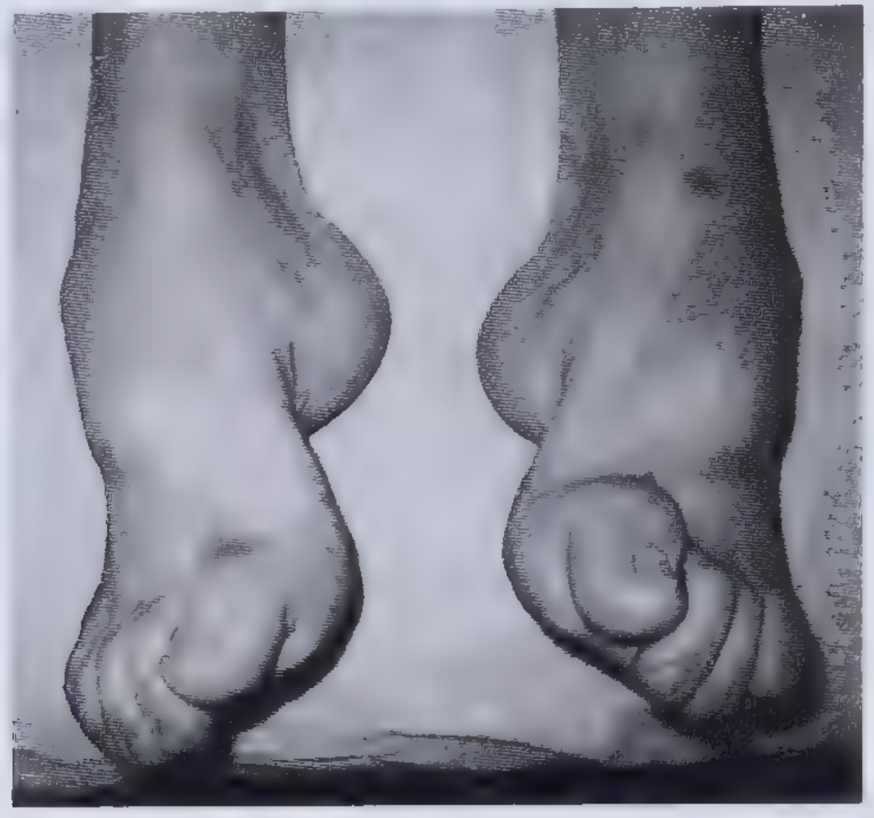
| Charcot-Marie-Tooth (CMT) | Young onset; family history; pes cavus + hammer toes (see image below); stork-leg deformity (distal wasting); palpably thickened nerves in some subtypes |
| Familial amyloidosis (TTR) | Carpal tunnel syndrome early; autonomic + sensorimotor; family history of neuropathy/heart disease |
| Porphyria (AIP) | Acute attacks; predominantly motor; abdominal pain, psychiatric symptoms, autonomic instability; urine discolours |
| HNPP | Hereditary neuropathy with liability to pressure palsies; episodic mononeuropathies at pressure points |
| Friedreich's ataxia | Spinocerebellar: ataxia + LMN neuropathy; pes cavus, scoliosis, cardiomyopathy |
| Mitochondrial disorders | Multi-system; hearing loss, ptosis, short stature, lactic acidosis |
Pes Cavus with Hammertoes - Charcot-Marie-Tooth (Hereditary Neuropathy)
STEP 4: MOST LIKELY DIAGNOSIS (By Probability)
| Rank | Diagnosis | Why Most Likely |
|---|---|---|
| #1 | Diabetic polyneuropathy | Most common cause of polyneuropathy in the industrialised world; middle-aged; distal symmetrical sensorimotor pattern is classic |
| #2 | Alcohol-related neuropathy | 2nd most common in UK; middle-aged man; look for liver disease signs + Dupuytren's |
| #3 | CIDP | Chronic, progressive; responds to treatment - important not to miss |
| #4 | B12 deficiency | Subacute combined degeneration - look for mixed UMN/LMN; anaemia, glossitis |
| #5 | CMT (hereditary) | If pes cavus, young-ish onset, family history, thickened nerves |
| #6 | Paraproteinaemia / myeloma | Middle-aged; check for bone pain, anaemia |
STEP 5: COMPLETING THE EXAMINATION IN PACES
"To complete my examination, I would like to: 1. Examine the upper limbs for distal weakness and sensory loss (glove distribution) - suggesting more severe or generalised polyneuropathy 2. Check for pes cavus and hammertoes - hereditary neuropathy 3. Palpate peripheral nerves for thickening - CMT, leprosy, amyloid 4. Look for systemic signs: spider naevi/Dupuytren's (alcohol), acanthosis/ulcers (diabetes), lymphadenopathy (malignancy/sarcoid), cushingoid features (steroid-induced myopathy DDx) 5. Check the spine for midline tenderness and do a rectal exam if cauda equina suspected 6. Gait assessment - high-stepping gait (foot drop) in peroneal/axonal neuropathy 7. Fundoscopy - diabetic retinopathy"
STEP 6: INVESTIGATIONS TO MENTION
| Investigation | What You're Looking For |
|---|---|
| FBS / HbA1c | Diabetes (most common cause) |
| Alcohol history + GGT/LFTs/MCV | Alcoholic neuropathy |
| TFTs | Hypothyroidism |
| Urea, creatinine, eGFR | Uraemic neuropathy |
| B12, folate, B1 | Nutritional deficiency |
| SPEP / UPEP + immunofixation | Paraproteinaemia (MGUS, myeloma) |
| FBC + film | Anaemia (B12, myeloma), macrocytosis |
| ANA, ANCA, anti-ENA | Connective tissue disease, vasculitis |
| ACE level + CXR | Sarcoidosis |
| HIV test | HIV neuropathy |
| Nerve conduction studies (NCS) + EMG | Axonal vs demyelinating; confirms polyneuropathy |
| CSF analysis | CIDP - raised protein, no cells |
| Genetic testing | CMT, if clinically suspected |
| Sural nerve biopsy | Amyloid, vasculitis, when diagnosis uncertain |
EXAMINER'S FAVOURITE QUESTIONS - Prepared Answers
"Distal symmetrical sensorimotor polyneuropathy - length-dependent, affecting the longest fibres first, causing stocking-and-glove sensory loss and distal > proximal weakness."
"The peripheral nerve forms the afferent and efferent limbs of the reflex arc. In polyneuropathy, damage to peripheral nerve fibres disrupts both limbs, leading to diminished or absent reflexes."
"Both cause weakness and sensory loss. Demyelinating neuropathies (GBS, CIDP, CMT-1) tend to cause more prominent reflex loss early and slowed nerve conduction velocities. Axonal neuropathies (diabetic, alcoholic) cause more prominent distal muscle wasting and reduced nerve action potential amplitudes on NCS. CIDP and GBS are important to identify as they are treatable with immunotherapy."
"Nerve conduction studies - they confirm the diagnosis of polyneuropathy, determine whether it is axonal or demyelinating, and quantify severity."
ok fine, in a similar case what points on examination will lead towards a possibility of much more likely cause as CIDP, only on physical examination as in short cases we can only examine the patient
Physical Examination Findings That Point Specifically to CIDP
THE SINGLE MOST IMPORTANT DISCRIMINATING FINDING
Proximal AND Distal Weakness - The Hallmark of CIDP
"Symmetric proximal and distal weakness is the hallmark of acquired immune demyelinating polyneuropathies, both the acute form (GBS) and the chronic form (CIDP). The importance of finding symmetric proximal AND distal weakness in a patient who presents with motor and sensory symptoms cannot be overstated."
"The occurrence of symmetric weakness in both proximal and distal muscles that progressively increases for more than 2 months is the pivotal symptom in the diagnosis of CIDP."
THE FULL SET OF CIDP-POINTING EXAMINATION FINDINGS
1. Proximal + Distal Weakness (Both Upper and Lower Limbs)
| Feature | CIDP | Diabetic/Alcoholic |
|---|---|---|
| Distal leg weakness | Yes | Yes |
| Proximal leg weakness (hip/knee) | Yes - bilateral | No / minimal |
| Upper limb weakness (hands + shoulders) | Yes - bilateral | Only in severe/late cases |
| Pattern | Symmetric, both proximal and distal | Symmetric, distal only |
Examination action: Test hip flexion (L1/2), knee extension (L3/4), shoulder abduction (C5), grip strength (C8/T1). CIDP will give you weakness at multiple levels including proximal. Diabetic neuropathy alone does not cause significant proximal weakness.
2. Upper Limb Involvement Relatively Early
3. Areflexia More Severe and Widespread Than Expected
- All four limb reflexes may be absent (arms + legs)
- Knee jerks often absent even when weakness appears mild
- Reflex loss is disproportionately severe relative to the degree of wasting
PACES tip: If you find absent knee AND ankle jerks AND absent biceps/triceps jerks in a patient with moderate (not severe) weakness, and no obvious metabolic cause staring at you (no Dupuytren's, no acanthosis) - think CIDP.
4. Large-Fibre Sensory Loss Predominates
| Modality | Fibre Type | CIDP Finding |
|---|---|---|
| Vibration sense | Large myelinated | Severely reduced/absent |
| Joint position sense (proprioception) | Large myelinated | Severely reduced/absent |
| Light touch | Large myelinated | Reduced |
| Pain and temperature | Small unmyelinated | Relatively preserved (or less affected) |
Contrast this with diabetic small-fibre neuropathy where pain/temperature loss (burning feet, neuropathic pain, reduced pinprick) often predominates early and vibration may be relatively preserved initially.
5. Sensory Ataxia (Romberg Positive)
- Romberg's sign is positive - the patient stands steadily with eyes open but sways/falls with eyes closed
- Gait is ataxic - broad-based, stamping gait (patient cannot sense where their feet are)
- Coordination testing (heel-shin) may be impaired due to proprioceptive loss, not cerebellar disease - the key distinction is that limb ataxia disappears when visual feedback is allowed
PACES action: Perform Romberg's test and observe the gait. A positive Romberg in the context of a polyneuropathy strongly supports large-fibre demyelination = CIDP.
6. Palpably Thickened Peripheral Nerves
- Great auricular nerve - runs over the sternocleidomastoid, visible and palpable as a cord in severe cases
- Ulnar nerve - at the elbow (behind the medial epicondyle)
- Common peroneal nerve - at the fibular head
- Radial nerve - in the spiral groove
- Sural nerve - at the lateral ankle
PACES tip: Run your fingers behind the medial epicondyle bilaterally and compare. A visibly prominent great auricular nerve over the SCM is dramatic and almost always noted by the examiner. This finding is present in CIDP, CMT-1, leprosy, and amyloid - but in a PACES context without tropical disease exposure, CIDP or CMT-1 is the answer.
7. No Systemic Signs of Metabolic Cause
| Absent finding | Excludes |
|---|---|
| No Dupuytren's, no parotid enlargement, no spider naevi | Alcohol |
| No acanthosis, no neuropathic ulcers, no lipohypertrophy marks | Diabetes |
| No dry skin, no bradycardia, no Queen Anne's sign | Hypothyroidism |
| No AV fistula, no pallor, no uraemic skin | CKD |
| No pes cavus, no family history clues, no scoliosis | CMT |
| No cachexia, no lymphadenopathy | Paraneoplastic |
8. Upper Limb Signs on Completing Examination
- Reduced grip strength bilaterally (distal hand weakness)
- Absent biceps/triceps/supinator reflexes
- Sensory loss in glove distribution (not just stocking)
- Positive Romberg when tested from standing
SUMMARY TABLE: PACES DISCRIMINATING FEATURES
| Examination Feature | Diabetic/Alcoholic | CIDP | CMT |
|---|---|---|---|
| Weakness distribution | Distal only | Proximal + Distal | Distal only |
| Upper limb involvement | Late/mild | Early/prominent | Early (hands) |
| Reflexes | Ankle jerks absent first | All reflexes lost, widespread | All reflexes lost |
| Sensory type predominant | Small fibre (pain/temp) | Large fibre (vibration/proprioception) | Variable |
| Romberg positive | Possible | Often positive | Possible |
| Nerve thickening | No | Yes (CIDP, some subtypes) | Yes (CMT-1) |
| Foot deformity (pes cavus) | No | No | Yes (CMT) |
| Systemic signs | Yes (diabetes/liver) | None | Family history |
| Temporal course | Chronic/insidious | Relapsing-remitting OR chronic progressive >2 months | Lifelong/stable |
THE MODEL PACES CANDIDATE STATEMENT FOR CIDP
"My examination findings reveal a bilateral sensorimotor polyneuropathy. Importantly, the weakness affects both proximal and distal muscle groups symmetrically in all four limbs, which is the hallmark of an acquired immune demyelinating polyneuropathy. Reflexes are absent throughout, including upper limbs, which is disproportionate to the degree of muscle wasting. Sensory loss preferentially affects large fibre modalities - vibration and joint position sense are severely impaired with a positive Romberg's sign, indicating sensory ataxia. I also note palpably thickened ulnar nerves at the elbows bilaterally. There are no stigmata of diabetes, alcohol-related liver disease, or hereditary foot deformity.These findings together most strongly suggest Chronic Inflammatory Demyelinating Polyneuropathy - CIDP. This is an important diagnosis to establish because it is potentially treatable with IVIg, plasma exchange, or corticosteroids. I would confirm with nerve conduction studies showing demyelinating pattern, and CSF showing elevated protein with normal cell count."
describe hyperkinetic movement disorders for MRCP (UK) PACES POV that candidates may get in short cases
Hyperkinetic Movement Disorders - MRCP PACES Short Case Guide
STEP 1: THE MASTER CLASSIFICATION TABLE
| Movement Type | Definition | Key Characteristic |
|---|---|---|
| Tremor | Rhythmic oscillation of a body part due to intermittent muscle contractions | Rhythmic, regular, oscillatory |
| Chorea | Rapid, semi-purposeful, graceful, dance-like non-patterned involuntary movements; distal or proximal | Random, flowing, non-repetitive |
| Ballism | Chorea of large amplitude, predominantly proximal | Violent, flinging, proximal |
| Dystonia | Involuntary, patterned, sustained or repeated muscle contractions; twisting movements and abnormal posture | Sustained, twisting, abnormal posture |
| Athetosis | Slow, distal, writhing involuntary movements; a form of dystonia with increased mobility | Slow, writhing, distal (hands) |
| Myoclonus | Sudden, brief (<100 ms), jerk-like, arrhythmic muscle twitches | Shock-like, lightning-fast jerks |
| Tic | Brief, repeated, stereotyped contractions - suppressible for a short time; simple or complex | Stereotyped, suppressible, urge-driven |
STEP 2: HOW TO DESCRIBE MOVEMENTS TO THE EXAMINER
- Distribution - which body parts? (face, hands, limbs, trunk, generalised)
- Symmetry - bilateral or unilateral?
- Relation to posture/action - present at rest, with posture, or only with movement?
- Rhythm - regular/rhythmic vs. random/arrhythmic?
- Speed - rapid, slow, sustained?
- Pattern - stereotyped and repetitive vs. random and flowing?
- Suppressibility - can the patient suppress it voluntarily?
DISORDER 1: TREMOR
Definition
Classification by Activation Condition
| Type | When Present | Classic Cause |
|---|---|---|
| Rest tremor | When limb fully supported, at rest; disappears with voluntary movement | Parkinson's disease |
| Postural tremor | When limb held against gravity (arms outstretched) | Essential tremor, enhanced physiologic tremor |
| Kinetic/Action tremor | During voluntary movement | Essential tremor, drug-induced |
| Intention tremor | Increases on approaching a target (finger-nose test) | Cerebellar disease |
PACES-Relevant Tremor Causes
A. Essential Tremor (ET) - Most Common Movement Disorder
- Affects ~1% of population, 5% over age 60
- 6-10 Hz high-frequency postural/action tremor
- Bilateral, predominantly upper limbs (hands)
- Head tremor in 30%, voice in 20%
- Improved by alcohol (pathognomonic clue - ask in history)
- Worsened by stress and caffeine
- Handwriting shows large, tremulous script (contrast with PD's micrographia)
- Family history in ~50% (autosomal dominant)
- Neurological exam otherwise normal (no bradykinesia, no rigidity)
- Treatment: propranolol, primidone
- Tremor present on arms outstretched - disappears at rest
- Tremor on finger-nose test (kinetic component) - no past-pointing
- Head nod (titubation)
- Normal tone, normal gait (or mild tandem gait difficulty)
- No cogwheel rigidity, no bradykinesia
B. Parkinson's Disease Tremor
- 4-6 Hz low-frequency resting tremor
- "Pill-rolling" - thumb rolling over fingers
- Disappears with voluntary movement (opposite to ET)
- Unilateral onset, later becomes bilateral
- Associated with bradykinesia, cogwheel rigidity, micrographia, shuffling gait, hypomimia
- (This is technically a hypokinetic disorder with a resting tremor - included here for differential)
C. Cerebellar Tremor
- Intention tremor - absent at rest, minimal with posture, maximal as approaching target
- Associated cerebellar signs: ataxia, nystagmus, dysarthria (scanning speech), dysdiadochokinesia, past-pointing, hypotonia
- Causes: MS, alcohol, paraneoplastic, cerebellar stroke
D. Enhanced Physiologic Tremor
- High-frequency (10-12 Hz), fine postural tremor
- Causes: hyperthyroidism, anxiety, alcohol withdrawal, caffeine, drugs (valproate, lithium, beta-agonists, steroids)
- Look for: thyroid signs, signs of alcohol dependence, drug history
DISORDER 2: CHOREA
Definition
PACES Examination Signs
- Milkmaid's grip - when asked to squeeze your fingers, alternating contractions cause grip to vary rhythmically (cannot sustain a constant squeeze)
- Pronator sign - patient cannot keep arms supinated when outstretched; hand pronates involuntarily
- Tongue impersistence - cannot keep tongue protruded steadily; it darts in and out
- Choreic gait - irregular, irregular steps, dancing quality
- The face, hands, and limbs all show random, flowing, non-repetitive movements
DDx of Chorea - PACES Causes
| Cause | Distinguishing Clues on Examination |
|---|---|
| Huntington's disease | Young-middle-aged; eye movement abnormalities (slowed saccades); cognitive decline (dementia); psychiatric features; positive family history; caudate atrophy on MRI (shown below) |
| Sydenham's chorea | Young patient; preceding streptococcal infection/rheumatic fever; may have mitral valve disease (cardiac murmur on auscultation); emotional lability |
| Drug-induced (levodopa, antipsychotics) | Medication history; dyskinesias in PD patients on levodopa - typically face/neck/trunk |
| Tardive dyskinesia | Chronic antipsychotic/metoclopramide use; orofacial movements (lip smacking, tongue protrusion, chewing) |
| SLE / antiphospholipid syndrome | Butterfly rash, arthritis, livedo reticularis, young woman |
| Pregnancy (chorea gravidarum) | History; associated with antiphospholipid or past rheumatic fever |
| Thyrotoxicosis | Goitre, tachycardia, tremor, sweating, weight loss |
| Polycythaemia rubra vera | Plethoric facies, splenomegaly |
| Wilson's disease | Young patient; Kayser-Fleischer rings on slit-lamp; liver disease; psychiatric features |
Huntington's Disease MRI (for PACES viva context)
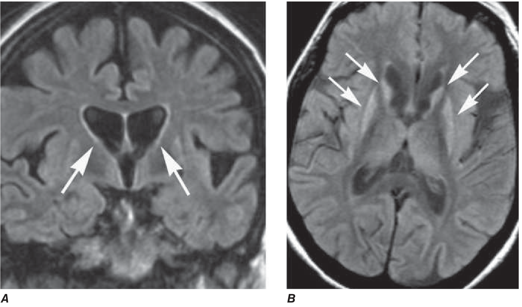
DISORDER 3: BALLISM / HEMIBALLISMUS
Definition
PACES Points
- Almost always unilateral (hemiballismus) → lesion of the contralateral subthalamic nucleus
- Most common cause: contralateral subthalamic nucleus infarct (lacunar stroke in hypertensive patient)
- Examine for: hypertension signs, contralateral signs of stroke
- Look for: Hemi-distribution (one side only), preserved consciousness, normal cognition
- Often self-limiting (resolves in weeks); treatment with tetrabenazine if disabling
DISORDER 4: DYSTONIA
Definition
Classification
| By Distribution | By Onset |
|---|---|
| Focal - one body part | Primary (idiopathic/genetic) |
| Segmental - two adjacent regions | Secondary (acquired) |
| Hemidystonia - one side | |
| Generalised |
PACES-Relevant Dystonia Scenarios
| Type | Clinical Features | Cause |
|---|---|---|
| Cervical dystonia (Torticollis) | Head turned/tilted to one side; neck muscles hypertrophied; may use "geste antagoniste" (touching chin relieves it) | Idiopathic, drug-induced |
| Writer's cramp | Task-specific hand dystonia; arm postures abnormally only when writing | Idiopathic |
| Blepharospasm | Forced, involuntary eye closure; bilateral; interferes with vision | Meige syndrome (+ oromandibular dystonia) |
| Oromandibular dystonia | Jaw opening/closing, grimacing, tongue protrusion | Tardive, idiopathic |
| Tardive dystonia | Axial predominant; chronic antipsychotic use; rocking trunk movements | Drug-induced |
| DYT1 (Oppenheim's) | Young onset; generalised; starts in one leg; autosomal dominant | Genetic |
| Wilson's disease | Young patient; dystonia + liver disease + KF rings + psychiatric | Copper metabolism |
DISORDER 5: MYOCLONUS
Definition
PACES Key Points
- Action myoclonus - jerks appear when patient moves (e.g., reaching for a cup)
- Stimulus-sensitive myoclonus - jerks triggered by touch, sound, or light
- Cortical myoclonus - focal, often stimulus-sensitive; associated with epilepsy
- Subcortical/spinal myoclonus - more generalised
PACES-Relevant Causes
| Cause | Clues on Examination |
|---|---|
| Physiologic (sleep starts, hiccups) | Normal otherwise |
| Essential myoclonus | Isolated, no other neurology |
| Post-hypoxic (Lance-Adams syndrome) | History of cardiac arrest or prolonged hypoxia; action-induced myoclonus |
| Metabolic (uraemia, hepatic encephalopathy, hyponatraemia) | Asterixis (negative myoclonus - flapping tremor); encephalopathy; systemic signs |
| Drug-induced (opioids, SSRIs, lithium, penicillin) | Medication history |
| Epileptic (juvenile myoclonic epilepsy) | Morning jerks; young person |
| Neurodegenerative (CJD, Alzheimer's) | Cognitive decline; CJD: rapid progression, startle myoclonus, cerebellar signs |
PACES tip: Asterixis (liver flap) is technically a negative myoclonus - brief lapses in sustained posture. Always look for it when you see flapping-type movements. Ask the patient to hold arms outstretched with wrists dorsiflexed. Causes: hepatic encephalopathy, uraemia, CO2 retention, drug toxicity.
DISORDER 6: TICS
Definition
PACES Points
- Patient can suppress but builds up an overwhelming urge to perform the movement
- After suppression: burst of tics when released
- Simple motor tics: eye blinking, facial grimacing, shoulder shrugging, head jerking
- Complex motor tics: jumping, touching, gesturing
- Vocal tics: throat clearing, sniffing, barking, coprolalia (in minority)
Tourette Syndrome
- Multiple motor tics + at least one vocal tic, onset <18 years, lasting >1 year
- Associated: OCD, ADHD, echolalia, coprolalia
- Worsens with stress, improves with sleep and concentration
- Examination: tics visible; otherwise neurologically normal
- Treatment: clonidine, risperidone, haloperidol, habit-reversal therapy
DISORDER 7: TARDIVE DYSKINESIA - HIGH YIELD PACES CASE
- Lip smacking / lip pursing
- Tongue protrusion / rolling
- Chewing movements (buccal-masticatory syndrome)
- Long-term antipsychotic use (haloperidol, chlorpromazine, older drugs)
- Also metoclopramide (>12 weeks - FDA black box warning)
- Prochlorperazine
- Also affects trunk, limbs, and respiratory muscles in severe cases
THE PACES CANDIDATE'S APPROACH - SUMMARY FLOW
PATIENT WITH INVOLUNTARY MOVEMENTS
│
DESCRIBE THE MOVEMENT FIRST
─────────────────────────────
Rhythmic? → TREMOR
Random/flowing? → CHOREA
Violent/flinging/proximal? → BALLISM (hemiballismus)
Sustained/twisting/posturing? → DYSTONIA
Lightning-fast jerks? → MYOCLONUS
Stereotyped/suppressible? → TICS
Orofacial/stereotyped on antipsychotics? → TARDIVE DYSKINESIA
│
THEN EXAMINE FOR CLUES TO THE CAUSE
(See table below)
MASTER TABLE: WHAT TO LOOK FOR ON EXAMINATION
| Movement | Examination Finding | Likely Cause |
|---|---|---|
| Rest tremor + rigidity + bradykinesia | Cogwheel rigidity, shuffling gait, hypomimia | Parkinson's disease |
| Action/postural tremor alone | Normal exam otherwise; improved with alcohol | Essential tremor |
| Intention tremor + cerebellar signs | Nystagmus, ataxia, dysarthria | Cerebellar disease |
| Postural tremor + thyroid signs | Goitre, tachycardia, lid lag | Thyrotoxicosis |
| Chorea + eye movement abnormalities + cognitive impairment | Slowed saccades, frontal dementia | Huntington's disease |
| Chorea + mitral murmur | Young patient, cardiac disease | Sydenham's / rheumatic fever |
| Chorea + butterfly rash + arthritis | SLE signs | SLE/antiphospholipid |
| Hemiballismus alone | Contralateral stroke signs | Subthalamic nucleus infarct |
| Cervical dystonia + geste antagoniste | Head tilt, neck hypertrophy, relieved by chin touch | Idiopathic cervical dystonia |
| Dystonia + KF rings + liver signs | Young, liver disease, psychiatric | Wilson's disease |
| Orofacial movements + antipsychotic use | Lip smacking, tongue rolling | Tardive dyskinesia |
| Myoclonus + flapping | Liver signs or uraemia signs | Metabolic encephalopathy (asterixis) |
| Myoclonus + rapid dementia | Global cognitive decline, cerebellar signs | CJD |
| Tics + stereotyped + suppressible | Young patient, OCD features | Tourette syndrome |
PACES PRESENTATION TEMPLATE
"On inspection of this patient, I observed [describe movement type and distribution]. The movements are [rhythmic/random/sustained/brief], affecting [body parts], present at [rest/posture/action], and [are/are not] suppressible.This is most consistent with [tremor/chorea/dystonia/myoclonus/tics].On examination, I additionally found [relevant positive and negative findings].The most likely underlying diagnosis is [specific cause], because [key distinguishing features].I would like to complete my examination by [checking tone, saccadic eye movements, fundoscopy, KF rings, cardiac auscultation, cognitive assessment] and would confirm with [genetic testing/slit-lamp/TFTs/copper studies/drug history]."
now give me DDx of Spastic Monoparesis, with normal examination findings in right leg but spastic findings in left leg, increased tone, reduced power, hyper-reflexia in knee, loss of vibration and proprioception but preserved soft touch and pain, I have not examined Upper limb, Cranial nerves and cerebellum, Give me a list of possible DDx that is relevant to MRCP (UK) PACES exam neurology short cases
- Left leg only - UMN pattern (spasticity, hyperreflexia, reduced power) = corticospinal tract lesion
- Sensory dissociation - vibration/proprioception LOST, soft touch/pain PRESERVED = dorsal column lost, spinothalamic intact
- This combination = Brown-Séquard syndrome pattern or a hemicord/unilateral cord lesion
- Right leg completely normal
MRCP PACES - Spastic Left Leg Monoparesis with Dorsal Column Loss
STEP 1: ANATOMICAL ANALYSIS - THE CRITICAL FIRST STEP
The Findings Mapped Anatomically
| Finding | Tract Involved | Location of Tract |
|---|---|---|
| Left leg: Increased tone, reduced power, hyper-reflexia (UMN pattern) | Left corticospinal tract | Ipsilateral in cord (descends uncrossed below foramen magnum) |
| Left leg: Vibration and proprioception LOST | Left dorsal column (fasciculus gracilis for leg) | Ipsilateral in cord |
| Left leg: Soft touch and pain PRESERVED | Left spinothalamic tract INTACT | Crosses contralateral in cord |
| Right leg: completely normal | Right cord spared |
The Anatomical Conclusion
- Left corticospinal tract (UMN signs left leg)
- Left dorsal column (vibration + proprioception lost left leg)
- Left spinothalamic tract is spared (pain/touch preserved)
"Brown-Séquard syndrome: ipsilateral pyramidal deficit, loss of ipsilateral tactile discrimination, position sense, and vibratory sensation, and loss of pain and temperature sensation on the contralateral aspect - one to two dermatomes below the level of injury."
PACES key insight: In a pure Brown-Séquard, you would expect right leg pain/temperature loss (contralateral spinothalamic). In this case pain/touch is preserved bilaterally - this is a partial/incomplete Brown-Séquard or an anterior-to-posterolateral cord lesion preferentially sparing the spinothalamic. This is actually the more common presentation in clinical practice.
STEP 2: WHERE IS THE LESION IN THE NEURAXIS?
- A cord lesion (thoracic or cervical - affecting lower limb representation)
- A parasagittal cortical/subcortical lesion (the leg area of motor homunculus lies in the parasagittal cortex - medial surface - and can give isolated leg weakness)
- An internal capsule lesion (though this would usually involve arm + leg + face)
- Dorsal columns are ipsilateral all the way to the medulla
- Combined UMN + dorsal column loss ipsilaterally = spinal cord (most likely) or medullary lesion
STEP 3: WHAT COMPLETING THE EXAMINATION WOULD REVEAL - AND WHY IT MATTERS
| Examination | What You're Looking For | Why It Matters |
|---|---|---|
| Upper limbs | UMN signs in left arm? (spasticity, hyperreflexia) | If yes → cervical cord or brain; if no → thoracic cord |
| Left arm vibration/proprioception | Lost? | If yes → cervical cord lesion |
| Sensory level on trunk | Where does sensory change start? | Defines the spinal cord level precisely |
| Cranial nerves | Any CN palsies? | If yes → brainstem involved; if no → cord or cortex |
| Left face involvement | UMN facial weakness? | If yes → supratentorial (brain) not cord |
| Lhermitte's sign | Electric shock down spine on neck flexion | Positive → cervical cord pathology (MS, spondylosis) |
| Bladder/bowel | Retention, incontinence | Cord compression |
| Spine | Midline tenderness, step deformity, scar | Structural cord pathology |
STEP 4: COMPLETE DDx - PACES RELEVANT
GROUP A: SPINAL CORD LESIONS (Most Likely Given Brown-Séquard Pattern)
1. Multiple Sclerosis (MS) - TOP DIAGNOSIS in PACES
- Relative afferent pupillary defect (RAPD) - previous optic neuritis
- Internuclear ophthalmoplegia (INO) - failure of adduction on lateral gaze (brainstem plaque)
- Lhermitte's sign - electric shock down spine on neck flexion (cervical cord plaque)
- Cerebellar signs (intention tremor, nystagmus, ataxia) - "DANISH" mnemonic: Dysarthria, Ataxia, Nystagmus, Intention tremor, Scanning speech, Hypotonia
- Upper limb signs if cervical cord plaque
- Uhthoff's phenomenon (worsening in heat) - ask in history
- No systemic signs of other disease - otherwise well-appearing patient
2. Spinal Cord Compression - Extradural
- Commonest cause of spinal cord compression in adults >50
- Gradual onset, often with neck pain radiating to arm
- Examination: neck stiffness, restricted neck movements, wasting of intrinsic hand muscles (C8/T1), Lhermitte's sign
- May have lower motor neurone signs at the level of compression (wasted hand) + UMN below
- Very high PACES yield - metastatic deposits in vertebrae compressing cord
- Look for: cachexia, lymphadenopathy, clubbing (bronchial), breast mass, midline vertebral tenderness on percussion, point tenderness
- Breast, lung, prostate, renal, thyroid are the common primaries ("BLT + KT" - "Bad Luck Tumours Killing Tissue")
- Thoracic cord most commonly compressed (longest segment, poorest vascular supply)
- Acute onset if pathological fracture; gradual if slow growth
- Fever + back pain + neurological deficit = emergency
- Examination: fever, tenderness over spine, may have source of infection (IV drug use track marks, skin infection, dental procedure)
- Staphylococcus aureus most common organism
3. Intradural Extramedullary Tumours
- Middle-aged to older women (female predominance)
- Thoracic cord most common site
- Slow progressive cord compression
- Examination: no systemic signs (benign), gradual UMN + sensory level
- Look for: neurofibromatosis stigmata (NF2 - multiple meningiomas + bilateral acoustic neuromas + café-au-lait spots)
- Can compress cord asymmetrically → Brown-Séquard
- NF1: café-au-lait spots ≥6, axillary freckling, neurofibromas, Lisch nodules (iris hamartomas)
- NF2: bilateral acoustic neuromas, spinal and cranial meningiomas
4. Intramedullary Cord Lesions
- Cavity within the cord, usually cervical
- Central cord syndrome classically: dissociated sensory loss (pain/temp lost, dorsal columns preserved early) - different from this case
- But when syrinx expands laterally → Brown-Séquard-like
- Examination: cape/vest distribution of pain/temperature loss over shoulders, wasted hands (lower motor neurone at level), UMN legs, Horner's syndrome (ciliospinal centre)
- Associated: Chiari malformation (cerebellar tonsillar descent), scoliosis, pes cavus, kyphoscoliosis
- Anterior spinal artery syndrome: bilateral UMN + bilateral pain/temperature loss, dorsal columns SPARED (this case has dorsal column loss, so ASA syndrome less likely - but partial presentations occur)
- Causes: aortic aneurysm repair, aortic dissection, atherosclerosis, emboli
- Sudden onset (seconds to minutes) - key history point
5. Inflammatory/Infectious Cord Lesions
- Acute/subacute inflammatory demyelination of cord
- Bilateral more common but can be asymmetric
- Causes: post-viral, MS, NMO (neuromyelitis optica), sarcoidosis, SLE
- NMO: associated with aquaporin-4 antibodies; predilection for optic nerves + long spinal cord segments; young women; Asian patients
- Important in immigrant/at-risk patients
- Tropical spastic paraparesis (HTLV-1): bilateral spastic legs, dorsal column loss, bladder involvement
- Examination: signs of HIV (if applicable)
- Dorsal column destruction by Treponema pallidum
- Loss of vibration + proprioception + Romberg positive
- Argyll Robertson pupils (small, irregular, accommodate but don't react to light - pathognomonic)
- UMN signs in legs
- History: past STI, risk behaviour (very relevant to our running patient case!)
GROUP B: BRAIN LESIONS (If Upper Limb Normal on Examination)
6. Parasagittal Cortical/Subcortical Lesion
- Parasagittal meningioma - classic PACES case; middle-aged woman; slowly progressive leg weakness; look for signs of raised ICP; bilateral leg weakness if crosses midline
- Superior sagittal sinus thrombosis (CVST) - young woman on OCP; headache + leg weakness; parasagittal cortical venous infarcts
- Falx meningioma - compresses medial motor cortex
- Parasagittal metastasis
- Cortical vein thrombosis with infarction
7. Anterior Cerebral Artery (ACA) Territory Infarct
- ACA supplies medial frontal and parietal cortex (leg area)
- Contralateral leg monoplegia + sensory loss
- Behavioural changes (frontal lobe involvement) - abulia, incontinence
- Usually bilateral if both ACAs affected (ACA territory infarct is rare but classic)
- Look for: atrial fibrillation, carotid bruit, risk factors for stroke
STEP 5: COMPLETE DDx TABLE FOR PACES
| Diagnosis | Key Distinguishing Examination Findings |
|---|---|
| Multiple sclerosis | Young adult; RAPD; INO; Lhermitte's; cerebellar signs; relapsing history; no systemic signs |
| Cervical disc / spondylotic myelopathy | Age >50; neck stiffness; restricted cervical movement; wasted hand muscles (LMN at level); Lhermitte's |
| Spinal metastasis (extradural) | Cachexia; vertebral tenderness on percussion; lymphadenopathy; primary tumour signs; older patient |
| Spinal meningioma | Middle-aged woman; no systemic signs; slow progression; NF2 stigmata if applicable |
| Spinal neurofibroma | Café-au-lait spots; axillary freckling; NF1 stigmata; palpable neurofibromas |
| Syringomyelia | Cape sensory loss (pain/temp); wasted hands; Horner's; scoliosis; Chiari signs (cerebellar) |
| Transverse myelitis / NMO | Subacute; young woman; prior optic neuritis (RAPD); Asian patient |
| Tabes dorsalis (syphilis) | Argyll Robertson pupils; sexual history; Romberg positive; lightning pains |
| Parasagittal meningioma | Slowly progressive; bilateral if large; signs of raised ICP; no spinal level |
| ACA territory infarct | Stroke risk factors; frontal lobe features; AF on pulse; carotid bruit |
| Spinal cord AVM | Young; sudden onset; bruit over spine (rare); high T2 signal on MRI |
| HTLV-1 myelopathy | Caribbean/South American/Japanese origin; spastic legs; bladder |
STEP 6: PACES CANDIDATE PRESENTATION
"On examination of this gentleman's lower limbs, I found a left-sided spastic monoparesis. Specifically, the left leg demonstrates increased tone - spasticity, with power reduced to [X]/5, and the left knee jerk is brisk/hyperreflexic with an extensor plantar response, consistent with an upper motor neurone lesion. The right leg is entirely normal.On sensory testing, there is loss of vibration and joint position sense in the left leg, indicating a left dorsal column deficit. Soft touch and pain sensation are preserved bilaterally, indicating the spinothalamic tracts are intact.This combination - ipsilateral UMN signs and ipsilateral dorsal column loss, with preserved pain and temperature - is the classic pattern of a Brown-Séquard syndrome or partial hemisection of the spinal cord, most likely at the thoracic or cervical level.I have not yet examined the upper limbs, cranial nerves, or cerebellum - I would like to do so to further localise the lesion. If the upper limbs are normal, this points to a thoracic cord lesion. If there are upper limb UMN or sensory signs, a cervical cord lesion is more likely. If cranial nerve signs are present, a brainstem lesion is possible.My leading differential diagnoses are: 1. Multiple sclerosis - most likely in a young adult with this pattern 2. Spinal cord compression - from a disc, meningioma, or metastatic deposit 3. A parasagittal brain lesion - if upper limbs are normalI would like to complete my examination and then confirm with MRI of the brain and whole spine with gadolinium."
STEP 7: THE EXAMINER'S LIKELY FOLLOW-UP QUESTIONS
"In a classic Brown-Séquard, the spinothalamic fibres crossing contralaterally are also injured, giving contralateral pain and temperature loss below the lesion. Here, pain and touch are preserved bilaterally, suggesting the spinothalamic tract is spared. This is a partial or incomplete Brown-Séquard, which is actually the more common presentation clinically."
"This would indicate a cervical cord lesion above the C5 level, with the leg representation more severely affected than the arms. This pattern is seen in cervical spondylotic myelopathy or a left-sided cervical cord MS plaque."
"The lesion is likely below C5 (thoracic cord) - or alternatively a parasagittal cortical lesion selectively affecting the leg area. A thoracic cord lesion would explain normal upper limbs + left leg UMN + left dorsal column loss."
"MRI of the spine (and brain) with gadolinium - to identify the level and nature of the cord lesion. This will distinguish demyelination (MS plaque - T2 bright, gadolinium enhancing in active disease), structural compression (disc, tumour), inflammatory (transverse myelitis), and syrinx."

This a Long Clinical Case Consultation (LCC) in MRCP (UK) PACES Give me detailed Hx questions and relevant physical examination schemes that I should perform according to the most imp DDx list for PACES
- Ms. Yasmeen, 34-year-old woman
- Pain in hands for past few months
- Minimal relief with painkillers + physiotherapy
- Vitals: Temp 38.2°C (low-grade fever), BP 130/60, PR 70, RR 18, SpO2 96% (borderline)
This a Long Clinical Case Consultation (LCC) in MRCP (UK) PACES Give me detailed Hx questions and relevant physical examination schemes that I should perform according to the most imp DDx list for PACES
it is tomorrow, why its not resetting