


1779121571570278309420560076682.jpg
+2 more
Explain visually
Reading File
The images cover Chapter 54 — Aminoglycoside Antibiotics from a pharmacology textbook. I'll create a comprehensive visual explanation covering all the content across the three pages.
Searching Images
aminoglycoside mechanism of action ribosome protein synthesis
Searching Images
aminoglycoside transport bacterial cell membrane
Loading Image
Here is a full visual breakdown of Chapter 54 — Aminoglycoside Antibiotics:
🦠 What Are Aminoglycosides?
Natural and semi-synthetic antibiotics with polybasic amino groups linked to aminosugar residues. All produced by soil actinomycetes.
| Category | Members |
|---|---|
| Systemic | Streptomycin, Gentamicin, Kanamycin, Tobramycin, Amikacin, Sisomicin, Netilmicin, Paromomycin |
| Topical | Neomycin, Framycetin |
⚙️ Mechanism of Action — Two Steps
Step 1 — Entry into the bacterial cell (multistep transport)
Outer coat (porin channels)
↓
Periplasmic space
↓ ← carrier-mediated, linked to electron transport chain
Cytoplasmic membrane ← Energy-Dependent Phase I (EDP₁)
↓
Inside cell
Why anaerobes are resistant: The carrier system is oxygen-dependent. Without O₂, the membrane is not polarized → drug can't get in. This also explains why abscesses (low O₂) reduce efficacy.
Why alkaline pH helps: Aminoglycosides are ~20× more active at alkaline pH. β-lactams and vancomycin (cell-wall agents) enhance entry by disrupting the outer barrier.
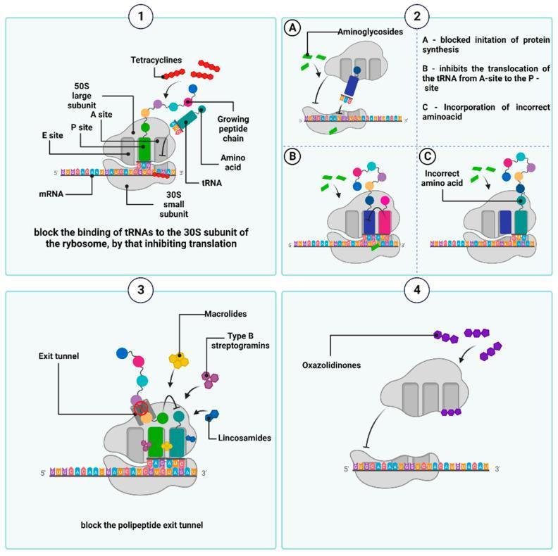
Step 2 — Ribosome binding → Protein synthesis inhibition
Aminoglycosides bind 30S ribosome (streptomycin) and also 50S subunit & 30S–50S interface (other members):
| Effect | Consequence |
|---|---|
| A Block initiation complex | Protein synthesis never starts |
| B Inhibit tRNA translocation (A→P site) | Elongation stops |
| C Distort mRNA codon recognition | Wrong amino acids incorporated → defective/toxic proteins |
These defective proteins insert into the cell membrane → membrane becomes permeable → ions, amino acids, proteins leak out → cell death (bactericidal, not just static).
This also triggers EDP₂ (Energy-Dependent Phase II) — more drug enters, reinforcing the lethal cycle.
🛡️ Mechanism of Resistance
3 Mechanisms:
1. Modifying enzymes (most important)
├─ Phosphorylate, adenylate, or acetylate the drug
├─ Modified drug can't bind ribosomes
└─ Encoded in plasmids → spread between bacteria
2. Ribosomal mutation
└─ Reduced affinity of ribosomal proteins → partial resistance
3. Decreased transport
├─ Outer coat porin channels become less permeable
└─ Active transport interfered → drug can't reach ribosome
Cross-resistance among aminoglycosides is variable (depends on which enzymes are present).
☠️ Shared Toxicities
Table 54.1 — Comparative Toxicity
| Drug | Vestibular Toxicity | Cochlear Toxicity | Nephrotoxicity |
|---|---|---|---|
| Streptomycin | ++ (high) | ± (low) | + |
| Gentamicin | ++ (high) | + | + |
| Kanamycin | + | ++ (high) | ++ |
| Tobramycin | +± | + | ++ |
| Amikacin | + | +± | +± |
| Sisomicin | +± | + | +± |
| Netilmicin | +± | + | +± |
1. 🔊 Ototoxicity (most important adverse effect)
Drug concentrates in labyrinthine fluid → slowly removed when plasma falls
↑ Plasma trough > threshold → concentration-dependent toxicity
For Gentamicin:
Trough > 2 μg/ml → vestibular damage (concentration-dependent)
Target trough < 1 μg/ml to avoid toxicity
Cochlear damage:
- Starts at base (high frequencies) → spreads to apex (low frequencies)
- Tinnitus → progressive hearing loss
- Sensory cells do NOT regenerate → damage is permanent
- Kanamycin & Amikacin = worst cochlear toxicity
Vestibular damage:
- Headache → nausea → dizziness → nystagmus → vertigo → ataxia
- Stop drug: enters chronic phase (6–10 weeks asymptomatic lying down, difficulty walking)
- Recovery over 1–2 years — often incomplete; elderly recover poorly
- Streptomycin & Gentamicin = worst vestibular toxicity
2. 🫘 Nephrotoxicity
Drug → high concentration in renal cortex (proximal tubules)
↓
Tubular damage → ↓ urinary concentrating power
↓
↓ GFR, nitrogen retention, albuminuria, casts
↓
↓ Drug clearance → ↑ blood levels → ↑ ototoxicity
- Single daily dosing causes less nephrotoxicity than thrice-daily dosing
- Worse in: elderly, pre-existing kidney disease
- If drug is stopped promptly → reversible
- Streptomycin = least nephrotoxic
3. 🧠 Neuromuscular Blockade
All aminoglycosides reduce ACh release at motor nerve endings by:
- Blocking synaptic vesicle fusion with terminal membrane (antagonize Ca²⁺)
- Reducing muscle end-plate sensitivity to ACh
Clinically silent usually, but apnoea and fatalities can occur if drug is put into peritoneal/pleural cavity after surgery (especially with curare-type muscle relaxants). Partially reversed by IV calcium salt. Myasthenic weakness is worsened.
⚠️ Precautions & Interactions
| Precaution | Reason |
|---|---|
| Avoid in pregnancy | Foetal ototoxicity |
| Avoid with NSAIDs, amphotericin B, vancomycin, cyclosporine, cisplatin | Additive nephrotoxicity |
| Caution with vancomycin, minocycline, furosemide | Potentially additive ototoxicity |
| Caution in patients >60 yrs or kidney disease | Reduced clearance, higher toxicity risk |
🔑 Common Properties (Memory Summary)
S-I-B-B-A-P-C-N-O
- Sulfate salts — highly water-soluble, stable for months
- Ionize in solution — not absorbed orally; no CNS/CSF penetration
- Bactericidal — more active at alkaline pH
- Bacteria targeted — aerobic gram-negative bacilli (not anaerobes)
- All excreted unchanged by glomerular filtration
- Partial cross-resistance only
- Concentration-distributed extracellularly only
- Narrow margin of safety
- Ototoxicity + nephrotoxicity in all members
This is a shared conversation. Sign in to Orris to start your own chat.