
Regional anatomy
I need a bit more context to give you the most useful answer. "Regional anatomy" is a broad topic that covers the entire body.
Asking for Preferences
Which region or topic are you interested in? · What do you need this for?
Reading File
Finding Sources
Finding Sources
Gray's Anatomy for Students is available. Let me pull the key Upper Limb content - bones, muscles, nerves, vessels, and joints - from multiple blocks simultaneously.
Finding Sources
Finding Sources
Finding Sources
Now let me read the key sections from block4 - the main Upper Limb chapter with bones/overview, brachial plexus, and shoulder content in parallel.
Reading File
Reading File
Reading File
Now let me read the arm/forearm, hand, and arterial supply sections simultaneously.
Reading File
Reading File
Reading File
Now let me read a few more key sections - the arm (humerus/compartments), hand overview, and fetch key diagrams.
Reading File
Reading File
Loading Image
I now have comprehensive content from Gray's Anatomy for Students. Let me also grab the key sections on the arm compartments, carpal tunnel, and fetch the brachial plexus schematic.
Finding Sources
Loading Image
Excellent - I have everything I need. Let me now compose the comprehensive Upper Limb Regional Anatomy study notes.
Upper Limb - Regional Anatomy (Exam Study Notes)
Source: Gray's Anatomy for Students
1. Overview and Bones
The upper limb is divided into five regions:
| Region | Key Bones |
|---|---|
| Shoulder girdle | Clavicle, scapula |
| Arm (brachium) | Humerus |
| Forearm (antebrachium) | Radius (lateral), Ulna (medial) |
| Wrist (carpus) | 8 carpal bones |
| Hand | 5 metacarpals + 14 phalanges |
Mnemonic for carpals (proximal to distal, lateral to medial):
- Proximal row: Scaphoid, Lunate, Triquetrum, Pisiform
- Distal row: Trapezium, Trapezoid, Capitate, Hamate
- "Some Lovers Try Positions That They Can't Handle"
2. Brachial Plexus
The most tested topic in upper limb anatomy.
Roots: Anterior rami of C5, C6, C7, C8, T1 - pass between anterior and middle scalene muscles, posterior to subclavian artery.

Schematic (Roots → Trunks → Divisions → Cords → Branches):

Trunks:
- Superior trunk = C5 + C6
- Middle trunk = C7 alone
- Inferior trunk = C8 + T1
Divisions: Each trunk divides into anterior and posterior divisions.
Cords (named by position relative to the 2nd part of the axillary artery):
- Lateral cord = anterior divisions of Superior + Middle trunks
- Medial cord = anterior division of Inferior trunk
- Posterior cord = all three posterior divisions
Key terminal branches:
| Cord | Terminal Nerves | Key Functions |
|---|---|---|
| Lateral | Musculocutaneous nerve, lateral root of median nerve | Arm flexors; forearm sensation |
| Medial | Ulnar nerve, medial root of median nerve | Intrinsic hand muscles; ring/little fingers |
| Posterior | Axillary nerve, radial nerve | Deltoid; all posterior compartment muscles |
The "M" over the axillary artery: The musculocutaneous nerve, lateral root of median nerve, median nerve, medial root of median nerve, and ulnar nerve form a visible M-shape over the 3rd part of the axillary artery - a landmark for identifying plexus components in surgery.
3. Axilla
The axilla is the gateway to the upper limb - a pyramidal space formed by the clavicle, scapula, upper thoracic wall, and humerus. It has four walls, an inlet (continuous with the neck), and a floor.
All major structures passing into/out of the upper limb pass through the axilla, including the axillary artery, axillary vein, and brachial plexus.
Key spaces in the posterior axillary wall:
- Quadrangular space: transmits axillary nerve + posterior circumflex humeral artery
- Triangular space: transmits circumflex scapular artery
- Triangular interval: transmits radial nerve + profunda brachii artery into the arm
4. Shoulder Region
Glenohumeral joint - ball-and-socket synovial joint, most mobile in body; least stable.
Rotator cuff muscles (SITS):
| Muscle | Action | Nerve |
|---|---|---|
| Supraspinatus | Abduction (initiates 0-15°) | Suprascapular (C5, C6) |
| Infraspinatus | Lateral rotation | Suprascapular (C5, C6) |
| Teres minor | Lateral rotation | Axillary (C5, C6) |
| Subscapularis | Medial rotation | Subscapular nerves (C5, C6) |
Axillary nerve exits the quadrangular space and innervates the deltoid and teres minor. It wraps around the surgical neck of the humerus - vulnerable in neck-of-humerus fractures and shoulder dislocations.
5. Arm (Humerus Region)
Two fascial compartments separated by medial and lateral intermuscular septa:
Anterior (flexor) compartment:
- Muscles: Biceps brachii, brachialis, coracobrachialis
- Nerve: Musculocutaneous nerve (C5, C6, C7)
- Biceps: long head from supraglenoid tubercle, short head from coracoid process → flexion + supination
- Brachialis: main elbow flexor (deep to biceps)
Posterior (extensor) compartment:
- Muscle: Triceps brachii (3 heads: long, medial, lateral)
- Nerve: Radial nerve (C7, C8)
- Radial nerve travels in the spiral groove (radial groove) of the humerus - vulnerable in mid-shaft humeral fractures → wrist drop (inability to extend wrist/fingers)
Main artery: Brachial artery (continuation of axillary) runs in the anterior compartment medial to the biceps tendon, bifurcating at the cubital fossa into radial and ulnar arteries.
6. Elbow Joint
- Hinge-type synovial joint allowing flexion/extension; also incorporates proximal radioulnar joint for pronation/supination
- Bones: Humerus (trochlea + capitulum) articulates with Ulna (trochlear notch) and Radius (head)
- Anular ligament of radius holds the radial head against the radial notch of the ulna; important in pronation/supination
- Fat pads lie over the olecranon, coronoid, and radial fossae - displacement ("sail sign") on X-ray indicates intra-articular effusion/fracture
- Medial (ulnar) collateral ligament & Lateral (radial) collateral ligament stabilize the joint
7. Forearm
Two compartments:
Anterior (Flexor) Compartment
Three layers, all mainly innervated by median nerve (except flexor carpi ulnaris + medial half of flexor digitorum profundus → ulnar nerve):
| Layer | Muscles |
|---|---|
| Superficial | Pronator teres, Flexor carpi radialis, Palmaris longus, Flexor carpi ulnaris |
| Intermediate | Flexor digitorum superficialis |
| Deep | Flexor digitorum profundus, Flexor pollicis longus, Pronator quadratus |
Posterior (Extensor) Compartment
All innervated by radial nerve (deep branch / posterior interosseous nerve):
| Layer | Muscles |
|---|---|
| Superficial | Brachioradialis, Extensor carpi radialis longus, ECRB, Extensor digitorum, Extensor digiti minimi, Extensor carpi ulnaris |
| Deep | Abductor pollicis longus, Extensor pollicis brevis, Extensor pollicis longus, Extensor indicis |
Note: Brachioradialis is innervated by the radial nerve before it divides (C5, C6) - flexes the elbow when forearm is in mid-pronation.
8. Carpal Tunnel & Wrist
The carpal tunnel is formed by the carpal bones posteriorly and the flexor retinaculum anteriorly.
Contents of the carpal tunnel (9 tendons + 1 nerve):
- Flexor digitorum superficialis (4 tendons)
- Flexor digitorum profundus (4 tendons)
- Flexor pollicis longus (1 tendon)
- Median nerve
The ulnar nerve and artery pass OUTSIDE the carpal tunnel, through Guyon's canal.
Carpal tunnel syndrome: Compression of the median nerve → pain/paresthesia in the lateral 3½ digits, thenar wasting, weak opposition of thumb.
9. Hand
Palmar Arches
| Arch | Source | Location |
|---|---|---|
| Superficial palmar arch | Mainly ulnar artery + radial superficial branch | Superficial to long flexor tendons |
| Deep palmar arch | Mainly radial artery + ulnar deep branch | Deep to long flexor tendons, over metacarpals |
Allen's test checks adequacy of anastomosis between radial and ulnar arteries.
Nerves of the Hand
| Nerve | Sensory | Motor |
|---|---|---|
| Median | Lateral 3½ fingers (palmar), distal dorsal tips of these fingers | Thenar muscles (LOAF: Lateral 2 lumbricals, Opponens pollicis, Abductor pollicis brevis, Flexor pollicis brevis) |
| Ulnar | Medial 1½ fingers (both surfaces) | All other intrinsic hand muscles (interossei, medial 2 lumbricals, hypothenar, adductor pollicis) |
| Radial (superficial branch) | Dorsum of lateral 3½ fingers (proximal) | None in hand |
Thenar vs. Hypothenar Eminences
- Thenar (base of thumb): median nerve - abductor pollicis brevis, opponens pollicis, flexor pollicis brevis
- Hypothenar (base of little finger): ulnar nerve - abductor digiti minimi, flexor digiti minimi, opponens digiti minimi
Anatomical Snuffbox
- Lateral border: Tendons of APL + EPB
- Medial border: Tendon of EPL
- Floor: Scaphoid + trapezium
- Contents: Radial artery (pulse palpable here), terminal branches of superficial radial nerve, origin of cephalic vein
- Clinical: Tenderness in snuffbox = scaphoid fracture until proven otherwise
10. Key Clinical Nerve Injuries
| Nerve | Injury Site | Deformity | Muscle Loss |
|---|---|---|---|
| Radial nerve | Spiral groove (mid-shaft humerus) | Wrist drop | Cannot extend wrist/fingers |
| Median nerve | Carpal tunnel | "Ape hand" (thenar wasting) | No thumb opposition |
| Ulnar nerve | Medial epicondyle (elbow) | "Claw hand" (medial 2 fingers) | Intrinsic hand muscles |
| Axillary nerve | Surgical neck of humerus | Rounded shoulder | Deltoid paralysis |
| Musculocutaneous nerve | Axilla | Weak elbow flexion | Biceps/brachialis |
Brachial plexus injuries:
- Erb's palsy (upper trunk C5-C6): "Waiter's tip" deformity - arm adducted, medially rotated, forearm pronated. Caused by forceful neck-shoulder separation (difficult delivery, motorcycle fall)
- Klumpke's palsy (lower trunk C8-T1): Intrinsic hand muscle paralysis + claw hand. May include Horner syndrome if T1 sympathetic fibers involved
11. Superficial Veins
| Vein | Course |
|---|---|
| Cephalic vein | Originates from lateral dorsal venous network → anatomical snuffbox → lateral forearm → deltopectoral groove → drains into axillary vein |
| Basilic vein | Medial dorsal network → medial forearm → pierces deep fascia → drains into brachial vein → forms axillary vein |
| Median cubital vein | Connects cephalic and basilic at the cubital fossa - standard IV access site |
Quick-Reference Summary Table
| Region | Key Nerve | Key Artery | High-Yield Clinical |
|---|---|---|---|
| Shoulder | Axillary (C5-C6) | Posterior circumflex humeral | Rotator cuff tears, shoulder dislocation |
| Arm | Musculocutaneous (ant), Radial (post) | Brachial | Radial nerve palsy - wrist drop |
| Elbow | Median, Radial, Ulnar | Brachial bifurcation | Lateral epicondylitis ("tennis elbow") |
| Forearm | Median (ant), Radial (post), Ulnar (FCU, FDP) | Radial + Ulnar | Pronator syndrome |
| Wrist/Carpal tunnel | Median | Radial artery | Carpal tunnel syndrome |
| Hand | Median (lateral), Ulnar (medial) | Superficial + deep palmar arches | Scaphoid fracture, dupuytren's, mallet finger |
Gray's Anatomy for Students, Chapter 7 - Upper Limb
Pulmonary edema Criteria Classification Clinical signs and symptoms Examination and evaluation Investigation Management
Reading File
This is a broad clinical topic. Running parallel searches across headings and paragraphs.
Finding Sources
Finding Sources
Finding Sources
Excellent finds. Now reading the key sections in parallel - Harrison's, Robbins, Tintinalli's, and Guyton for the full picture.
Reading File
Reading File
Reading File
Reading File
Excellent. Now reading more of Harrison's treatment section, Robbins classification table, and the Murray & Nadel detailed content in parallel.
Reading File
Reading File
Reading File
Now let me get the Killip classification, radiology/CXR findings, and investigation details in parallel.
Finding Sources
Finding Sources
Finding Sources
Now reading the Killip classification detail, CXR radiology findings, and the Tintinalli BNP/lab section in parallel.
Reading File
Reading File
Reading File
Let me fetch the key CXR image of pulmonary edema and get the Rosen's Emergency Medicine clinical signs section.
Loading Image
Reading File
I now have comprehensive content from Harrison's, Robbins, Guyton, Tintinalli, and Murray & Nadel. I have everything needed for a thorough set of exam notes. Let me compile the full answer.
Pulmonary Edema - Complete Exam Study Notes
Sources: Harrison's Principles of Internal Medicine 22E, Robbins & Cotran Pathologic Basis of Disease, Guyton & Hall Medical Physiology, Tintinalli's Emergency Medicine, Murray & Nadel's Respiratory Medicine
1. Definition & Pathophysiology
Pulmonary edema is the abnormal accumulation of fluid in the pulmonary interstitium and/or alveoli, impairing gas exchange and lung mechanics.
Fluid accumulates when factors increasing filtration out of the pulmonary capillaries outweigh lymphatic clearance, causing pulmonary interstitial fluid pressure to rise from the normal negative range into positive territory.
Starling forces governing fluid movement:
- Increased hydrostatic pressure in capillaries → pushes fluid out
- Decreased colloid osmotic pressure (hypoalbuminemia) → less fluid retained in vessels
- Increased capillary permeability → protein-rich exudate leaks across alveolar wall
- Lymphatic obstruction → impaired fluid removal
Pulmonary Edema "Safety Factor" (Guyton):
- Normal pulmonary capillary pressure = ~7 mmHg
- Plasma colloid osmotic pressure = ~28 mmHg
- Edema does not develop until capillary pressure exceeds ~28 mmHg (acute safety factor of ~21 mmHg)
- In chronic elevation (e.g., mitral stenosis), lymphatics hypertrophy 10-fold → patients can tolerate capillary pressures of 40-45 mmHg without lethal edema
2. Classification & Causes
(Robbins & Cotran, Table 15.1)
Type 1 - Hemodynamic (Cardiogenic) Pulmonary Edema
| Mechanism | Causes |
|---|---|
| Increased hydrostatic pressure (most common) | Left-sided heart failure, volume overload, pulmonary vein obstruction |
| Decreased oncotic pressure | Hypoalbuminemia, nephrotic syndrome, liver disease, protein-losing enteropathy |
| Lymphatic obstruction (rare) | Lymphangitic carcinomatosis, post-transplant |
Type 2 - Non-Cardiogenic (Increased Permeability) Pulmonary Edema
Caused by injury to alveolar walls (microvascular endothelium or alveolar epithelium):
Direct lung injury:
- Infections: bacterial pneumonia, viral (SARS-CoV-2), fungal
- Inhaled toxins: high-concentration oxygen, smoke, chlorine gas, phosgene
- Aspiration: gastric contents, near-drowning
- Radiation, lung trauma
Indirect (systemic) injury:
- Sepsis/SIRS, burns, pancreatitis, extensive trauma
- Blood transfusion-related (TRALI)
- Drugs: heroin, cocaine, bleomycin, methadone, amphotericin B, hydrochlorothiazide, paraquat
Type 3 - Edema of Uncertain/Mixed Origin
| Cause | Notes |
|---|---|
| High-altitude pulmonary edema (HAPE) | Hypoxic pulmonary vasoconstriction, rapid ascent |
| Neurogenic pulmonary edema | CNS trauma/hemorrhage → massive sympathetic surge |
| Re-expansion pulmonary edema | After rapid drainage of pneumothorax or pleural effusion |
| Flash pulmonary edema | Acute MI, hypertensive crisis with RAS |
3. Killip Classification (Cardiogenic - MI Context)
(Harrison's 22E)
| Class | Features | Hospital Mortality (1967) | Modern Mortality |
|---|---|---|---|
| I | No signs of pulmonary or venous congestion | 0-5% | ~1-2% |
| II | Moderate HF: basal rales, S3 gallop, tachypnea, right-sided failure signs | 10-20% | ~5% |
| III | Severe HF: frank pulmonary edema | 35-45% | ~10-15% |
| IV | Cardiogenic shock: SBP <90 mmHg, peripheral cyanosis, confusion, oliguria | 85-95% | ~50-60% |
4. Clinical Signs and Symptoms
Symptoms
| Symptom | Details |
|---|---|
| Dyspnea | Rapid onset at rest; most prominent feature |
| Orthopnea | Difficulty breathing when lying flat (increased venous return) |
| Paroxysmal nocturnal dyspnea (PND) | Waking at night, relieved by sitting upright |
| Cough | May be productive of pink, frothy sputum (alveolar flooding) |
| Anxiety/air hunger | Due to hypoxemia and sympathetic activation |
| Fatigue, reduced exercise tolerance | In subacute presentations |
Signs on Examination
Respiratory:
- Tachypnea (>20 breaths/min), use of accessory muscles
- Bibasal crackles (fine crepitations) - fluid in alveoli; "wet crackles"
- Wheeze - "cardiac asthma" due to peribronchial cuffing and airway compression
- Reduced air entry at bases if pleural effusion present
- SpO2 low; central cyanosis in severe cases
Cardiovascular:
- Tachycardia
- Hypertension (catecholamine release) OR hypotension (cardiogenic shock)
- S3 gallop (ventricular volume overload)
- S4 gallop (diastolic dysfunction)
- Murmurs (mitral regurgitation, aortic stenosis as precipitating cause)
- Elevated JVP (right heart failure / volume overload)
Peripheral:
- Pitting pedal/ankle edema
- Cold, clammy peripheries (low cardiac output)
- Hepatomegaly (right heart congestion)
Positional sign: Patient sits upright, leaning forward - cannot lie flat.
5. Examination and Evaluation
Bedside Assessment
- Vitals: HR, RR, BP, SpO2 - tachycardia, tachypnea, hypoxia are hallmarks
- Position: Patient typically upright, distressed
- Respiratory exam: Crackles, wheeze, percussion dullness (effusions)
- Cardiac exam: Murmurs, gallops (S3/S4), JVP
- Peripheral exam: Edema, perfusion, signs of chronic heart failure
- Weight: Recent gain >2 kg is a clue to volume overload
Hemodynamic Parameters (ICU/PAC)
| Parameter | Cardiogenic PE | Non-Cardiogenic PE |
|---|---|---|
| PCWP | >18 mmHg | Normal (≤18 mmHg) |
| Cardiac output | Low | Normal or high |
| Pulmonary vascular resistance | Elevated | Normal/elevated |
| Protein content of edema fluid | Low (transudate) | High (exudate) |
Pulmonary artery catheterization (PAC) is indicated when etiology is uncertain, edema is refractory to therapy, or accompanied by refractory hypotension.
6. Investigations
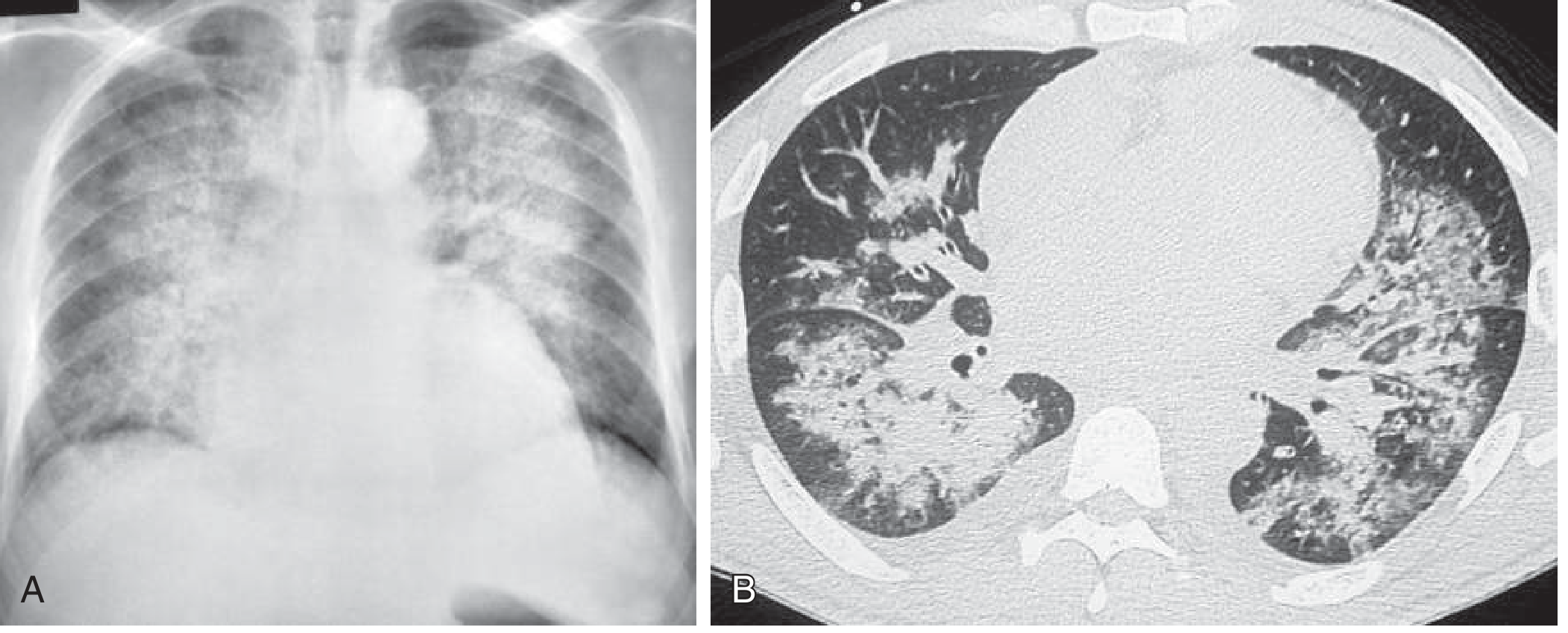
1. Chest X-Ray (CXR) - Most Important Initial Investigation
Classic CXR findings in cardiogenic pulmonary edema:
| Stage | CXR Finding | Description |
|---|---|---|
| Stage 1: Interstitial | Upper lobe vascular diversion (cephalization) | Pulmonary venous pressure 12-18 mmHg |
| Stage 2: Interstitial edema | Kerley B lines (short horizontal lines at bases), peribronchial cuffing, haziness | PCWP 18-25 mmHg |
| Stage 3: Alveolar edema | "Bat-wing" / "butterfly" pattern - bilateral perihilar alveolar consolidation; air bronchograms | PCWP >25 mmHg |
| + | Cardiomegaly (CTR >0.5), bilateral pleural effusions |
In non-cardiogenic PE (ARDS): Bilateral diffuse alveolar infiltrates without cardiomegaly or upper lobe vascular diversion; Kerley B lines absent.
2. ECG
- Evidence of acute MI (ST elevation, Q waves) → prompt revascularization
- Arrhythmias (AF, VT) as precipitants
- LVH (hypertensive/valvular disease)
- Sinus tachycardia
3. Blood Tests
| Test | Purpose / Findings |
|---|---|
| BNP / NT-proBNP | BNP <100 pg/mL excludes heart failure; BNP >500 pg/mL supports cardiac cause; NT-proBNP <300 pg/mL excludes HF |
| Troponin (I or T) | Detects ACS as precipitant; elevated in cardiogenic shock |
| ABG (Arterial Blood Gas) | Hypoxia (↓PaO2), respiratory alkalosis early, metabolic/respiratory acidosis late |
| Serum electrolytes, U&E, creatinine | Renal function (guides diuretic dosing); hyponatremia = poor prognosis |
| FBC | Anaemia (precipitant), leukocytosis (infection) |
| LFTs + albumin | Hypoalbuminemia → reduced oncotic pressure |
| Serum glucose, HbA1c | Hyperglycaemia in decompensation |
| Thyroid function (TFTs) | Hypo/hyperthyroidism as precipitant |
| Blood cultures | If sepsis-triggered non-cardiogenic PE |
4. Echocardiography
- Identifies systolic vs. diastolic LV dysfunction
- Valvular lesions (mitral stenosis, mitral regurgitation, aortic stenosis)
- Estimates LVEF (preserved vs. reduced)
- Pericardial effusion/tamponade
- Wall motion abnormalities (acute MI)
5. Bedside Lung Ultrasound (POCUS)
- B-lines (comet-tail artifacts): ≥3 B-lines per intercostal space = interstitial syndrome
- More sensitive and faster than CXR for detecting pulmonary edema
- Differentiates cardiac from non-cardiac causes of dyspnea (especially useful in emergency)
6. Pulmonary Artery Catheterization (PAC)
- Gold standard for differentiating cardiogenic from non-cardiogenic PE
- PCWP >18 mmHg = cardiogenic; ≤18 mmHg = non-cardiogenic
- Reserved for refractory, uncertain, or complex cases
7. Management
The goal is simultaneous support of oxygenation + treat the underlying cause.
A. Position and Immediate Measures
- Sit patient upright with legs dangling - reduces venous return and preload
- IV access, continuous monitoring (SpO2, ECG, BP)
- Identify and treat precipitants: arrhythmia, MI, infection, non-compliance with medications
B. Oxygenation and Ventilatory Support
| Modality | Indication | Details |
|---|---|---|
| Supplemental O2 | All hypoxic patients | Target SpO2 ≥92% (avoid >98%) |
| High-Flow Nasal Cannula (HFNC) | Acute hypoxic RF, normal PaCO2 | Better outcomes than BiPAP in non-cardiogenic setting |
| NIV - BiPAP / CPAP | Cardiogenic PE, respiratory fatigue | Reduces preload, improves oxygenation, reduces intubation need; beneficial in cardiogenic PE |
| Mechanical ventilation (intubation) | Failure of NIV, severe respiratory failure | PEEP reduces preload and afterload; redistributes lung water; improves gas exchange |
C. Preload Reduction
| Drug | Mechanism | Dose/Route |
|---|---|---|
| Furosemide (loop diuretic) | Immediate venodilation then diuresis | IV: 0.5 mg/kg (up to 1 mg/kg in renal insufficiency or chronic use); first-line diuretic |
| Bumetanide / Torsemide | Alternative loop diuretics | Useful in furosemide resistance |
| Sublingual GTN (nitroglycerin) | Venodilator + coronary vasodilator; immediate | 0.4 mg × 3 doses every 5 min; first-line for cardiogenic PE |
| IV GTN | Sustained preload/afterload reduction | 5-10 μg/min, titrate up if no hypotension |
| IV Nitroprusside | Potent venous + arterial vasodilator | 0.1-5 μg/kg/min; for hypertensive PE; needs arterial line; avoid in reduced coronary perfusion |
| Nesiritide (IV BNP) | Arterial + venous vasodilation + diuresis | Reserved for refractory cases; avoid in ACS/MI |
| Morphine | Venodilation, anxiolysis, ↓catecholamines | 2-4 mg IV boluses; use cautiously - some registry data show increased mortality |
| ACE inhibitors | ↓Preload and afterload | Especially in hypertensive patients + MI; short-acting agent first |
D. Inotropic Support (Reduced Cardiac Output / Cardiogenic Shock)
| Drug | Class | Notes |
|---|---|---|
| Dobutamine | Sympathomimetic β1-agonist | First-line inotrope; reduces PCWP, increases CO |
| Dopamine | Catecholamine | Higher doses: vasopressor effect |
| Milrinone | PDE-3 inhibitor (inodilator) | 50 μg/kg loading dose → 0.25-0.75 μg/kg/min; vasodilates + inotrope; avoid in hypotension |
E. Renal Replacement Therapy
- Indicated when refractory volume overload + metabolic acidosis (pH <7.15-7.25), hypoxemia, or persistent hyperkalemia
- Continuous RRT (CRRT) preferred over intermittent HD in hemodynamically unstable patients
- In ESRD: urgent dialysis is definitive treatment
F. Treat Underlying Cause (Simultaneous)
| Precipitant | Specific Treatment |
|---|---|
| Acute MI | PCI/revascularization; thrombolytics if PCI unavailable |
| Arrhythmia | Cardioversion (AF), rate control |
| Hypertensive emergency | Nitroprusside, IV labetalol |
| Acute MR / valvular disease | Intra-aortic balloon pump + surgical repair |
| Infection/sepsis | Broad-spectrum antibiotics |
| ARDS | Lung-protective ventilation (low tidal volume 6 mL/kg), prone positioning, conservative fluid strategy |
G. Long-Term Management (Cardiogenic PE)
- ACE inhibitors / ARBs (reduce mortality in HF)
- Beta-blockers (carvedilol, bisoprolol) - once stabilized
- Mineralocorticoid receptor antagonists (spironolactone/eplerenone)
- SGLT-2 inhibitors (empagliflozin, dapagliflozin) - reduce HF hospitalization
- Oral loop diuretics for maintenance
- Treat underlying cause (revascularization, valve replacement, AF management)
8. Cardiogenic vs. Non-Cardiogenic PE - Key Differentiating Features
| Feature | Cardiogenic | Non-Cardiogenic (ARDS) |
|---|---|---|
| History | HF, MI, valvular disease | Sepsis, aspiration, trauma |
| JVP | Elevated | Normal or low |
| S3 gallop | Present | Absent |
| Peripheral edema | Present | Absent |
| CXR heart size | Cardiomegaly | Normal heart size |
| Kerley B lines | Present | Usually absent |
| Distribution | Perihilar "bat-wing" | Diffuse, peripheral |
| PCWP | >18 mmHg | ≤18 mmHg |
| BNP/NT-proBNP | Markedly elevated | May be mildly elevated (sepsis, PE) |
| Edema fluid protein | Low (transudate) | High (exudate) |
| Response to diuretics | Good | Poor |
Quick-Reference Summary
PULMONARY EDEMA AT A GLANCE
━━━━━━━━━━━━━━━━━━━━━━━━━━━━━━━━━━━━━━━━━━━━━━━━━━
Cardiogenic (↑PCWP):         Non-Cardiogenic (normal PCWP):
  LVF, MI, MR, AS               ARDS, sepsis, TRALI
  Volume overload                Aspiration, drugs

Symptoms: Dyspnoea, PND, orthopnoea, pink frothy sputum
Signs:    Tachypnoea, crackles, wheeze, S3, ↑JVP, oedema

Investigations:
  CXR: bat-wing, Kerley B, cardiomegaly, effusions
  BNP: >500 = cardiac; <100 = excludes HF
  ABG: hypoxia ± hypercapnia
  ECG: MI, arrhythmia, LVH
  Echo: LV function, valves
  PCWP (PAC): >18 = cardiogenic

Management:
  1. Sit upright + O2 → target SpO2 ≥92%
  2. NIV (BiPAP/CPAP)
  3. Sublingual GTN → IV GTN
  4. IV Furosemide (0.5-1 mg/kg)
  5. Inotropes if low CO (dobutamine)
  6. Treat cause (MI → PCI, AF → rate control)
  7. Renal replacement if refractory
━━━━━━━━━━━━━━━━━━━━━━━━━━━━━━━━━━━━━━━━━━━━━━━━━━
Harrison's Principles of Internal Medicine 22E, pp. 2373-2374 | Robbins & Cotran Pathologic Basis of Disease, pp. 630-631 | Guyton & Hall Medical Physiology, pp. 508-509 | Tintinalli's Emergency Medicine | Murray & Nadel's Respiratory Medicine
This is a shared conversation. Sign in to Orris to start your own chat.