
Chorionic villus sampling For 3 rd year pg final exams
Reading File
Finding Sources
Finding Sources
Searching Images
Chorionic villus sampling technique transabdominal transcervical diagram
Reading File
Reading File
Reading File
Chorionic Villus Sampling (CVS)
Definition
CVS is an invasive prenatal diagnostic procedure that samples the developing placenta (chorionic frondosum/trophoblastic tissue) rather than the amniotic fluid, allowing first-trimester genetic diagnosis - Creasy & Resnik's Maternal-Fetal Medicine, p. 680.
Timing
- Performed between 10 and 13 weeks of gestation (ideally not before 10 weeks - earlier sampling is associated with increased risk of limb-reduction defects/oromandibular-limb hypogenesis).
- Key exam point: CVS gives results weeks earlier than amniocentesis (done at 15-16 weeks), permitting earlier reassurance or earlier, safer termination if abnormal - The Developing Human: Clinically Oriented Embryology, p. 317.
Indications
- Advanced maternal age / increased risk of aneuploidy
- Previous child with chromosomal abnormality
- Parental balanced translocation/chromosomal rearrangement
- Known single-gene disorder in family (needs DNA-based diagnosis) - e.g., thalassemia, hemophilia, muscular dystrophy, inborn errors of metabolism
- Abnormal first-trimester screening (NT, biochemical markers)
- Rapid karyotype needed for early decision-making
Technique
1. Transcervical CVS
- Ultrasound first confirms fetal viability, size, and placental location; bladder moderately filled to help straighten an anteverted uterus (overfilling is avoided as it lengthens the sampling path).
- Patient in lithotomy position; vulva, vagina, and cervix prepped with povidone-iodine.
- A malleable catheter (distal 3-5 cm curved) is passed transcervically under continuous ultrasound guidance to the distal edge of the placenta, parallel to the chorionic membranes.
- Stylet removed, a 20 mL syringe with nutrient medium attached, negative pressure applied while withdrawing the catheter to aspirate villi.
- Uterine contractions can obstruct catheter passage - the procedure is delayed until the contraction resolves (Fig. 30.11 in Creasy & Resnik).
2. Transabdominal CVS
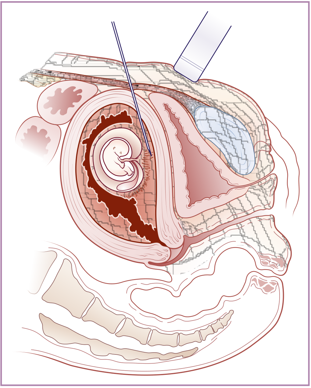
- A needle is inserted through the maternal abdominal and uterine walls under continuous ultrasound guidance into the chorionic frondosum, kept parallel to the chorionic plate.
- Sample obtained by back-and-forth needle movement while maintaining negative pressure, needle tip kept in view throughout - Creasy & Resnik, p. 681; The Developing Human, p. 317.
- Preferred when the placenta is posterior/fundal or transcervical access is difficult (e.g., cervical stenosis, active infection, myomas).
5-20 mg of trophoblastic tissue is obtained, sufficient for karyotype, chromosomal microarray, FISH, or molecular/DNA-based testing.
Advantages over amniocentesis
- Earlier diagnosis (first trimester vs second trimester)
- Larger tissue yield allows faster cytogenetic/molecular turnaround
- Avoids amniotic cavity puncture (no risk of early oligohydramnios-related clubfoot, which is linked to fluid leakage after early amniocentesis)
Limitations / Pitfalls (favorite exam points)
- Confined placental mosaicism (CPM): mosaic result on CVS that does not reflect the true fetal karyotype - requires confirmatory amniocentesis in ~1-2% of cases.
- Maternal cell contamination: can give false results if maternal decidual tissue is inadvertently sampled instead of pure villi.
- Cannot detect neural tube defects (no AFP/acetylcholinesterase assay possible, unlike amniocentesis) - maternal serum AFP or later ultrasound is still needed.
- Not useful for assessing fetal lung maturity or bilirubin/Rh isoimmunization studies.
Complications
- Pregnancy loss: comparable to amniocentesis when performed by experienced operators. The Canadian Collaborative trial showed 7.6% loss (CVS) vs 7.0% (amniocentesis) after adjusting for background first-trimester loss (RR 1.10, 95% CI 0.92-1.30). A meta-analysis by Salomon et al. (cited in Creasy & Resnik) confirms similar procedure-related loss rates between CVS and amniocentesis once background loss is accounted for.
- Repeated catheter passes significantly raise miscarriage risk (10.8% loss with ≥3 passes vs 2.9% with a single pass) - Creasy & Resnik's Maternal-Fetal Medicine, p. 681.
- Limb reduction defects/oromandibular hypogenesis if performed before 10 weeks.
- Vaginal spotting (more common with transcervical route), chorioamnionitis (rare), Rh sensitization (Rh-negative women need anti-D prophylaxis).
Diagnostic yield
- Fetal loss rate overall ~0.5-1%, comparable to amniocentesis.
- Can diagnose chromosomal abnormalities, inborn errors of metabolism, and X-linked disorders (Langman's Medical Embryology, The Developing Human, p. 317-318).
Here is an illustration of the transabdominal technique for reference:
High-yield one-liner for exams: "CVS = first-trimester (10-13 weeks) biopsy of chorionic villi via transcervical catheter or transabdominal needle under USG guidance, for early karyotyping/DNA diagnosis; main pitfalls are confined placental mosaicism and maternal cell contamination; procedure-related loss rate is comparable to amniocentesis (~1%) when done after 10 weeks by an experienced operator."
References: Creasy & Resnik's Maternal-Fetal Medicine, p. 680-681; The Developing Human: Clinically Oriented Embryology, p. 317-318; Langman's Medical Embryology; Emery's Elements of Medical Genetics and Genomics.
This is a shared conversation. Sign in to Orris to start your own chat.