
Double barrel aorta in short explain
Double barrel aorta refers to a condition where the aorta appears to have two lumens (channels) running parallel to each other, resembling a double-barreled shotgun on cross-sectional imaging. This occurs in the context of aortic dissection, where an intimal tear allows blood to enter the aortic wall and create a false lumen alongside the true lumen.
Searching sources...
Reading File
Finding Sources
Searching the Web
double barrel aorta aortic dissection radiology pathology
Finding Sources
Reading File
Here is a concise explanation of double barrel aorta:
Double Barrel Aorta
What it is
"Double barrel aorta" is a descriptive term used in aortic dissection, where a tear in the intima allows blood to enter the medial layer of the aortic wall. This creates two parallel channels (lumens) running side by side:
| Lumen | Description |
|---|---|
| True lumen | The original aortic channel; typically smaller, higher flow |
| False lumen | The newly created channel within the media; typically larger, lower flow |
These two lumens are separated by an intimomedial flap (the dissection flap), giving the aorta a "double barrel" or two-tube appearance - similar to a double-barreled shotgun.
How it forms
- An intimal tear occurs (often due to hypertension, Marfan syndrome, or atherosclerosis)
- Blood forces its way into the medial layer, splitting the inner 2/3 from the outer 1/3 of the media
- The cleavage plane extends proximally or distally, creating the false lumen
- Re-entry tears may form, where blood re-enters the true lumen
Where it is seen
- On CT angiography (most common imaging modality) - the two lumens are clearly visible in cross-section
- On aortography, MRI, or transesophageal echocardiography (TOE/TEE)
- The false lumen typically lies along the outer curvature of the aorta
Key distinguishing features
| Feature | True Lumen | False Lumen |
|---|---|---|
| Size | Smaller | Larger |
| Flow | Faster (brighter on CT) | Slower |
| Shape | Circular | Crescentic |
| Signs | - | Cobweb sign, beak sign |
| Thrombosis | Rare | Common over time |
Clinical significance
- The false lumen can compress the true lumen, causing malperfusion of branch vessels (renal, mesenteric, spinal arteries)
- Multi-barreled dissection (3+ lumens) occurs in ~9% of acute Type B dissections and is associated with higher mortality
- Classification: Stanford Type A (involves ascending aorta - surgical emergency) vs Stanford Type B (beyond left subclavian artery - medical management initially)
The term "double barrel" is a gross pathology and imaging descriptor, most commonly encountered in exam MCQs in the context of aortic dissection.
- Grainger & Allison's Diagnostic Radiology, Aortic Dissection section
What is dissecting aneurysm
A dissecting aneurysm is essentially aortic dissection where blood splits through the layers of the aortic wall, creating a false channel. The term is somewhat of a misnomer - it is not a true aneurysm but rather a dissection of the vessel wall layers.
Searching sources...
Finding Sources
Searching the Web
dissecting aneurysm definition pathology aorta
Reading File
Reading File
Loading Image
Dissecting Aneurysm
Definition
A dissecting aneurysm is formed when blood enters through a tear in the tunica intima (inner lining of the artery) and is forced between the layers of the arterial wall - specifically between the inner and outer coats of the tunica media - creating a blood-filled channel within the vessel wall itself.
"A dissecting aneurysm is formed as a result of splitting or dissection of an arterial wall by blood entering or exiting through a tear."
- Bradley and Daroff's Neurology in Clinical Practice
Is it a True Aneurysm?
Technically, a dissecting aneurysm is classified as a type of true aneurysm (involves all three arterial wall layers), but it is not a true dilatation in the classical sense. It is more accurately described as an aortic dissection - the term "dissecting aneurysm" is older terminology still commonly used.
Types of true aneurysm (by shape):
| Type | Description |
|---|---|
| Fusiform | Uniform expansion of entire circumference |
| Saccular | Expansion of only part of circumference (often traumatic) |
| Dissecting | Blood forces through intima into tunica media layers |
Mechanism (Step by Step)
- Trigger: Tunica intima ruptures - usually beneath an atheromatous plaque (or due to cystic medial degeneration in Marfan syndrome)
- Entry: High-pressure blood is forced through the intimal tear into the media
- Propagation: Blood dissects longitudinally along the media, splitting it into two layers - creating the false lumen
- Extension: The dissection can propagate proximally (toward the heart) or distally (toward the legs), spiraling around the aorta
- Result: Two outcomes depending on direction of wall displacement:
- Inward displacement → narrows the true lumen (stenosis/malperfusion)
- Outward displacement → creates the visible aneurysmal bulge (the "double barrel" appearance)
What it Looks Like on CT
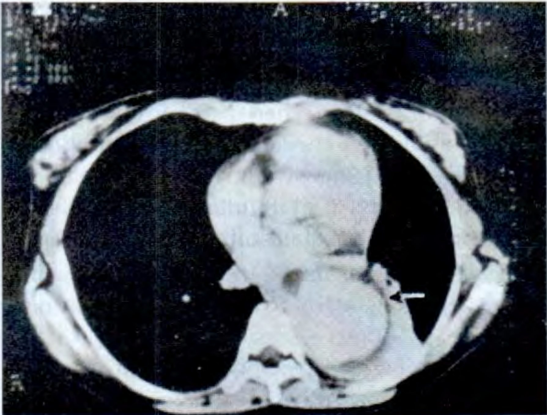
CT cross-section of the thoracic aorta showing a dissecting aneurysm - note the two distinct lumens (the "double barrel" appearance)
Common Causes
| Category | Examples |
|---|---|
| Degenerative | Hypertension (most common), atherosclerosis |
| Connective tissue | Marfan syndrome, Ehlers-Danlos syndrome |
| Congenital | Bicuspid aortic valve, aortic coarctation |
| Trauma | Blunt chest injury (especially with pre-existing atherosclerosis) |
| Other | Pregnancy, aortitis, syphilis |
Common Site
Most commonly affects the thoracic aorta (especially the ascending aorta), but can occur in any artery. Intracranial dissecting aneurysms commonly affect the vertebral artery and PICA (posterior inferior cerebellar artery).
Key Complications
- Aortic rupture → hemopericardium, hemothorax, sudden death
- Malperfusion syndrome → renal failure, bowel ischemia, limb ischemia, stroke
- Aortic regurgitation (if dissection involves aortic valve root)
- Haemopericardium → cardiac tamponade
Dissecting Aneurysm vs Aortic Dissection
These terms are often used interchangeably. "Aortic dissection" is the modern preferred term; "dissecting aneurysm" is the classic/older term for the same condition.
- S Das - A Manual on Clinical Surgery, 13th Edition
- Bradley and Daroff's Neurology in Clinical Practice
This is a shared conversation. Sign in to Orris to start your own chat.