
Mcd
It looks like your message got cut off! Could you complete your thought? Are you asking about:
- McDonald's (the fast food chain)
- MCD (a stock ticker or financial term)
- MCD as a medical abbreviation
- Something else entirely?
Minimal change disease
Reading File
Quick answer: Minimal change disease (MCD) is the most common cause of nephrotic syndrome in children. It gets its name from the fact that light microscopy shows virtually normal glomeruli - the only abnormality is effacement (fusion) of podocyte foot processes visible on electron microscopy. It responds well to steroids and carries a good prognosis.
Searching sources...
Finding Sources
Searching PubMed
"minimal change disease" nephrotic syndrome
Good - multiple excellent sources found. Let me read the key ones in parallel.
Reading File
Reading File
Reading File
Excellent content retrieved. Now let me get the histology images and treatment details.
Loading Image
Finding Sources
Finding Sources
Let me get the treatment details from the Clinical Nephrology textbook and the Brenner & Rector EM section.
Reading File
Reading File
I went a bit off-target for treatment details. Let me get treatment info directly from the Comprehensive Clinical Nephrology and Primer on Kidney Diseases textbooks.
Reading File
Reading File
I now have comprehensive content to write an excellent answer on Minimal Change Disease.
Minimal Change Disease (MCD)
Definition & Overview
Minimal change disease is a form of nephrotic syndrome named for the fact that the glomeruli appear virtually normal on light microscopy - the "minimal changes" are only apparent on electron microscopy. It is the most common cause of nephrotic syndrome in children (70-90% of childhood nephrotic syndrome) but accounts for only 10-15% of nephrotic syndrome in adults, with peak incidence between 2-6 years of age.
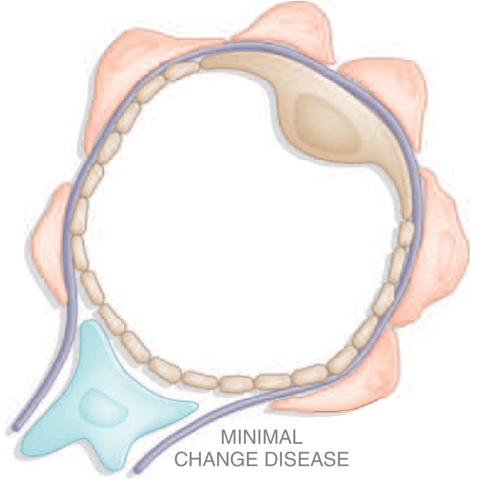
The diagram below illustrates the glomerular schematic in MCD:
Etiology
Primary (idiopathic): Most cases.
Secondary associations include:
- Hodgkin lymphoma (classic association) and other lymphomas
- NSAIDs and lithium use
- Infections (viral upper respiratory tract infections often precede a relapse)
- Allergic/atopic states (atopy in ~40% of children, 30% of adults)
- Bee sting, vaccinations
Pathogenesis
The exact mechanism remains incompletely understood, but current evidence points to immune dysfunction - originally thought to be purely T-cell mediated, now with evidence for a mixed B-cell/T-cell/autoantibody process:
-
T-cell hypothesis: Dysregulated T cells release circulating glomerular permeability factors (cytokines such as IL-13 and IL-4) that alter the charge/structural barrier of the glomerular capillary wall. Evidence includes: response to steroids, association with Hodgkin disease, remissions during viral infections (measles) that depress cell-mediated immunity.
-
CD80 (B7.1) overexpression: Podocytes in MCD show increased urinary and tissue CD80, normally expressed only by dendritic cells and B cells. Urinary CD80 correlates with disease activity. CD80 on podocytes promotes dysregulated interactions with T lymphocyte antigen-4 (CTLA-4).
-
Anti-nephrin antibodies (newer discovery): A subset of MCD patients have circulating antibodies against nephrin, a transmembrane podocyte protein and essential slit diaphragm component. These antibodies cause podocyte injury by interfering with nephrin crosslinking, leading to breakdown of the slit diaphragm. IgG staining on immunofluorescence colocalizes with clustered nephrin.
-
Angiopoietin-like-4 overexpression: Associated with the proteinuric response; reduced by corticosteroids.
-
Charge barrier defect: Loss of glomerular polyanions contributes to proteinuria.
- Harrison's Principles of Internal Medicine 22E, p. 1317-1322
- Robbins, Cotran & Kumar Pathologic Basis of Disease, p. 846
- Comprehensive Clinical Nephrology, 7th Ed., p. 257
Morphology
| Microscopy | Findings |
|---|---|
| Light microscopy | Glomeruli appear completely normal |
| Immunofluorescence | Negative, or occasional small amounts of IgM in mesangium; subtle fine granular IgG over podocytes (anti-nephrin antibody pattern) |
| Electron microscopy | Diffuse, uniform effacement (flattening/retraction) of podocyte foot processes with loss of slit diaphragms; microvillous transformation; NO electron-dense deposits; GBM normal |
The term "fusion" of foot processes is technically a misnomer - it actually represents simplification of podocyte architecture with collapse of the central actin cytoskeleton.
Proximal tubular cells are often laden with lipid and protein (from reabsorption of filtered lipoproteins) - hence the old name "lipid nephrosis."
Key diagnostic point: Foot process effacement occurs in many proteinuric states (membranous nephropathy, diabetic nephropathy). MCD is diagnosed only when effacement is seen alongside normal glomeruli by light microscopy - it is a diagnosis by exclusion.
- Robbins, Cotran & Kumar Pathologic Basis of Disease, p. 846
- Brenner and Rector's The Kidney, p. 1342
Clinical Features
- Onset: Abrupt onset of full-blown nephrotic syndrome
- Edema: Periorbital (especially in children), peripheral, ascites, pleural effusion
- Proteinuria: Massive - average 24-hour urine protein ~10 g; predominantly albumin (selective proteinuria)
- Hypoalbuminemia: Severe
- Hypertension: Less common - 30% of children, 20-50% of adults
- Hematuria: Microscopic in 20% of children, 33% of adults
- Renal function: Usually preserved despite massive proteinuria - a characteristic feature
- Atopy/allergic symptoms: 40% of children, 30% of adults
- AKI: Can occur in adults, often with low serum albumin and interstitial edema (nephrosarca); must distinguish from hypovolemia
- Urine sediment: Acellular (no RBC casts, no cellular casts)
Treatment
First-line: Corticosteroids
- Children: Prednisone 60 mg/m²/day (max 80 mg) for 4-6 weeks, then alternate-day dosing with gradual taper
-
90% achieve complete remission within 8 weeks
- Up to 30% have a spontaneous remission, but most are treated
-
- Adults: Response is slower but 80-90% ultimately achieve complete remission; adults are not considered steroid-resistant until after 4 months of therapy
Response Categories
| Category | Definition |
|---|---|
| Complete remission | Proteinuria <0.2 mg/24h after a single course of prednisone |
| Steroid-dependent | Relapse as steroid dose is tapered |
| Frequent relapsers | ≥2 relapses within 6 months of taper |
| Steroid-resistant | Failure to respond to steroid therapy (in children ~5% of cases) |
- Relapses occur in 70-75% of children after first remission; frequency decreases after puberty
- Patients with steroid resistance may have FSGS on repeat biopsy (sampling error or true progression)
Second-line agents (steroid-dependent/frequently relapsing/steroid-resistant)
-
Cyclophosphamide: Alkylating agent; useful for frequently relapsing disease
-
Cyclosporine / Tacrolimus (CNIs): 3-5 mg/kg/day in divided doses; up to 90% achieve partial or complete remission, but relapse after discontinuation is common
-
Mycophenolate mofetil (MMF): Used after CNI therapy
-
Rituximab (anti-CD20): Effective for steroid-dependent disease in both children and adults; evidence also supports a B-cell role in pathogenesis
-
Pulse IV methylprednisolone: May be tried in truly steroid-resistant children
-
Brenner and Rector's The Kidney, p. 1342-1352
Prognosis
- Long-term prognosis is excellent, even in steroid-dependent disease
- The disease may fully resolve when children reach puberty
- Adults have a similar good long-term prognosis
- Persistent MCD does not typically progress to end-stage renal disease unless there is underlying/unrecognized FSGS
- Podocyte changes are completely reversible after corticosteroid therapy, along with disappearance of circulating anti-nephrin antibodies
Key Distinguishing Features vs. FSGS
| Feature | MCD | FSGS |
|---|---|---|
| Light microscopy | Normal glomeruli | Segmental sclerosis in some glomeruli |
| EM | Diffuse foot process effacement | Diffuse foot process effacement |
| Steroid response | >90% respond | Variable, often resistant |
| Prognosis | Excellent | Worse |
| Progression | Rarely to ESRD | Can progress |
MCD and FSGS may represent a disease spectrum ("podocytopathy spectrum"), with MCD at the milder, more steroid-sensitive end.
Recent evidence note: A 2025 systematic review (PMID 41357184) examined histopathological subtypes in steroid-resistant nephrotic syndrome in children - FSGS was found to be the predominant pattern, supporting the clinical overlap between MCD and FSGS in steroid-resistant cases. A separate 2025 review (PMID 39352607) highlighted the association of MCD with solid neoplasms, not just hematological malignancies, expanding the secondary causes to monitor.
This is a shared conversation. Sign in to Orris to start your own chat.