
How can I know that I have bhagandar ...what's the pic of bhagandar pidika
anal fistula fistula in ano perianal abscess clinical photo
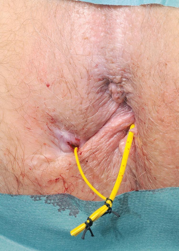
Clinical photograph of the perianal region demonstrating the management of a fistula-in-ano with a loose seton. The image shows a flexible, yellow vascular sling or vessel loop inserted through an external fistula opening located lateral to the anus and exiting through an internal opening within the anal canal. The two ends of the yellow tubing are secured together externally using black non-absorbable sutures to create a continuous, non-constricting loop (loose seton). The surrounding perianal skin shows mild erythema and minor blood spotting consistent with a recent surgical procedure. This clinical finding is characteristic of the surgical treatment for complex or high anal fistulas, where a seton is utilized to promote drainage, prevent recurrent abscess formation, and facilitate the gradual fibrous maturation of the fistula tract while minimizing the risk of fecal incontinence.
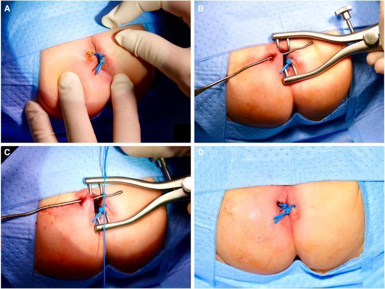
A series of four clinical photographs (A–D) illustrating a sphincter-sparing surgical technique for treating fistula-in-ano (FIA) in a pediatric patient. Panel A: Initial clinical presentation of a perianal abscess and fistula opening at the 10 o’clock position, with a previously placed blue silicone seton already visible at the 3 o’clock position. Panel B: Identification and exploration of the fistulous tract using a metal bulb-headed lacrimal probe while a surgical retractor provides exposure of the anal verge. Panel C: Placement of a new silicone vessel loop (seton) by attaching it to the tip of the lacrimal probe to be pulled through the tract. Panel D: Post-procedural view showing the silicone vessel loop ligated loosely outside the anus. This procedure demonstrates a non-cutting seton placement aimed at promoting drainage and controlled fibrosis while avoiding damage to the anal sphincter, particularly relevant in neonatal and infant proctology.
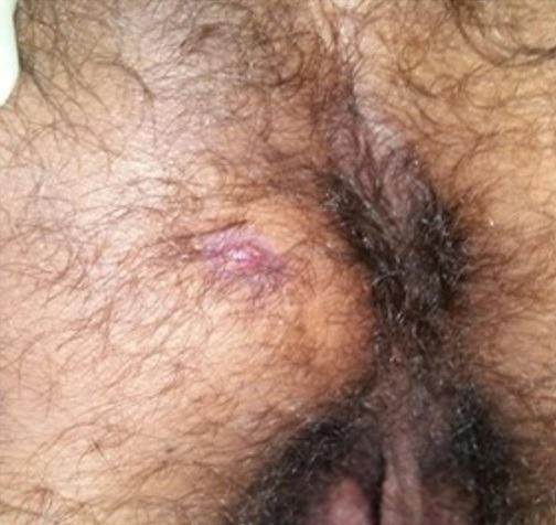
This clinical photograph shows a close-up view of the perianal region in a patient with significant local hirsutism. The primary finding is a small, erythematous skin lesion located approximately 2 cm from the anal margin. The lesion presents as a raised, inflammatory papule or nodule with a central punctum or orifice, suggestive of an external opening for a fistula (fistula in ano). The surrounding skin exhibits a reddish-purple hue and a moist texture, indicating active inflammation and potential purulent discharge. The anatomical context involves the anal sphincter complex and perianal soft tissues. In the provided clinical case, this represents a manifestations of extra-pulmonary tuberculosis (tuberculous anal fistula). Key educational concepts illustrated include the visual presentation of perianal abscess/fistula disease, the identification of inflammatory skin orifices in proctology, and the clinical signs of chronic localized infections.
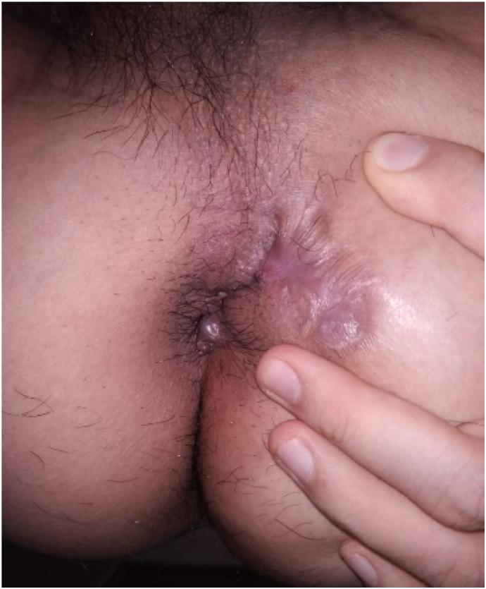
This clinical photograph shows the perianal region of a patient 8 weeks following a surgical procedure for a complex anal fistula. The image demonstrates the anal orifice and the surrounding skin. Notable features include a prominent area of pale, hypertrophic scarring and fibrotic tissue extending from the right perianal side towards the gluteal fold, indicative of a healing surgical tract or previously active fistula site. There is mild residual erythema and skin irritation immediately surrounding the anus, though the tissue appears largely stable with no visible active purulent discharge or acute abscess formation at this stage. The surrounding skin shows signs of re-epithelialization and maturation of granulation tissue. This image serves as a clinical example of the mid-term healing phase after fistula-in-ano treatment, illustrating the progression of secondary intention healing and scar remodeling in the colorectal/proctological specialty.
भगन्दर (Bhagandar / Anal Fistula) - पूरी जानकारी
भगन्दर क्या है?
भगन्दर के लक्षण (Symptoms) - कैसे पहचानें?
| लक्षण | विवरण |
|---|---|
| दर्द (Pain) | गुदे के पास तेज, धड़कन जैसा दर्द |
| सूजन (Swelling/Pidika) | गुदे के पास गांठ या फोड़ा जो बार-बार होता है |
| मवाद/पस निकलना | बाहरी छेद (external opening) से बदबूदार पस या खून मिला द्रव निकलना |
| खुजली | गुदे के आसपास खुजली और जलन |
| बुखार | अगर फोड़ा (abscess) हो तो बुखार भी आता है |
| बाहरी छेद दिखना | गुदे के पास 1-2 cm दूर त्वचा पर एक छोटा छेद दिखता है जिससे पस निकलती है |
मुख्य पहचान: गुदे के पास बार-बार फोड़ा होना, फटना, पस निकलना और फिर कुछ राहत मिलना - यह भगन्दर का सबसे आम चक्र है।
- Bailey and Love's Short Practice of Surgery, 28th Ed.
भगन्दर पिडिका (Bhagandar Pidika) - कैसी दिखती है?
नैदानिक चित्र (Clinical Photos):
भगन्दर के प्रकार (Parks' Classification)
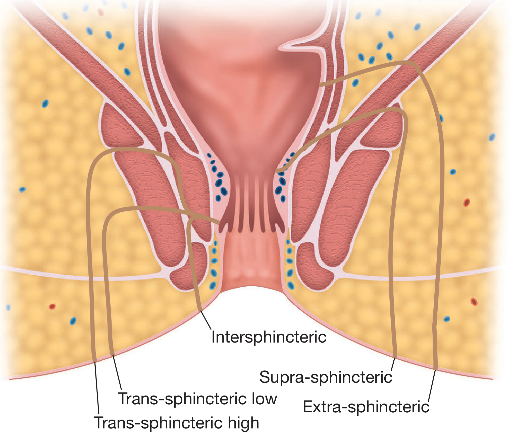
| प्रकार | विवरण |
|---|---|
| Intersphincteric | सबसे आम प्रकार, sphincter के बीच से जाता है |
| Trans-sphincteric | sphincter को काटता हुआ जाता है |
| Supra-sphincteric | sphincter के ऊपर से जाता है |
| Extra-sphincteric | sphincter से बाहर से जाता है (सबसे जटिल) |
भगन्दर कैसे होता है? (Cause)
- गुदे के पास फोड़ा (Anorectal abscess) - सबसे मुख्य कारण
- Crohn's disease
- Tuberculosis (TB)
- Ulcerative Colitis
- मधुमेह (Diabetes)
डॉक्टर कैसे जांच करते हैं?
- निरीक्षण (Inspection): गुदे के पास बाहरी छेद देखते हैं
- उंगली से जांच (Bidigital palpation): एक उंगली अंदर और अंगूठा बाहर रखकर नली को महसूस करते हैं
- Proctoscopy: अंदरूनी छेद देखने के लिए
- MRI: जटिल भगन्दर में सटीक नक्शा बनाने के लिए
इलाज (Treatment)
- Surgery (Fistulotomy): साधारण (low) भगन्दर को काटकर खोल देते हैं
- Seton: जटिल (high) भगन्दर में धागा डालकर धीरे-धीरे काटते हैं
- Ayurvedic Kshar Sutra: आयुर्वेद में औषधीय धागे से इलाज किया जाता है
⚠️ महत्वपूर्ण: अगर आपको ये लक्षण हैं तो खुद से इलाज न करें। किसी Colorectal Surgeon या General Surgeon से मिलें। भगन्दर का सही इलाज सर्जरी से ही होता है।