
When do you suspect pneumonia in paediatrics based on history and examination tell me briefly
Reading File
Now let me search the library for paediatric pneumonia clinical features.
Finding Sources
Finding Sources
Finding Sources
Finding Sources
Finding Sources
Good, found relevant content in Park's. Let me read the key sections.
Reading File
Reading File
I now have enough information to provide a thorough, brief answer. Let me compile it.
Suspecting Pneumonia in Paediatrics: History & Examination
History (Key Questions to Ask)
- Cough - duration and character
- Difficulty breathing / fast breathing - main symptom to pursue
- Inability to feed (infants <2 months: stopped feeding well; older children: unable to drink/eat)
- Fever - how long, how high
- Altered sensorium - excessive drowsiness, difficult to wake, convulsions
- Turning blue (cyanosis episodes) or irregular breathing / apnoeic spells
- Antecedent illness - recent measles increases pneumonia risk significantly
- Risk factors - malnutrition, prematurity, crowding, passive smoke exposure, day-care attendance
Physical Examination (What to Look For)
1. Fast Breathing (Tachypnoea) - Most Important Sign
Age-specific thresholds (WHO / IMCI):
| Age | Fast Breathing Cut-off |
|---|---|
| < 2 months | ≥ 60 breaths/min |
| 2 months - < 12 months | ≥ 50 breaths/min |
| 12 months - < 5 years | ≥ 40 breaths/min |
- Count for a full minute with the child calm; chest/abdomen exposed.
- Repeat in infants <2 months if borderline (their rate is often erratic).
2. Chest Indrawing (Lower Chest Wall Retractions)
- Lower chest wall draws in on inspiration - indicates increased work of breathing.
- Present alone = severe pneumonia; more ominous than fast breathing alone.
3. Danger Signs (suggest Very Severe Disease)
- Unable to drink or breastfeed
- Vomits everything
- Convulsions
- Abnormally sleepy / unconscious / difficult to wake
- Stridor at rest
4. Other Examination Findings
- Nasal flaring
- Grunting (especially in neonates)
- Cyanosis - check in good light; sign of hypoxia
- Auscultation: reduced air entry, bronchial breathing, crackles (crepitations)
- Fever or hypothermia (hypothermia is a warning sign in neonates)
- Pulse oximetry: SpO2 <94% supports diagnosis and indicates severity
IMCI Classification Summary
| Classification | Key Signs |
|---|---|
| Very Severe Disease | Any danger sign (unable to drink, convulsions, cyanosis, stridor at rest, altered consciousness) |
| Severe Pneumonia | Chest indrawing (with or without fast breathing) |
| Pneumonia (not severe) | Fast breathing only, no chest indrawing, no danger signs |
| No Pneumonia: Cough/Cold | No fast breathing, no chest indrawing, no danger signs |
Note: In severely malnourished children, fast breathing and chest indrawing may be absent or blunted despite significant pneumonia - these children need extra careful evaluation.
(Source: Park's Textbook of Preventive & Social Medicine; WHO/IMCI guidelines)
Risk factors for asthma give me 10 main points
Searching the medical library for asthma risk factors.
Finding Sources
Reading File
Finding Sources
Reading File
Excellent - I have comprehensive content. Here are the 10 main risk factors:
10 Main Risk Factors for Asthma
1. Atopy (Most Important)
The single most important predisposing factor. Atopic individuals have a genetic tendency to produce IgE against common environmental allergens. Asthma is classified as atopic (extrinsic) - driven by allergen sensitization - or non-atopic (intrinsic). Allergens also cause subclinical airway inflammation that heightens airway responsiveness.
2. Genetic Predisposition
Asthma runs strongly in families. It is a polygenic disease with an interplay between multiple susceptibility genes and environmental factors. Having a first-degree relative with asthma significantly raises individual risk.
3. Allergen Exposure
- Indoor: house dust mite, cockroach, animal dander, indoor fungi
- Outdoor: pollens, outdoor fungi Chronic allergen exposure leads to sensitization, persistent airway inflammation, and remodelling.
4. Tobacco Smoke (Active & Passive)
Both active smoking and passive (secondhand) smoke exposure are established risk factors. Maternal smoking during pregnancy and early childhood passive smoke exposure are particularly significant in paediatric asthma development and severity.
5. Respiratory Infections in Early Life
Early childhood viral lower respiratory tract infections - especially RSV and rhinovirus - are strongly associated with later asthma development. The risk is compounded in the presence of atopy; viral infections alone (without atopy) are less likely to cause asthma.
6. Obesity
A major independent risk factor. Both BMI (general obesity) and waist circumference (abdominal obesity) correlate with new-onset asthma. Obese asthmatics tend to have worse symptom control and reduced response to corticosteroids.
7. Occupational Sensitizers
Accounts for ~5% of adult asthma cases. Common agents include isocyanates (spray painting, foam), flour/grain dust, latex, wood dust, and laboratory animal proteins. Prolonged exposure can cause sensitization and irreversible asthma.
8. Gender & Hormonal Factors
- In childhood: asthma is more common in boys
- After puberty: prevalence shifts - women are more affected
- Hormonal fluctuations (menstrual cycle, pregnancy, menopause) can influence asthma severity
9. Socioeconomic Status & Environment
Lower socioeconomic status is associated with higher asthma prevalence and worse outcomes - via increased allergen exposure, air pollution, damp housing, reduced access to care, and higher psychosocial stress.
10. Drugs
Certain medications can precipitate or worsen asthma:
- Beta-blockers (including eye drops)
- Aspirin / NSAIDs (aspirin-exacerbated respiratory disease - AERD)
- ACE inhibitors (cause cough, can trigger bronchospasm)
Endogenous factors: atopy, airway hyperresponsiveness, genetics, gender, ethnicity Environmental factors: allergens, tobacco, infections, obesity, occupational exposure, socioeconomic status
(Source: Fishman's Pulmonary Diseases and Disorders)
In accordance with paediatrics
Searching for paediatric-specific asthma risk factors.
Finding Sources
Finding Sources
Good sources found. Let me read the most relevant paediatric sections from Harrison's and Fishman's.
Reading File
Reading File
Finding Sources
Finding Sources
I have enough paediatric-specific content from Harrison's and Fishman's. Here are the 10 main risk factors reframed specifically for paediatrics:
10 Risk Factors for Asthma in Paediatrics
1. Atopy / Personal or Family History of Allergic Disease
The strongest single risk factor in children. A personal or family history of atopic dermatitis (eczema), allergic rhinitis, or food allergy ("atopic march") strongly predicts asthma development. Boys have higher atopy prevalence in childhood, explaining why asthma is more common in boys before puberty.
2. Family History of Asthma
Having a parent with asthma (especially the mother) is an independent predictor of persistent wheezing and asthma in children. This reflects both genetic susceptibility and shared environmental exposures.
3. Maternal Smoking & In Utero Smoke Exposure
Maternal cigarette smoking during pregnancy is one of the most important modifiable risk factors. It impairs fetal lung development, reduces lung function at birth, and strongly predicts persistent wheezing. Postnatal passive smoke exposure further compounds the risk.
4. Early-Life Viral Respiratory Infections
Respiratory syncytial virus (RSV) bronchiolitis and rhinovirus wheezing illnesses in infancy are closely linked to later asthma, especially in atopic children. The combination of viral infection + pre-existing atopy carries the highest risk of progression to asthma.
5. Abnormal Lung Function at Birth / Prematurity
Children born with reduced lung function (even before any wheeze) are at higher risk for persistent asthma. Prematurity and associated conditions (bronchopulmonary dysplasia) independently increase asthma risk. Preeclampsia in the mother is also associated with increased risk in the child.
6. Male Sex
Asthma is significantly more common in boys throughout childhood. Boys have relatively smaller airways compared to lung size, increasing susceptibility to wheeze and airway hyperresponsiveness. This sex disparity reverses after puberty.
7. Indoor Allergen Exposure
Early and sustained exposure to house dust mite, cockroach allergen, pet dander (cat/dog), and indoor mould is a key environmental trigger and sensitiser. Sensitisation before age 3 years is particularly predictive of persistent asthma.
8. Caesarean Section Delivery
Babies born by C-section have a higher risk of asthma, likely because they miss exposure to maternal vaginal and gut microbiota, which is important for healthy immune programming. This relates to the hygiene hypothesis - reduced microbial diversity in early life shifts immune responses toward atopy.
9. Obesity / Rapid Early Weight Gain
Childhood obesity is an established risk factor for new-onset asthma. Adipokines (e.g., leptin, IL-6) promote airway inflammation. Obese children with asthma tend to have poorer symptom control and a non-eosinophilic (non-type 2) phenotype that responds poorly to inhaled corticosteroids.
10. Prenatal & Perinatal Nutritional Factors
- Vitamin D deficiency in the mother increases asthma risk in the child
- High maternal sugar intake is associated with increased childhood asthma
- Maternal supplementation with vitamins C & E, zinc, and omega-3 fatty acids may be protective
- Breastfeeding reduces early wheezing, though its effect on long-term asthma is less clear
Key paediatric concept: The Tucson Children's Respiratory Study identified 4 wheeze phenotypes in children. "Persistent wheezers" (the true future asthmatics) were predicted by: young maternal age, elevated IgE at 9 months, parental asthma, in-utero smoke exposure, reduced lung function at birth, and male sex.
(Sources: Harrison's Principles of Internal Medicine 22E; Fishman's Pulmonary Diseases and Disorders)
Risk factors for pneumonia give me importannt points
Based on the previous conversation context, I'll search for paediatric pneumonia risk factors specifically.
Finding Sources
Finding Sources
Finding Sources
Finding Sources
Finding Sources
Reading File
Reading File
Finding Sources
Reading File
I have comprehensive content. Here are the important risk factors for paediatric pneumonia:
Risk Factors for Pneumonia in Paediatrics
HOST FACTORS
1. Young Age
Infants and children under 5 years are the most vulnerable. Case fatality rates are highest in young infants due to immature immune systems, smaller airways, and underdeveloped cough reflexes. Boys under 3 years are affected more often and more severely than girls.
2. Malnutrition / Low Birth Weight
The most important modifiable risk factor in developing countries. Malnourished children have impaired cell-mediated immunity, reduced secretory IgA, and a blunted response to hypoxia. Low birth weight babies have underdeveloped lungs and immune systems, dramatically increasing case fatality from pneumonia.
3. Lack of Breastfeeding
Breast milk provides secretory IgA, lactoferrin, and immune cells that protect the respiratory mucosa. Non-breastfed infants have significantly higher rates of respiratory infections and pneumonia.
4. Immunocompromised State
- HIV infection - Pneumocystis jirovecii pneumonia accounts for at least 25% of all pneumonia deaths in HIV-infected infants
- Immunoglobulin deficiency (primary or secondary)
- Chemotherapy / stem cell transplant
- Chronic underlying diseases (congenital heart disease, cystic fibrosis, sickle cell disease)
5. Incomplete Vaccination
Non-vaccination or incomplete vaccination against Streptococcus pneumoniae (PCV) and Haemophilus influenzae type b (Hib) - the two most common bacterial causes of childhood pneumonia - greatly increases risk.
ENVIRONMENTAL FACTORS
6. Indoor Air Pollution / Biomass Smoke
Intense indoor smoke from burning biomass fuels (wood, dung, coal) is a major risk factor, particularly in developing countries. It damages respiratory epithelium and impairs mucociliary clearance, making children far more susceptible to lower respiratory tract infections.
7. Household Crowding & Daycare/School Attendance
Overcrowded dwellings and close contact (child care centres, school-age siblings at home) facilitates droplet transmission of respiratory pathogens. Younger siblings of school-going children are at particularly high risk as infections are introduced into the household.
8. Maternal Cigarette Smoking
Maternal smoking - both in utero and postnatal passive exposure - is directly linked to increased respiratory tract infections in the first year of life. It impairs lung development and mucosal defences.
9. Low Socioeconomic Status & Poor Parental Education
Children from low-income households face multiple compounding risks: poor nutrition, damp/overcrowded housing, indoor pollution, delayed care-seeking, and reduced vaccine uptake. Low parental educational levels are independently associated with increased risk.
10. Climatic Conditions & Seasonality
Cold weather, high altitude, and seasonal influenza epidemics drive significant spikes in pneumonia incidence and mortality in children. Urban communities tend to have higher rates than rural ones (paradoxically due to crowding and pollution).
Quick Summary Table
| Category | Key Risk Factors |
|---|---|
| Host | Age <5 yrs, malnutrition, low birth weight, no breastfeeding, HIV/immunodeficiency, unvaccinated |
| Environment | Indoor smoke, crowding, passive smoking, daycare |
| Socioeconomic | Poverty, low parental education, poor housing |
| Seasonal/Climate | Cold season, influenza epidemics |
Note: In severely malnourished children, classical signs of pneumonia (fast breathing, chest indrawing) may be absent or blunted, making clinical assessment particularly challenging.
(Source: Park's Textbook of Preventive and Social Medicine)
2 catogeries of treatment in tb
Searching the medical library for TB treatment categories.
Finding Sources
Finding Sources
Finding Sources
Finding Sources
Finding Sources
Reading File
Finding Sources
Finding Sources
Reading File
Now let me search specifically for the WHO/RNTCP Category I and Category II classification used in the Indian context (Park's).
Finding Sources
Finding Sources
I have enough from the textbooks. The "2 categories of treatment in TB" refers to the classic WHO/RNTCP framework - Category I (new cases) and Category II (retreatment cases). Let me provide a comprehensive answer from my knowledge combined with the textbook content on the 2-phase treatment structure.
2 Categories of TB Treatment (WHO / RNTCP Framework)
The Revised National Tuberculosis Control Programme (RNTCP) / WHO classifies TB patients into two treatment categories based on whether the patient is a new case or has been previously treated.
CATEGORY I - New Cases
Who qualifies:
- New sputum smear-positive pulmonary TB
- New sputum smear-negative pulmonary TB (with X-ray evidence)
- New cases of severe extrapulmonary TB (e.g., TB meningitis, TB spine, miliary TB, pericardial TB, abdominal TB)
- New severe concomitant HIV disease
Regimen:
2HRZE / 4HR| Phase | Duration | Drugs |
|---|---|---|
| Intensive Phase | 2 months | Isoniazid (H) + Rifampicin (R) + Pyrazinamide (Z) + Ethambutol (E) |
| Continuation Phase | 4 months | Isoniazid (H) + Rifampicin (R) |
Total duration: 6 months
CATEGORY II - Retreatment Cases
Who qualifies:
- Relapse - previously treated, declared cured/treatment completed, now sputum positive again
- Treatment failure - sputum smear still positive at 5 months or beyond
- Treatment after default (TAD) - interrupted treatment for ≥2 months, returned with sputum positive
- Others - previously treated cases, smear-positive (unknown or unclassified outcome)
Regimen:
2HRZES / 1HRZE / 5HRE| Phase | Duration | Drugs |
|---|---|---|
| Intensive Phase (with Streptomycin) | 2 months | H + R + Z + E + Streptomycin (S) |
| Intensive Phase (without Streptomycin) | 1 month | H + R + Z + E |
| Continuation Phase | 5 months | H + R + E |
Total duration: 8 months
Key Differences at a Glance
| Feature | Category I | Category II |
|---|---|---|
| Patient type | New cases | Previously treated |
| Duration | 6 months | 8 months |
| Intensive phase | 2 months (4 drugs) | 2 months (5 drugs) + 1 month (4 drugs) |
| Streptomycin | Not used | Used in first 2 months |
| Continuation phase | 4 months (HR) | 5 months (HRE) |
Important note (Modern update): WHO's 2022 guidelines and India's National TB Elimination Programme (NTEP) now prefer daily fixed-dose combinations (FDCs) under DOTS over intermittent therapy. Category II with Streptomycin is being phased out in favour of DST (drug susceptibility test)-guided individualised regimens for retreatment cases. MDR-TB (resistant to H + R) is managed separately with longer regimens (6-9 or 18-20 months) using second-line drugs.
(Source: Tintinalli's Emergency Medicine; Park's Textbook of Preventive & Social Medicine)
Diagnostic yest for tb plus ccr findings
Searching the medical library for TB diagnostics and CXR findings simultaneously.
Finding Sources
Finding Sources
Reading File
Reading File
Reading File
Reading File
Excellent - rich content from Murray & Nadel's and Harrison's. Let me also fetch the CXR images to display them.
Loading Image
Loading Image
Loading Image
I now have comprehensive diagnostic and CXR content. Here is the full answer:
Diagnostic Tests for TB + CXR Findings
A. DIAGNOSTIC TESTS
1. Sputum Smear Microscopy (ZN Stain / Auramine-Rhodamine)
- First-line, most widely available test
- Shows Acid-Fast Bacilli (AFB) - Ziehl-Neelsen stain turns AFB red on blue background
- Sensitivity: 40-60% (poor in smear-negative TB, HIV co-infection, children)
- Requires 3 samples (early morning preferred)
- A positive smear = infectious case - high epidemiological importance
2. Sputum Culture (Gold Standard)
- Lowenstein-Jensen (LJ) medium (solid) - results in 4-8 weeks
- MGIT (Mycobacteria Growth Indicator Tube) liquid medium - results in 1-3 weeks
- Most sensitive and specific test
- Allows drug susceptibility testing (DST)
- Definitive confirmation of active TB
3. CBNAAT / GeneXpert MTB/RIF (Molecular - WHO Recommended First Test)
- Cartridge-Based Nucleic Acid Amplification Test
- Simultaneously detects M. tuberculosis AND rifampicin resistance (proxy for MDR-TB)
- Results in 2 hours
- Sensitivity ~88% (smear positive), ~67% (smear negative)
- Now recommended by WHO as the initial diagnostic test where available
- Very useful in children, HIV co-infected, extrapulmonary TB
4. Tuberculin Skin Test / Mantoux Test (TST)
- Intradermal injection of 5 TU of PPD on volar aspect of forearm
- Read at 48-72 hours - measure induration (not redness) in mm
- Interpretation:
- ≥5 mm: HIV+, recent TB contact, immunosuppressed
- ≥10 mm: High-risk groups (healthcare workers, immigrants, diabetics)
- ≥15 mm: Low-risk individuals
- Limitations: False positive with BCG vaccination & NTM infections; false negative in immunosuppressed, miliary TB, severe malnutrition
- Tests infection, not necessarily active disease
5. IGRA - Interferon Gamma Release Assay (QuantiFERON-TB Gold / T-SPOT.TB)
- Measures IFN-γ release by T cells in response to TB-specific antigens (ESAT-6, CFP-10)
- More specific than TST - not affected by BCG vaccination
- Useful in BCG-vaccinated populations, HIV patients, children
- Cannot distinguish latent TB from active TB
- QFT-Plus is the latest version with improved sensitivity
6. New TB-Specific Skin Tests (TBSTs)
- Uses same ESAT-6/CFP-10 antigens as IGRA but administered like Mantoux
- Combines simplicity of TST + specificity of IGRA
- WHO has assessed accuracy as similar to IGRA, superior to TST
7. Bronchoscopy + BAL (Bronchoalveolar Lavage)
- For smear-negative patients unable to produce sputum
- BAL sent for AFB smear, culture, GeneXpert
- Also useful for endobronchial TB
8. Biopsy (Lymph node, Pleural, Bone, Liver)
- Caseating granuloma with Langhans giant cells = histological hallmark
- Used for extrapulmonary TB diagnosis
9. Adenosine Deaminase (ADA)
- Elevated in TB pleural effusion (>40 U/L), TB meningitis, TB peritonitis
- Simple, cheap, supportive test
10. Blood Tests (Supportive)
- ESR - elevated (non-specific)
- CBC - lymphocytosis, anaemia of chronic disease
- LFT - baseline before treatment (INH hepatotoxicity risk)
- HIV test - mandatory in all TB patients
B. CHEST X-RAY (CXR) FINDINGS IN TB
Primary TB (Children / First Infection)
Key feature: Lower/mid zone + lymphadenopathy
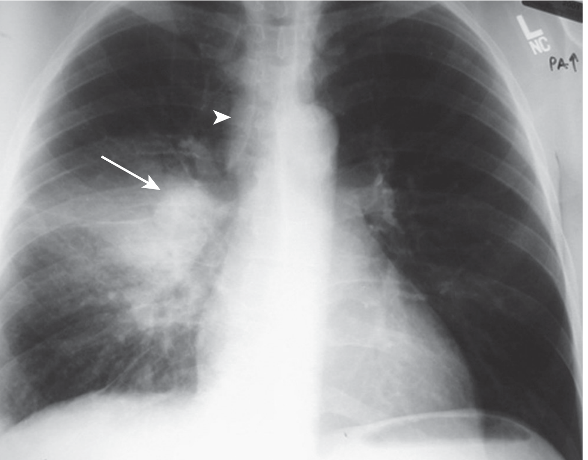
- Ghon focus - small parenchymal opacity (mid/lower zone)
- Ghon complex = Ghon focus + ipsilateral hilar lymphadenopathy
- Hilar / paratracheal lymphadenopathy - hallmark of primary TB
- Enlarged nodes can compress bronchi → collapse/consolidation or ball-valve hyperinflation
- Pleural effusion (especially in adolescents/young adults)
- Miliary pattern if disseminated (see below)
Post-Primary / Reactivation TB (Adults)
Key feature: Upper lobe + cavitation
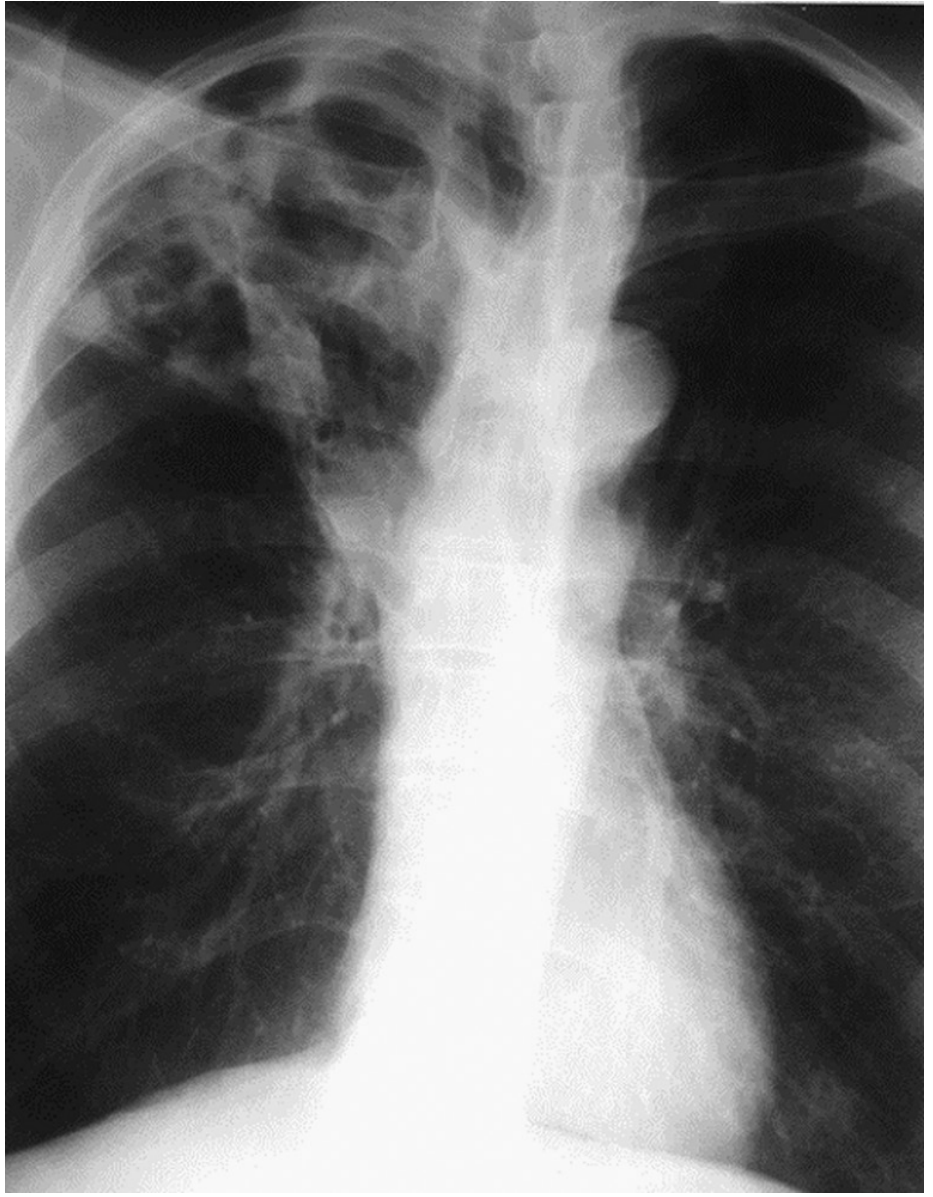
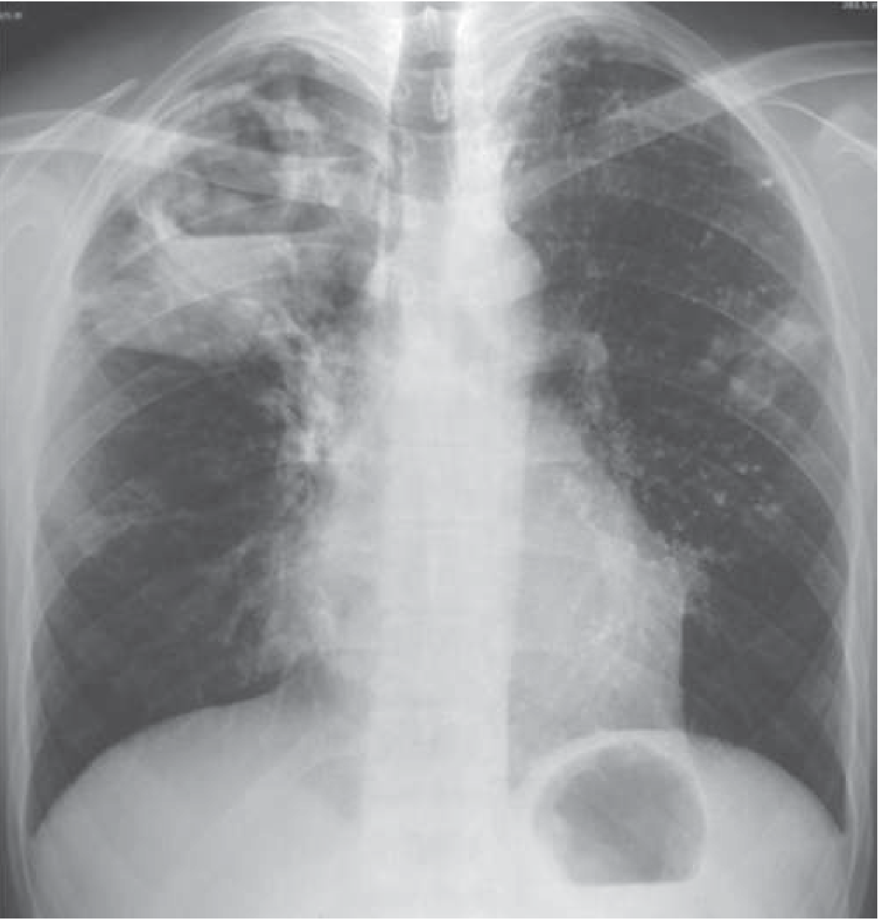
| Finding | Details |
|---|---|
| Upper lobe infiltrates | Apical & posterior segments of RUL most common; apico-posterior LUL also affected |
| Cavitation | Thick-walled cavities - due to liquefaction necrosis; highly infectious |
| Air-fluid level in cavity | Indicates active liquefaction |
| Bronchogenic spread | "Satellite" nodules/consolidation in lower lobes from aspiration of cavity contents |
| Fibrosis & volume loss | Upper lobe shrinkage, tracheal deviation toward affected side |
| Calcification | Healed TB - calcified Ghon complex = Ranke complex |
| No/minimal hilar adenopathy | Distinguishes reactivation from primary TB in immunocompetent adults |
Miliary TB
- Bilateral fine 1-3 mm millet-seed nodules uniformly distributed throughout both lungs
- Results from haematogenous dissemination
- Seen in immunocompromised, young children, HIV patients
TB in HIV Co-infection
| CD4 Count | CXR Pattern |
|---|---|
| High CD4 (early HIV) | Typical - upper lobe infiltrate + cavitation |
| Low CD4 (<200) | Atypical - lower zone opacities, diffuse infiltrates, hilar adenopathy, no cavitation |
| Very low CD4 | May have normal CXR despite active disease |
Paediatric note: CXR findings in children with TB are often subtle. Hilar adenopathy may be the only finding. Sputum smear is often negative in children - gastric lavage or induced sputum + GeneXpert are preferred diagnostic approaches.
(Sources: Harrison's Principles of Internal Medicine 22E; Murray & Nadel's Textbook of Respiratory Medicine)
Give me cxr findings as 4-5 points alone
Based on the textbook content already in context from the previous response, here are the CXR findings in TB as 5 concise points:
CXR Findings in TB (5 Key Points)
1. Upper Lobe Infiltrates (Reactivation TB)
Opacities predominantly in the apical and posterior segments of the right upper lobe and apico-posterior segment of the left upper lobe. This is the most classic and characteristic finding in post-primary/reactivation TB.
2. Cavitation
Thick-walled cavities, often in the upper lobes, due to liquefactive necrosis. May show an air-fluid level inside. Cavitation = high bacillary load = highly infectious patient.
3. Hilar / Paratracheal Lymphadenopathy
Classic feature of primary TB (especially in children). The Ghon focus + ipsilateral hilar lymphadenopathy = Ghon complex. Enlarged nodes can compress bronchi causing collapse or hyperinflation.
4. Bronchogenic Spread / Satellite Lesions
When cavity contents are aspirated via airways, nodular opacities and consolidation appear in the lower lobes or opposite lung - the "tree-in-bud" pattern. Indicates active disease spread.
5. Miliary Pattern
Bilateral fine 1-3 mm millet-seed nodules uniformly scattered throughout both lungs - indicates haematogenous dissemination. Seen in immunocompromised patients, young children, and HIV co-infection.
Bonus: Healed TB leaves calcification + fibrosis + upper lobe volume loss (tracheal deviation toward affected side). In HIV with low CD4, cavitation is absent and lower zone diffuse opacities predominate.
(Source: Murray & Nadel's Textbook of Respiratory Medicine; Harrison's 22E)
5 main causes of wheezing
Searching the medical library for causes of wheezing.
Finding Sources
Finding Sources
Finding Sources
Finding Sources
Reading File
Reading File
I have all the content needed. Here are the 5 main causes:
5 Main Causes of Wheezing
1. Asthma
Most common cause of wheeze. Caused by reversible airway inflammation, bronchospasm, and mucosal oedema from allergen/trigger exposure. Wheeze is typically expiratory, bilateral, polyphonic, worse at night and with exercise, and relieved by bronchodilators. The classic "must not miss and must not forget" diagnosis.
2. COPD (Chronic Obstructive Pulmonary Disease)
Chronic wheeze in a smoker >40 years old with progressive dyspnoea. Caused by fixed/irreversible airflow obstruction from chronic bronchitis and emphysema. Wheeze is persistent and accompanied by chronic productive cough and reduced exercise tolerance. Partially reversible with bronchodilators.
3. Acute Bronchiolitis / Bronchitis
- Bronchiolitis - most common cause of wheeze in infants <2 years, usually due to RSV. Causes inflammation and oedema of small airways with widespread expiratory wheeze and hyperinflation.
- Acute bronchitis - proximal airway inflammation, typically viral, causing transient wheeze with cough in older children and adults.
4. Cardiac Failure / Pulmonary Oedema ("Cardiac Asthma")
Left ventricular failure causes pulmonary venous congestion and interstitial oedema, which compresses small airways and produces wheeze. This is "cardiac asthma." Key distinguishing features: orthopnoea, raised JVP, basal crackles, S3 gallop, and bilateral perihilar oedema on CXR.
5. Foreign Body Aspiration / Airway Obstruction
A sudden-onset unilateral wheeze in a child (especially toddler) must raise suspicion for inhaled foreign body until proven otherwise. The obstruction produces a ball-valve effect - air enters but cannot fully exit, causing localised hyperinflation and wheeze. Other causes of obstruction: tumour, mucus plug, extrinsic compression by lymph nodes.
Other causes to keep in mind: Pulmonary embolism, bronchiectasis, anaphylaxis, vocal cord dysfunction (PVCM - mimics asthma, produces inspiratory stridor more than wheeze), and sarcoidosis.
(Source: Symptom to Diagnosis 4th Ed.; Murray & Nadel's Textbook of Respiratory Medicine)
For paediatrics
Searching for paediatric-specific wheezing causes.
Finding Sources
Finding Sources
Reading File
Finding Sources
Reading File
Excellent - I have precise paediatric content including the age-based breakdown. Here is the focused paediatric answer:
5 Main Causes of Wheezing in Paediatrics
Key rule from Tintinalli's: Age is the most important guide to the likely cause.
- < 1 year → Bronchiolitis most common
- 1-2 years → Mixed (either possible)
- > 2 years → Asthma most common
1. Bronchiolitis (Infants < 2 years - Most Common in This Age)
- Caused predominantly by RSV (also rhinovirus, human metapneumovirus, parainfluenza)
- Inflammation and oedema of small bronchioles → widespread bilateral expiratory wheeze + hyperinflation
- Preceded by URTI symptoms (nasal congestion, poor feeding, low-grade fever)
- First episode, age <1 year, sick contact during viral season = classic bronchiolitis picture
- Infants are particularly vulnerable due to smaller, more collapsible airways
2. Asthma (Most Common in Children > 2 years)
- Recurrent, episodic wheeze with identifiable triggers (allergens, viral URTI, exercise, cold air)
- Positive family history of atopy / wheeze makes asthma more likely
- Responds to bronchodilators - this is a key diagnostic clue
- Worse at night; symptom-free intervals between episodes
- ~50% of children have a history of wheezing by age 6
3. Foreign Body Aspiration
- Classic in toddlers (6 months - 4 years)
- Sudden onset wheeze after a choking/gagging episode - no preceding URTI
- Typically unilateral / focal wheeze (rather than diffuse bilateral)
- Ball-valve effect → ipsilateral hyperinflation on CXR, mediastinal shift away
- History of playing with small objects or eating nuts/seeds is key
4. Viral-Induced Wheeze / Recurrent Virus-Associated Wheeze
- Distinct from asthma in preschool children (age 1-5 years)
- Wheeze occurs only during viral URTI episodes with no interval symptoms
- No atopy, no triggers between illnesses
- Often resolves by school age
- Related to transient airway hyperresponsiveness post-viral infection
5. Pneumonia / Lower Respiratory Tract Infection
- Bacterial or viral pneumonia can cause wheeze, especially in younger children
- Associated with fever, tachypnoea, chest indrawing, focal crackles
- Wheeze here is due to airway inflammation and secretions, not bronchospasm
- Does not respond well to bronchodilators - key distinguishing feature from asthma
Other Important Paediatric Causes to Exclude
| Cause | Clue |
|---|---|
| Cystic fibrosis | Failure to thrive, recurrent chest infections, steatorrhoea |
| Vascular ring / tracheal compression | Persistent wheeze from birth, not responding to treatment, stridor |
| Congenital heart disease | Cyanosis, poor weight gain, cardiac murmur, pulmonary oedema |
| Gastro-oesophageal reflux (GORD) | Post-feed wheeze, arching, recurrent aspiration |
| Tracheomalacia | Monophonic wheeze from birth, worse with crying/feeds |
(Source: Tintinalli's Emergency Medicine; Swanson's Family Medicine Review)
What is atypical pneumonia and drug of choice
Searching the medical library for atypical pneumonia.
Finding Sources
Finding Sources
Reading File
Reading File
Reading File
Comprehensive content obtained. Here is the full answer:
Atypical Pneumonia
What is it?
Atypical pneumonia refers to a pneumonia syndrome caused by pathogens that:
- Are not visualised on routine Gram stain or ZN stain
- Cannot be cultured on standard media
- Are resistant to beta-lactam antibiotics (penicillins, cephalosporins)
- Produce a milder, more insidious clinical picture than "typical" bacterial pneumonia
The term was first coined in the 1930s when physicians noticed some patients had a milder illness than the typical S. pneumoniae picture.
Typical vs. Atypical Pneumonia - Key Differences
| Feature | Typical | Atypical |
|---|---|---|
| Onset | Acute, abrupt with rigors | Gradual, insidious |
| Cough | Productive, purulent/bloody | Dry, non-productive |
| Fever | High, with chills | Low-grade |
| Pleuritic chest pain | Common | Rare |
| WBC | High neutrophilia | Normal / mildly raised |
| CXR | Lobar consolidation, air bronchograms | Patchy bilateral interstitial / reticulonodular infiltrates |
| Extrapulmonary features | Rare | Prominent - malaise, headache, myalgia, diarrhoea |
| Response to penicillin | Good | None |
| Procalcitonin | Elevated (>0.5) | Low (≤0.1 μg/L) |
Causative Organisms
| Organism | Key Clinical Clue |
|---|---|
| Mycoplasma pneumoniae | Most common; school-age children & young adults; bullous myringitis, cold agglutinins, haemolytic anaemia, erythema multiforme |
| Chlamydophila pneumoniae | Mild illness; obligate intracellular; common in young adults |
| Chlamydophila psittaci (Psittacosis) | Contact with birds (parrots, pigeons) |
| Legionella pneumophila (Legionnaires') | Relative bradycardia despite high fever; diarrhoea; hyponatraemia; air-conditioning/water cooling towers; cruise ships |
| Coxiella burnetii (Q fever) | Contact with farm animals (cattle, sheep); can cause endocarditis |
| Francisella tularensis (Tularaemia) | Hunting / skinning rabbits; resistant to macrolides |
| Viral (RSV, influenza, adenovirus, metapneumovirus) | No antibiotic response; PCR diagnosis |
Mycoplasma pneumoniae is by far the most common - accounts for up to 15% of outpatient CAP cases and is the leading cause of atypical pneumonia in school-age children and young adults.
Drug of Choice
| Pathogen | Drug of Choice |
|---|---|
| Mycoplasma, Chlamydia, Legionella | Macrolide (Azithromycin - 1st choice, especially in children) |
| Alternative (adults) | Doxycycline |
| Severe Legionella / Fluoroquinolone preferred | Levofloxacin / Moxifloxacin |
| Francisella tularensis | Aminoglycoside (Streptomycin) - macrolides do NOT work |
| Viral pneumonia (Influenza) | Oseltamivir (most effective when started early) |
In Paediatrics Specifically:
- Azithromycin is the drug of choice for Mycoplasma and Chlamydia pneumonia in children
- Doxycycline is avoided in children <8 years (tooth discolouration)
- Fluoroquinolones are generally not used in young children
Key exam point: Atypical pneumonia = dry cough + extrapulmonary features + normal WBC + patchy CXR + no response to penicillin → treat with Azithromycin (macrolide)
(Sources: Fishman's Pulmonary Diseases; Murray & Nadel's Respiratory Medicine; Frameworks for Internal Medicine)
Imnci classification of pneumonia
Searching the medical library for IMNCI classification of pneumonia.
Finding Sources
Finding Sources
Finding Sources
Finding Sources
Reading File
Reading File
I have the complete IMNCI classification from Park's. Here is the comprehensive answer:
IMNCI Classification of Pneumonia
IMNCI (Integrated Management of Neonatal and Childhood Illness) classifies respiratory illness in children into two age groups with separate classification systems.
A. Child Aged 2 Months to 5 Years
Four classifications:
I. Very Severe Disease
Signs (any one):
- Not able to drink
- Convulsions
- Abnormally sleepy or difficult to wake
- Stridor in a calm child
- Severe malnutrition
Action: Refer URGENTLY to hospital + first dose antibiotic + treat fever/wheeze if present
II. Severe Pneumonia
Signs:
- Chest indrawing (lower chest wall goes IN on inspiration)
- ± Fast breathing may or may not be present (exhausted child may have slow breathing but still has indrawing)
A child with chest indrawing is at higher risk of death than a child with fast breathing alone.
Action: Refer urgently to hospital + first dose antibiotic
III. Pneumonia (Not Severe)
Signs:
- Fast breathing only - no chest indrawing, no danger signs
Fast breathing thresholds:
| Age | Fast Breathing |
|---|---|
| < 2 months | ≥ 60 breaths/min |
| 2-12 months | ≥ 50 breaths/min |
| 1-5 years | ≥ 40 breaths/min |
Action: Treat at home with oral antibiotic (cotrimoxazole/amoxicillin) + follow up in 2 days
IV. No Pneumonia: Cough or Cold
Signs:
- No fast breathing
- No chest indrawing
- No danger signs
Action: Home care - soothe throat, clear nose, continue feeding, return if worsens
B. Young Infant (Birth to 2 Months)
Only two classifications - any pneumonia in a young infant is considered severe:
I. Very Severe Disease
Signs (any one):
- Stopped feeding well (takes <½ usual amount)
- Convulsions / abnormally sleepy / difficult to wake
- Stridor in calm child
- Wheezing
- Fever (≥38°C) OR low body temperature (hypothermia)
Action: Refer URGENTLY + keep warm + first dose antibiotic
II. Severe Pneumonia
Signs:
- Severe chest indrawing, OR
- Fast breathing (≥60/min)
Note: In young infants, mild chest indrawing can be normal (soft chest wall). Only severe chest indrawing is significant.
Action: Refer urgently + keep warm + first dose antibiotic
(If referral not feasible: treat with antibiotic + follow closely)
III. No Pneumonia: Cough or Cold
- No severe chest indrawing
- No fast breathing (<60/min)
Action: Home care - keep warm, breastfeed frequently, clear nose, return quickly if breathing becomes difficult/fast or feeding worsens
Summary Table
| Classification | Age 2m-5yrs Signs | Age <2m Signs | Action |
|---|---|---|---|
| Very Severe Disease | Can't drink, convulsions, sleepy, stridor, severe malnutrition | Poor feeding, convulsions, sleepy, stridor, wheeze, fever/hypothermia | Urgent referral |
| Severe Pneumonia | Chest indrawing | Severe chest indrawing OR RR ≥60 | Urgent referral + antibiotics |
| Pneumonia (not severe) | Fast breathing only | - (not applicable) | Oral antibiotics at home |
| No Pneumonia | No fast breathing, no indrawing | No fast breathing, no severe indrawing | Home care |
(Source: Park's Textbook of Preventive and Social Medicine - WHO/IMNCI guidelines)
This is a shared conversation. Sign in to Orris to start your own chat.