
COPD MANAGEMENT according to Malaysian guidelines
Malaysian COPD guidelines CPG management 2024 2025
"COPD" AND "Malaysia" AND "management"
http://www1.mts.org.my/Portals/0/Images/2nd%20Edition%20of%2…
https://www.iqvia.com/-/media/iqvia/pdfs/asia-pacific/white-…
COPD management algorithm stepwise treatment flowchart
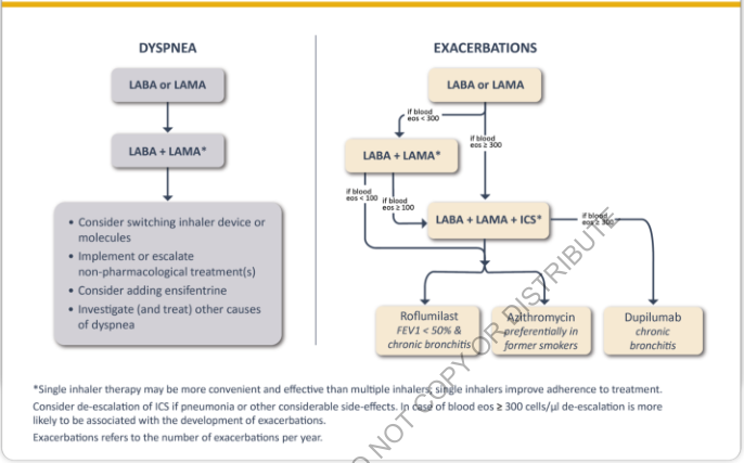
Summary : This figure presents two flowcharts outlining the pharmacological management of chronic obstructive pulmonary disease (COPD) based on the primary symptom: dyspnea or exacerbations. It details stepwise treatment escalation, decision points based on blood eosinophil counts, and additional therapeutic options. flowchart: # Dyspnea Management : Nodes : • LABA or LAMA (rounded rectangle) • LABA + LAMA* (rounded rectangle) • Consider switching inhaler device or molecules; Implement or escalate non-pharmacological treatment(s); Consider adding ensifentrine; Investigate (and treat) other causes of dyspnea (large rounded rectangle) Connectors : • Downward arrows from LABA or LAMA → LABA + LAMA* → management options box Layout : • Vertical, single branch, two main steps followed by a box with four bullet points # Exacerbations Management : Nodes : • LABA or LAMA (rounded rectangle) • LABA + LAMA* (rounded rectangle) • LABA + LAMA + ICS* (rounded rectangle) • Roflumilast (rounded rectangle, “FEV1 < 50% & chronic bronchitis”) • Azithromycin (rounded rectangle, “preferentially in former smokers”) • Dupilumab (rounded rectangle, “chronic bronchitis”) Connectors : • LABA or LAMA → LABA + LAMA* (if blood eos < 300) • LABA or LAMA → LABA + LAMA + ICS* (if blood eos ≥ 300) • LABA + LAMA* → LABA + LAMA + ICS* (if blood eos ≥ 100) • LABA + LAMA + ICS* → Roflumilast, Azithromycin, Dupilumab (branching arrows) • LABA + LAMA + ICS* → Dupilumab (if blood eos ≥ 300) Layout : • Vertical with multiple branches based on blood eosinophil count thresholds (<100, <300, ≥100, ≥300) • Final step branches into three parallel options # Additional Information : • Footnote: Single inhaler therapy may be more convenient and effective than multiple inhalers; single inhalers improve adherence to treatment. • Consider de-escalation of ICS if pneumonia or other considerable side-effects. In case of blood eos ≥ 300 cells/μl de-escalation is more likely to be associated with the development of exacerbations. • Exacerbations refers to the number of exacerbations per year. # Analysis : • The dyspnea pathway is linear, focusing on escalation and device/molecule switching, with consideration for non-pharmacological treatments and other causes. • The exacerbation pathway is more complex, with branching based on blood eosinophil counts, leading to different combinations of inhaled therapies and add-on treatments for specific patient subgroups (chronic bronchitis, former smokers, high eosinophil counts). • The figure emphasizes personalized treatment escalation and the importance of blood eosinophil count in guiding therapy for COPD exacerbations.
Summary : This flowchart outlines the stepwise management and treatment algorithm for osteoarthritis (OA) of the hip and/or knee, guiding clinicians from diagnosis through non-surgical and surgical interventions based on patient response. flowchart: # Nodes : • Start (rectangle): "Patient diagnosed with OA of the hip and/or knee" • Step 2 (rectangle): "Assess pain, severity, and functional limitations" • Step 3 (rectangle): "Develop individualized treatment plan with core non-surgical management approaches (see Sidebar 1)" • Step 4 (diamond): "Has the patient’s pain or functional limitation improved on re-evaluation?" • Step 5 (rectangle): "Continue long-term management of OA and reassess annually or as needed" • Step 6 (rectangle): "Offer referral to physical therapy and/or combination pharmacotherapy (see Sidebars 2 and 3)" • Step 7 (diamond): "Has the patient’s pain or functional limitation improved on re-evaluation?" • Step 8 (rectangle): "Referral for additional physical therapy" • Step 9 (diamond): "Has the patient’s pain or functional limitation improved on re-evaluation?" • Step 10 (rectangle): "Obtain weight-bearing plain radiographs prior to referral for surgical consultation (see Appendix K)" • Step 11 (rectangle): "Refer for surgical consultation" # Connectors : • Downward arrows connect each step in sequence unless otherwise specified. • From Step 4 (diamond): – "Yes" arrow to Step 5. – "No" arrow to Step 6. • From Step 7 (diamond): – "Yes" arrow to Step 5. – "No" arrow to Step 8. • From Step 9 (diamond): – "Yes" arrow to Step 5. – "No" arrow to Step 10. • Step 10 leads to Step 11. • Step 5 loops back to Step 7 for reassessment as needed. # Layout : • The flowchart is arranged vertically, with decision diamonds branching to the right or left for "Yes"/"No" answers, and merging back into the main flow. • There are three main decision points (Steps 4, 7, 9) that determine whether to continue current management, escalate therapy, or proceed to surgical evaluation. • The process allows for repeated reassessment and escalation based on patient response. # Analysis : • The algorithm emphasizes a stepwise, evidence-based approach, prioritizing non-surgical management and physical therapy before considering surgery. • Multiple reassessment points ensure that treatment is responsive to patient progress. • Only after failure of conservative measures and additional physical therapy, and after obtaining appropriate imaging, is surgical consultation recommended. • The flowchart supports individualized care and annual or as-needed reassessment for long-term management.
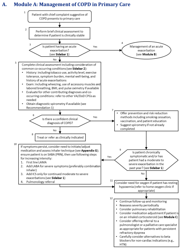
Summary : This flowchart outlines the stepwise management of Chronic Obstructive Pulmonary Disease (COPD) in primary care, including initial assessment, diagnosis, risk reduction, medication adjustment, and referral decisions. flowchart: # Nodes : • Start (rectangle): "Patient with chief complaint suggestive of COPD presents to primary care" • Rectangle: "Perform brief clinical assessment to determine if patient is clinically stable" • Diamond: "Is patient having an acute exacerbation? (see Sidebar 1)" • Ellipse: "Management of an acute exacerbation (see Module B)" • Rectangle: "Complete clinical assessment including consideration of common co-occurring conditions (see Sidebar 2): History, Exam, Evaluate for other contributing diagnoses, Obtain diagnostic spirometry if available" • Diamond: "Is there a confident clinical diagnosis of COPD?" • Rectangle: "Offer prevention and risk reduction methods including smoking cessation, vaccination, and patient education; Suggest spirometry if not already completed" • Rectangle: "Treat or refer as clinically indicated" • Diamond: "Is patient chronically symptomatic and/or has patient had a moderate to severe exacerbation in the past year? (see Sidebar 1)" • Rectangle: "If symptoms persist, consider need to initiate/adjust medication and assess inhaler technique (see Appendix G); ensure patient is on SABA (PRN), then use following steps for increasing intensity: 1. First line LAMA, 2. Add LABA for severe symptoms (preferably combination inhaler), 3. Add ICS only for continued moderate to severe exacerbations (see Sidebar 1), 4. Pulmonology referral" • Rectangle: "Consider need for oxygen if patient has resting hypoxemia (refer to home oxygen clinic if appropriate)" • Rectangle: "Continue follow-up and monitoring; Reassess severity periodically; Consider pulmonary rehabilitation; Consider medication adjustment if patient is on an inhaled corticosteroid (see Module C); Consider offering referral to a pulmonologist or a palliative care specialist as appropriate for patients with persistent refractory dyspnea; Carefully consider alternatives to beta blockers for non-cardiac indications (e.g., HTN)" # Connectors : • Arrows connect nodes in a top-down sequence, with diamonds representing decision points. • From "Is patient having an acute exacerbation?" (diamond): Yes → "Management of an acute exacerbation (ellipse)"; No → "Complete clinical assessment" (rectangle). • From "Is there a confident clinical diagnosis of COPD?" (diamond): Yes → "Offer prevention and risk reduction methods" (rectangle); No → "Treat or refer as clinically indicated" (rectangle). • From "Offer prevention and risk reduction methods" (rectangle) → "Is patient chronically symptomatic and/or has patient had a moderate to severe exacerbation in the past year?" (diamond). • From "Is patient chronically symptomatic..." (diamond): Yes → "If symptoms persist, consider need to initiate/adjust medication..." (rectangle); No → "Consider need for oxygen if patient has resting hypoxemia" (rectangle). • Both "If symptoms persist..." and "Consider need for oxygen..." nodes lead to "Continue follow-up and monitoring..." (rectangle). # Layout : • The flowchart is arranged vertically, with decision diamonds branching to different management paths. • Merges occur after medication adjustment and oxygen consideration, leading to a unified follow-up and monitoring node. • Sidebars and appendices are referenced for further detail at several steps. # Analysis : • The workflow prioritizes acute exacerbation management first, then proceeds to comprehensive assessment and diagnosis. • Preventive strategies and risk reduction are emphasized early for confirmed COPD cases. • Medication adjustment and specialist referral are considered for persistent symptoms or recent exacerbations. • Oxygen therapy is considered for patients with resting hypoxemia. • Ongoing follow-up, severity reassessment, and consideration of rehabilitation or specialist referral are recommended for all patients. • The flowchart supports a systematic, evidence-based approach to COPD management in primary care, with clear decision points and escalation pathways.
COPD Management: Malaysian Clinical Practice Guidelines (CPG)
Source: Malaysian COPD CPG, 2nd Edition (Malaysian Thoracic Society / MOH Malaysia). Note: As of 2024-2026, this CPG has not been updated since its 2nd edition (~2009 base). Local experts now cross-reference GOLD 2025 for updated ABCE grouping and triple therapy guidance. Key gaps vs. GOLD: Groups C & D have since merged into Group E, and upfront triple therapy (LABA+LAMA+ICS) for high-risk patients is now recommended by GOLD but not reflected in the older Malaysian CPG.
1. Definition
2. Diagnosis
Clinical Features
- Chronic cough (with or without sputum)
- Progressive breathlessness (dyspnea)
- History of significant exposure to risk factors
Key Risk Factors in the Malaysian Context
- Tobacco smoking (>80% of Malaysian COPD patients are smokers/ex-smokers)
- Vaping and e-cigarette use
- Occupational exposure (dust, fumes, chemicals)
- Indoor air pollution and environmental haze (a key local factor)
- Biomass fuel smoke
Spirometry - Confirmatory Test
- Post-bronchodilator FEV1/FVC < 0.7 confirms airflow limitation
- GOLD Grading of Airflow Obstruction:
| GOLD Grade | FEV1 (% predicted) | Severity |
|---|---|---|
| 1 | ≥ 80% | Mild |
| 2 | 50-79% | Moderate |
| 3 | 30-49% | Severe |
| 4 | < 30% | Very severe |
Symptom Assessment Tools
- mMRC Dyspnoea Scale (0-4)
- CAT (COPD Assessment Test) - scores ≥ 10 = significant symptom burden
Patient Classification (GOLD ABE, current international standard)
| Group | Exacerbation History | Symptoms |
|---|---|---|
| A | 0-1/year (not hospitalised) | mMRC 0-1 or CAT < 10 |
| B | 0-1/year (not hospitalised) | mMRC ≥ 2 or CAT ≥ 10 |
| E | ≥ 2/year OR ≥ 1 hospitalisation | Any symptom level |
3. Reducing Risk Factors
Smoking Cessation (Most Effective Intervention)
- All COPD patients who smoke must be strongly advised to quit
- Options: brief counselling, NRT (nicotine replacement therapy), pharmacotherapy (varenicline, bupropion)
- Reduces rate of FEV1 decline
Other Risk Reduction
- Avoid occupational dust/fume/chemical exposure
- Minimise indoor biomass smoke and haze exposure
- Encourage regular physical activity
4. Vaccination
| Vaccine | Recommendation |
|---|---|
| Influenza | Annually (Level I evidence) - reduces AECOPD frequency and mortality |
| Pneumococcal | At least once in a lifetime; repeat ≥ 5 years later (max 2 doses lifetime) |
5. Management of Stable COPD
5A. Pharmacological Treatment
Malaysian CPG Treatment Steps (Adapted by Severity):
- Smoking cessation, exercise, ideal BMI, vaccinations
- Short-acting bronchodilator PRN: SABA (salbutamol) or SAMA (ipratropium) as needed
- Consider alternative causes
- SABA + SAAC combination regularly + ICS + theophylline
- Consider referral to tertiary centre for long-acting bronchodilators (LABA/LAMA)
- SABA + SAAC combination + ICS + theophylline regularly
- Long-term oxygen therapy (LTOT)
- Consider lung transplantation / LVRS (Lung Volume Reduction Surgery)
Current GOLD 2025 Pharmacological Algorithm (to supplement the older Malaysian CPG):
- Blood eos < 300: LABA+LAMA
- Blood eos ≥ 300: LABA+LAMA+ICS (triple therapy)
- Add-ons (if still exacerbating): roflumilast (FEV1<50% + chronic bronchitis), azithromycin (former smokers), dupilumab (chronic bronchitis)
Drug Classes Summary
| Class | Examples | Role |
|---|---|---|
| SABA | Salbutamol, terbutaline | Reliever; PRN or before exercise |
| SAMA | Ipratropium | Reliever; often combined with SABA |
| LABA | Formoterol, salmeterol, indacaterol | Maintenance |
| LAMA | Tiotropium, glycopyrronium, umeclidinium | Maintenance; preferred over LABA as monotherapy |
| ICS | Fluticasone, budesonide | Add-on for frequent exacerbators; NOT routine monotherapy |
| LABA+LAMA | Indacaterol/glycopyrronium, etc. | Dual bronchodilation |
| LABA+ICS | Formoterol/budesonide, salmeterol/fluticasone | For exacerbation-prone with eosinophilia |
| Triple (LABA+LAMA+ICS) | Fluticasone/umeclidinium/vilanterol | High-risk Group E with eos ≥ 300 |
| Theophylline | Oral | Adjunct when inhaled therapy unavailable; narrow therapeutic index |
| Roflumilast | Oral PDE4 inhibitor | FEV1 < 50% + chronic bronchitis + frequent exacerbations |
| Macrolide | Azithromycin | Former smokers with recurrent exacerbations |
5B. Non-Pharmacological Treatment
Pulmonary Rehabilitation
- Strongly recommended (Level I evidence) for moderate-to-severe COPD
- Aims: reduce symptoms, decrease disability, increase participation in physical/social activities
- Components: exercise training (aerobic + resistance), education, nutritional advice, psychosocial support
- Minimum 6-8 weeks duration
- Malaysia-specific challenge: Only a handful of district hospitals have structured pulmonary rehabilitation programs; the Malaysian CPG recommends expanding access
Domiciliary Oxygen Therapy (LTOT)
- Indications (≥ 15 hours/day):
- PaO2 ≤ 7.3 kPa (55 mmHg) or SaO2 ≤ 88%
- PaO2 7.3-8.0 kPa (55-60 mmHg) with pulmonary hypertension, cor pulmonale, or polycythaemia
- Goal: PaO2 ≥ 8 kPa (60 mmHg) or SpO2 ≥ 90%
- Improves survival in severe COPD with chronic hypoxemia
Nutrition
- Malnutrition worsens respiratory muscle function and prognosis
- Low BMI is an independent risk factor for mortality
- Nutritional supplementation recommended for underweight patients (BMI < 21)
Lung Volume Reduction Surgery (LVRS)
- Selected patients with upper-lobe emphysema + low exercise capacity
- Improves FEV1, exercise tolerance, quality of life
- Option for centres with expertise
Lung Transplantation
- Reserved for very severe COPD (GOLD 4) refractory to maximal medical therapy
- Referral criteria: BODE index > 5, FEV1 < 20%, severe hypoxemia, rapid decline
Patient Education (Self-Management)
- Recognition of exacerbation symptoms and when to seek help
- Correct inhaler technique
- Strategies to minimise dyspnoea
- Breathing exercises (diaphragmatic, pursed-lip)
- Nutritional guidance and exercise
- Role of vaccination
- Self-management plan (action plan) for acute situations
6. Management of Acute Exacerbations (AECOPD)
Definition
Common Causes
- Respiratory infections (bacterial: H. influenzae, S. pneumoniae, M. catarrhalis; viral: rhinovirus, influenza)
- Air pollution / haze
- Non-compliance with maintenance therapy
- Pulmonary embolism, heart failure, pneumothorax (differential diagnoses to exclude)
Assessment of Severity
- Severe dyspnea not responding to initial treatment
- SpO2 < 90% or significant hypoxemia
- Acute confusion/altered consciousness (likely hypercapnia)
- Inability to eat, sleep, or care for self
- Rapid onset, high-risk features
- Failure of outpatient management
- Uncertain diagnosis
- Significant comorbidities
6A. Home/Community Management
- Increase frequency of SABA/SAMA nebulisation
- Add/increase short course oral corticosteroids (prednisolone 30-40 mg/day x 5-7 days)
- Antibiotics if purulent sputum or signs of infection (amoxicillin-clavulanate, doxycycline, macrolide)
- Self-management action plan
6B. Hospital Management
- Target: PaO2 ≥ 8 kPa (60 mmHg) or SpO2 88-92% (NOT higher - risk of hypercapnia)
- Deliver via 24-28% oxygen Venturi mask (preferred over nasal cannula for precision)
- Monitor ABG 30-60 minutes after initiation; adjust if hypercapnia worsens
- Nebulised SABA (salbutamol 2.5-5 mg) + SAMA (ipratropium 0.5 mg) every 4-6 hours
- Can be co-nebulised
- Switch to MDI + spacer when stable
- Prednisolone 30-40 mg/day orally x 5-7 days
- IV methylprednisolone if cannot take orally
- Reduces recovery time, improves FEV1, shortens hospital stay
- Purulent sputum, signs of pneumonia, or ≥ 2 of: increased dyspnea, increased sputum volume, increased sputum purulence
- First-line: amoxicillin, doxycycline, or macrolide
- If at risk for Pseudomonas (severe COPD, recent antibiotics, bronchiectasis): ciprofloxacin or antipseudomonal beta-lactam
- Indications: respiratory acidosis (pH < 7.35 + PaCO2 > 6 kPa) despite medical treatment
- Reduces need for intubation, mortality, and hospital stay (Level I evidence)
- Use IPAP 12-20 cmH2O / EPAP 4-8 cmH2O; titrate to improve pH and PaCO2
- If NIV fails or contraindicated
- Consider goals of care and patient wishes (especially in very severe/end-stage COPD)
7. COPD in Primary Care (Malaysian Context)
- Early diagnosis through targeted spirometry in at-risk individuals (smokers ≥ 40 years, chronic cough, recurrent respiratory infections)
- Spirometry should be available at all primary care facilities
- Tier-based management:
- Primary care: mild-moderate COPD, smoking cessation, SABDs, vaccinations, education
- Secondary care: moderate-severe, initiate LABDs (LABA/LAMA), manage comorbidities
- Tertiary care: severe, complex, NIV, surgical options, pulmonary rehabilitation
- Referral criteria to specialist: FEV1 < 50%, frequent exacerbations (≥ 2/year), diagnostic uncertainty, onset < 40 years, rapid decline, cor pulmonale, suspected malignancy
8. Monitoring and Follow-up
| Parameter | Frequency |
|---|---|
| Spirometry | At diagnosis; annually for moderate-severe; after significant change |
| Inhaler technique check | Every visit |
| Symptom scores (mMRC/CAT) | Every 3-6 months |
| Exacerbation history | Every visit |
| Smoking status | Every visit |
| BMI and nutrition | Annually |
| Oxygen saturation | Every visit (severe COPD) |
| ABG | When SpO2 < 92% or assessing LTOT |
9. Comorbidity Management
- Cardiovascular disease (most common; do not withhold cardioselective beta-blockers if needed)
- Metabolic syndrome / diabetes
- Osteoporosis (ICS use + low BMI + physical inactivity; baseline DEXA in high-risk patients)
- Depression and anxiety - screen routinely; impacts adherence and outcomes
- Lung cancer (COPD is independent risk factor; consider annual LDCT in eligible patients)
10. Key Differences: Malaysian CPG vs. GOLD 2025
| Aspect | Malaysian CPG (2nd Ed.) | GOLD 2025 |
|---|---|---|
| Patient grouping | ABCD | ABE (C+D merged to E) |
| First-line therapy | SABA/SAMA PRN for mild | LAMA monotherapy preferred |
| ICS use | Combined with SABA/SAAC in moderate-severe | Eosinophil-guided (eos ≥ 100-300 threshold) |
| Triple therapy | Not specified | Recommended upfront for Group E with eos ≥ 300 |
| Dupilumab | Not mentioned | Approved for COPD with chronic bronchitis |
| Roflumilast | Mentioned as adjunct | Specifically for FEV1 < 50% + chronic bronchitis |
| Ensifentrine | Not mentioned | New dual PDEV/PDIII inhibitor; add-on for dyspnea |