
Hangman 's fracture.
Reading File
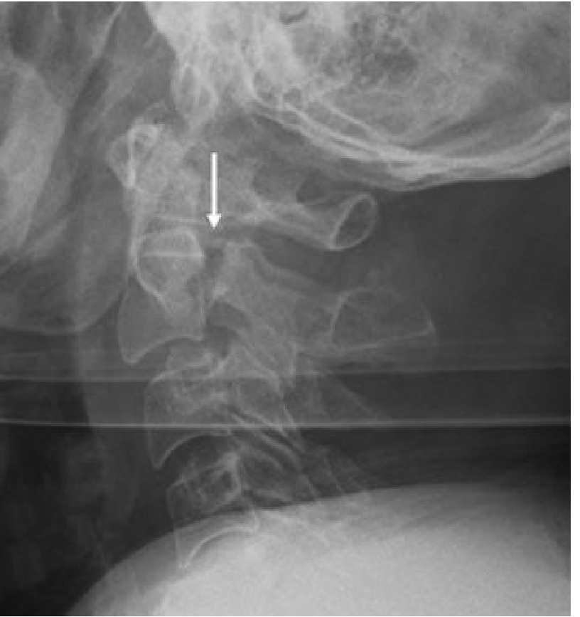
Hangman's fracture is a traumatic spondylolisthesis of C2 - bilateral fractures through the pedicles/pars interarticularis of C2, caused by hyperextension + axial loading (classically from hanging, but now more often seen in high-speed MVAs).
Searching sources...
Finding Sources
Finding Sources
Searching PubMed
"hangman fracture" AND "C2" AND classification
Good - I have strong sources. Now let me pull the detailed content from the most authoritative sources in parallel.
Reading File
Reading File
Reading File
Excellent content gathered. Let me also pull the Campbell's Operative Orthopaedics and Miller's Review sections, and fetch the key X-ray image.
Reading File
Reading File
Loading Image
I now have comprehensive content from multiple authoritative sources. Here is the complete answer:
Hangman's Fracture (Traumatic Spondylolisthesis of C2)
Definition
A Hangman's fracture is a bilateral fracture through the pars interarticularis (isthmus/posterior arch) of C2, causing traumatic spondylolisthesis of C2 on C3. The C2 vertebral body subluxes anteriorly relative to C3 while the posterior elements remain normally aligned.
Etymology - A Historical Misnomer
The name "hangman's fracture" is actually a misnomer. First described by Haughton in the 19th century, postmortem examination of corpses following judicial hanging showed this fracture pattern was a rare occurrence - most victims showed no fracture at all. The term was later applied by analogy to a characteristic radiographic pattern from high-speed trauma. - Rockwood & Green's Fractures in Adults, 10th ed. 2025
Mechanism of Injury
The classic mechanism is hyperextension + axial loading (e.g., unrestrained passenger hitting a windshield, MVA). However, different force vectors produce different fracture subtypes:
- Type I: axial load + extension
- Type II: extension, then flexion-rebound
- Type IIa: predominantly flexion (important - NO traction!)
- Type III: severe flexion with rebound extension
Today, high-speed motor vehicle accidents are the most common cause, not hanging.
Levine and Edwards Classification (Most Widely Used)
| Type | Displacement | Angulation | Mechanism | C2-C3 Disc | Stability |
|---|---|---|---|---|---|
| I | <3 mm, no translation | None | Axial load + extension | Intact | Stable |
| Ia (Starr-Eismont) | Fracture extends into posterior C2 body | Variable | Extension | Variable | Unstable - higher neuro risk |
| II | >3 mm translation | + Angulation | Extension, then flexion | Disrupted | Unstable |
| IIa | Minimal translation | Marked angulation | Flexion | Disrupted | Unstable - NO traction |
| III | + Facet dislocation C2-C3 | + Angulation | Severe flexion + extension | Disrupted | Most unstable |
Type I is the most common. Types II and III are rare.
Why Neurological Injury is Uncommon
Because the fracture effectively widens the AP diameter of the spinal canal at the level of slip - the posterior arch separates from the vertebral body, decompressing the canal. This paradox is known as the "self-decompressing" fracture. However, Type Ia (Starr-Eismont variant) causes posterior displacement of the posterior arch-body fragment complex, which actually narrows the canal and is associated with a higher incidence of neurological deficit.
Radiology
- X-ray (lateral cervical spine): Fracture line through both pedicles of C2; anterior subluxation of C2 body on C3; the spinolaminar line of C2 remains behind that of C1 and C3 (differentiates from physiological subluxation)
- CT: Best for defining fracture morphology and degree of comminution
- MRI: For assessing C2-3 disc injury, ligamentous damage (ALL, PLL), and spinal cord status - mandatory pre-operatively and in neurologically compromised patients
Treatment
Treatment is guided by fracture type and stability:
Nonoperative
| Fracture Type | Management |
|---|---|
| Type I | Rigid cervical collar |
| Type Ia | Cervical orthosis (unless spinal cord injury) |
| Type II | Cervical traction → halo vest immobilization |
| Type IIa | NO TRACTION - extension + compression via halo; traction accentuates deformity |
A trial of external rigid orthosis is appropriate for patients with <4 mm translation and <10 degrees of angulation (Miller's Review of Orthopaedics, 9th ed.).
Operative Indications
- Type III (facet dislocation) - always surgical; usually posterior stabilization
- Neurological deficit
- Failed closed reduction / reduction not maintained
- Significant instability (required traction to reduce - won't hold in collar/halo)
- Starr-Eismont (Ia) variant with spinal cord injury
- Significantly displaced fractures → internal fixation + bone grafting C2-C3 (anterior or posterior approach)
Surgical Options
- Anterior: C2-C3 discectomy and fusion (ACDF)
- Posterior: C2 pedicle screw fixation ± C1-C3 fusion
- Posterior surgery preferred for type III
Key Points for Exams
- Bilateral pars/pedicle fracture of C2 (not unilateral)
- Levine-Edwards classification - 4 types (I, Ia/Starr-Eismont, II, IIa, III)
- Type IIa - never traction (flexion mechanism; traction increases deformity)
- Neurological deficit is rare due to self-decompressing canal widening
- Type III always requires surgery (facet dislocation)
- Most common fracture pattern in clinical practice: Type I
Sources: Rockwood & Green's Fractures in Adults, 10th ed. 2025 | Grainger & Allison's Diagnostic Radiology | Campbell's Operative Orthopaedics, 15th ed. 2026 | Bailey & Love's Short Practice of Surgery, 28th ed. | Miller's Review of Orthopaedics, 9th ed.
This is a shared conversation. Sign in to Orris to start your own chat.