
can you explain me difference between parasympathetic and sympathetic nervous system in simple language possible
sympathetic parasympathetic nervous system comparison diagram
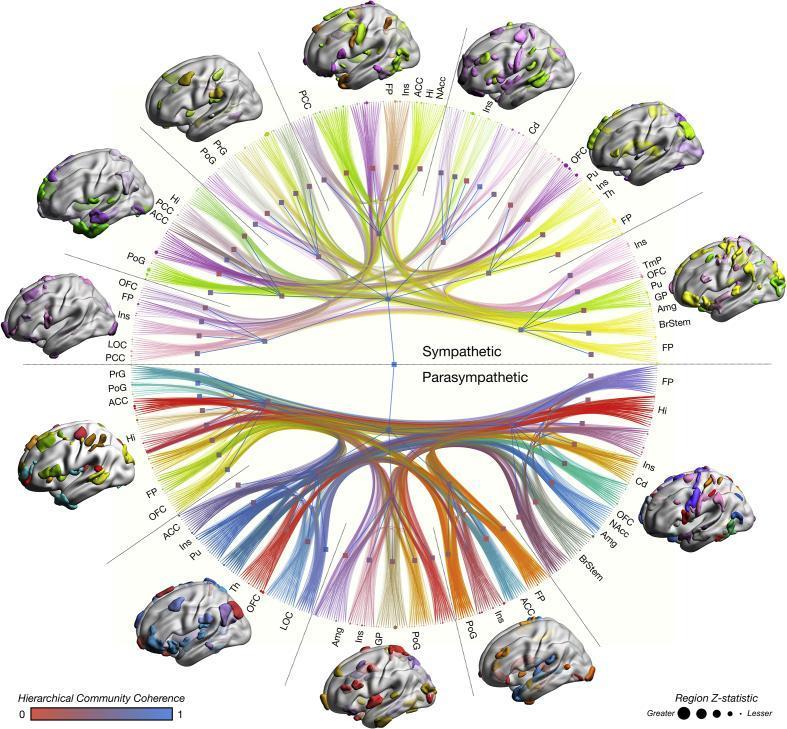
This multimodal anatomical and functional diagram illustrates the human 'autonomic connectome,' detailing the neural infrastructure of sympathetic and parasympathetic regulation. The central element is a circular network graph partitioned into two hemispheres: the upper represents the sympathetic nervous system and the lower the parasympathetic nervous system. Surrounding the center are colorful lines (edges) representing network-based statistics from functional MRI, gray matter morphometry, and white matter tractography. Edge width indicates connection effect size. The outer perimeter contains nodes corresponding to specific brain regions, such as the anterior cingulate cortex (ACC), insula (Ins), amygdala (Amg), and brainstem (BrStem). Node size is proportional to the Z-statistic importance of the region. Surrounding the central connectome are several 3D cortical surface reconstructions displaying color-coded parcellations that visualize hierarchical community structures. A color scale at the bottom indicates hierarchical community coherence (ranging from red to blue), while a size legend defines the regional Z-statistics. This educational visual explains the high-dimensional integration of disparate cortical and subcortical structures involved in autonomic homeostasis and sympathovagal balance.
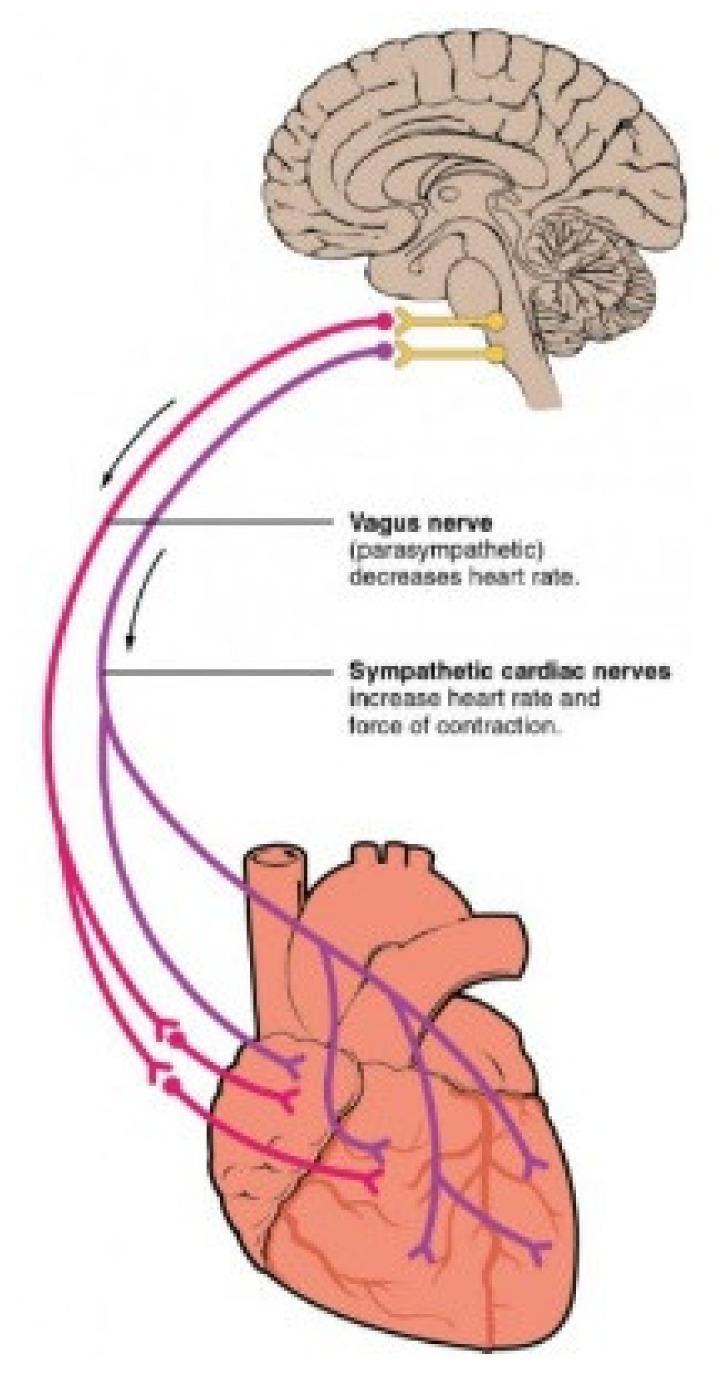
This medical illustration depicts the Autonomic Nervous System (ANS) pathways connecting the brainstem to the heart. A sagittal section of the human brain shows the medulla oblongata as the origin for both parasympathetic and sympathetic innervation. The Vagus nerve (parasympathetic), color-coded in purple, is shown descending from the brainstem to the heart, where it branches to innervate the atria and ventricles; text annotations indicate its role in decreasing heart rate. The sympathetic cardiac nerves, color-coded in red, originate from the same region and follow a parallel descending path, branching across the cardiac tissue; text labels specify their role in increasing both heart rate and the force of contraction. The diagram illustrates the functional antagonism between the two branches of the ANS in cardiac regulation, highlighting the primary innervation sites at the sinoatrial node, atrioventricular node, and the myocardium.
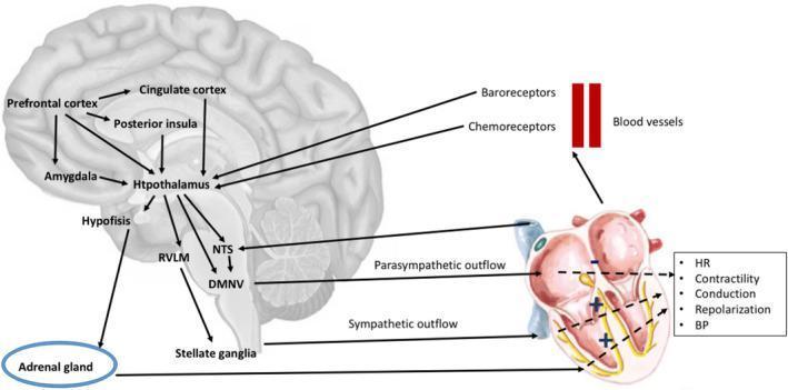
This pathophysiology diagram illustrates the neuroanatomical pathways connecting the central nervous system to the cardiovascular system, emphasizing the brain-heart axis. The schematic shows cortical and subcortical structures, including the prefrontal cortex, cingulate cortex, posterior insula, and amygdala, which provide inputs to the hypothalamus. The hypothalamus serves as a central hub, influencing brainstem nuclei such as the Rostral Ventrolateral Medulla (RVLM), Nucleus Tractus Solitarius (NTS), and Dorsal Motor Nucleus of the Vagus (DMNV). The autonomic pathways are bifurcated into parasympathetic outflow (originating from the DMNV/NTS) and sympathetic outflow, which descends through the RVLM to the stellate ganglia and adrenal gland. These pathways culminate in the heart, modulating clinical parameters including heart rate (HR), contractility, conduction, repolarization, and blood pressure (BP). Additionally, a feedback loop is depicted where baroreceptors and chemoreceptors in blood vessels transmit signals back to the NTS. The diagram highlights the integrated control of cardiovascular function through neural and endocrine (Hypophysis/Adrenal gland) mechanisms.
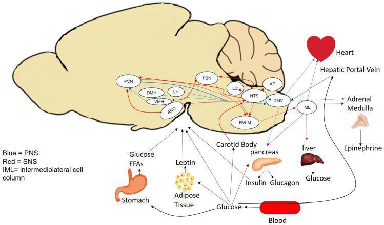
This pathophysiology diagram illustrates the glucose-sensing pathways and neuroendocrine feedback loops involved in metabolic homeostasis. A central anatomical brain silhouette highlights key regulatory nuclei, including the hypothalamus (PVN, DMH, LH, VMH, ARC) and brainstem (PBN, LC, AP, NTS, DMV, RVLM). Red arrows indicate Sympathetic Nervous System (SNS) pathways, while blue arrows represent Parasympathetic Nervous System (PNS) pathways, showing the integration of autonomic signals. Peripheral inputs are depicted from the stomach (FFAs), adipose tissue (leptin), pancreas (insulin, glucagon), liver, carotid body, and blood (glucose). These organs communicate via afferent signals to the brain's glucose-sensing regions. The diagram also shows efferent pathways from the DMV and the Intermediolateral cell column (IML) to target organs like the heart, liver, and adrenal medulla, with the latter releasing epinephrine to stimulate the hepatic portal vein. This educational visual maps the complex communication between the central nervous system and peripheral metabolic organs to maintain glucose levels through counterregulatory responses.
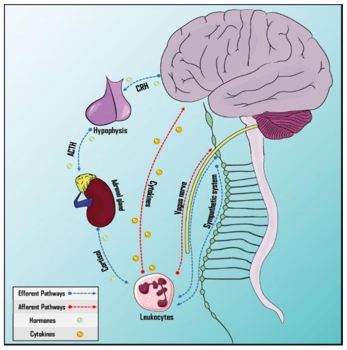
This pathophysiology diagram illustrates the bidirectional neuro-immune communication pathways of the psychoneuroimmunology (PNEI) system. It depicts the afferent (red dotted arrows) and efferent (blue dashed arrows) connections between the central nervous system and the immune system. The humoral efferent pathway highlights the Hypothalamus-Pituitary-Adrenal (HPA) axis, showing the sequence of CRH from the brain to the hypophysis, ACTH to the adrenal gland, and the subsequent release of cortisol. The neural pathways demonstrate the autonomic nervous system's involvement, specifically the vagus nerve (parasympathetic) and the sympathetic nervous system chain, which provide innervation to leukocytes and lymphoid structures. Afferent signaling is represented by cytokines (yellow spheres) crossing the blood-brain barrier and neural feedback via the vagus nerve. The diagram serves as an educational tool for understanding the molecular and anatomical basis of neuroimmunological integration, including the physiological mediators like hormones and cytokines that regulate the body's response to stress and internal stimuli.
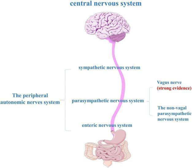
An anatomical diagram illustrating the components of the gut-brain axis and the peripheral autonomic nervous system (ANS). At the top, a human brain represents the central nervous system (CNS), which is connected via a long neural pathway to the gastrointestinal tract (stomach and intestines) at the bottom. The peripheral autonomic nervous system is categorized into three major divisions: the sympathetic nervous system, the parasympathetic nervous system, and the enteric nervous system (ENS). The diagram highlights the role of the vagus nerve within the parasympathetic division, noted with 'strong evidence' as a primary bidirectional communication route between the brainstem and the gut. It also distinguishes between vagal and non-vagal parasympathetic pathways. This illustration is used to explain the neuroanatomical basis of the gut-brain axis, particularly in the context of neurodegenerative research like Parkinson's disease, where alpha-synuclein may propagate from the ENS to the CNS via these autonomic pathways.
Sympathetic vs. Parasympathetic Nervous System
The Simple Analogy: A Car
- Sympathetic = the GAS PEDAL - speeds things up, prepares for action
- Parasympathetic = the BRAKE PEDAL - slows things down, helps you recover
Sympathetic: "Fight or Flight" 🏃
| What happens | Why |
|---|---|
| Heart beats faster | Pumps more blood to muscles |
| Blood pressure rises | Better perfusion of vital organs |
| Pupils dilate (widen) | Let more light in, see better |
| Airways open wider | Take in more oxygen |
| Blood sugar rises | More energy available |
| Skin blood vessels narrow | Limits bleeding from wounds |
| Digestion slows down | Energy redirected to muscles |
| Sweat glands activate | Cool the body during exertion |
Parasympathetic: "Rest and Digest" 😌
| What happens | Why |
|---|---|
| Heart rate slows | No need to pump hard |
| Blood pressure drops | Relaxed state |
| Pupils constrict (narrow) | Normal light levels |
| Digestion speeds up | Time to absorb food |
| Saliva production increases | Helps break down food |
| Bladder contracts | Helps empty the bladder |
| Airways narrow slightly | Less air needed at rest |
Side-by-Side Comparison
| Feature | Sympathetic | Parasympathetic |
|---|---|---|
| Nickname | Fight or Flight | Rest and Digest |
| When it activates | Stress / danger | Rest / relaxation |
| Heart rate | Increases | Decreases |
| Blood pressure | Increases | Decreases |
| Pupils | Dilate (wide) | Constrict (narrow) |
| Digestion | Slows down | Speeds up |
| Airways | Opens wide | Slight narrowing |
| Chemical messenger | Noradrenaline (mainly) | Acetylcholine |
| Origin in spine | Thoracic & lumbar (T1-L2) | Cranial nerves & sacral (S2-S4) |
Do They Always Oppose Each Other?
- Antagonistic (opposites): Heart rate, digestive activity, airway size
- Complementary: Salivary glands - both produce saliva, but sympathetic makes it thick and viscous, parasympathetic makes it watery
- Synergistic (work together): Sexual function - parasympathetic causes erection, sympathetic causes ejaculation
Some Organs Only Have ONE System
- Only sympathetic: Blood vessels, sweat glands, hair-raising muscles (goosebumps), adrenal glands
- Only parasympathetic: Tear glands (lacrimal), ciliary muscle (eye focusing), nasopharyngeal glands