
Functions of all the plasma proteins
plasma proteins classification functions albumin globulin fibrinogen diagram
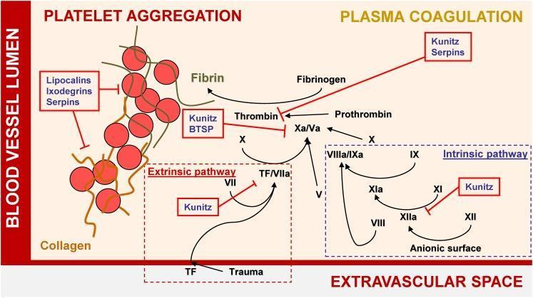
A pathophysiology diagram illustrating the mechanisms of hemostasis and the inhibitory effects of tick salivary proteins on platelet aggregation and plasma coagulation. The diagram is divided into the 'Blood Vessel Lumen' and the 'Extravascular Space.' On the left, 'Platelet Aggregation' is shown as a cluster of red spheres representing platelets cross-linked by fibrin strands; this process is inhibited by Lipocalins, Ixodegrins, and Serpins. On the right, the 'Plasma Coagulation' cascade is detailed, featuring both the Extrinsic pathway (initiated by Tissue Factor/TF due to trauma) and the Intrinsic pathway (activated via anionic surfaces). The diagram highlights key enzymatic conversions, such as Factor X to Xa/Va, Prothrombin to Thrombin, and Fibrinogen to Fibrin. Red annotation boxes identify specific tick protein families—including Kunitz-type inhibitors, BTSPs, and Serpins—that target and inhibit various coagulation factors like Thrombin, Factor Xa, and the XIIa/XIa complex. This visual resource effectively demonstrates the interference of exogenous biological agents with human blood clotting mechanisms.
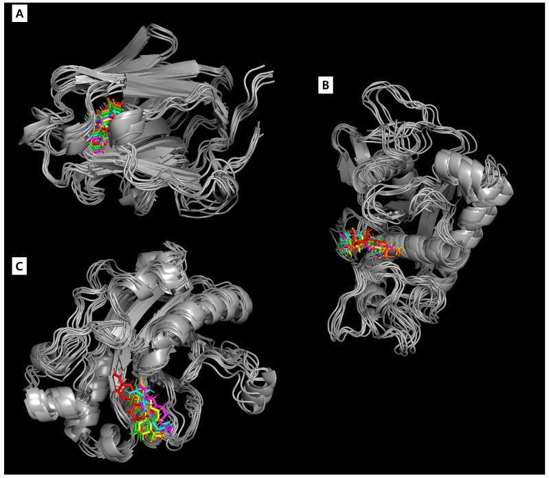
This diagnostic diagram illustrates the results of a 100-nanosecond (ns) molecular dynamics (MD) simulation, showcasing the structural stability and temporal interaction between the compound acanthoic acid and three key human proteins: sex hormone-binding globulin (SHBG, Panel A), ADAM metallopeptidase domain 17 (ADAM17, Panel B), and Deoxyribonuclease I (DNase I, Panel C). The proteins are rendered as grey ribbon and tube models, representing their three-dimensional folds and binding pockets. Superimposed within these pockets are multicolored representations of acanthoic acid, highlighting its spatial trajectory at discrete time intervals: 0 ns (red), 20 ns (green), 40 ns (yellow), 60 ns (magenta), 80 ns (cyan), and 100 ns (orange). The tight clustering of these colored ligand orientations demonstrates a stable binding mode and consistent molecular positioning throughout the simulation. This visualization serves as a computational model in pharmaceutical research related to male fertility, specifically analyzing the potential for acanthoic acid to modulate these proteins' biological functions.
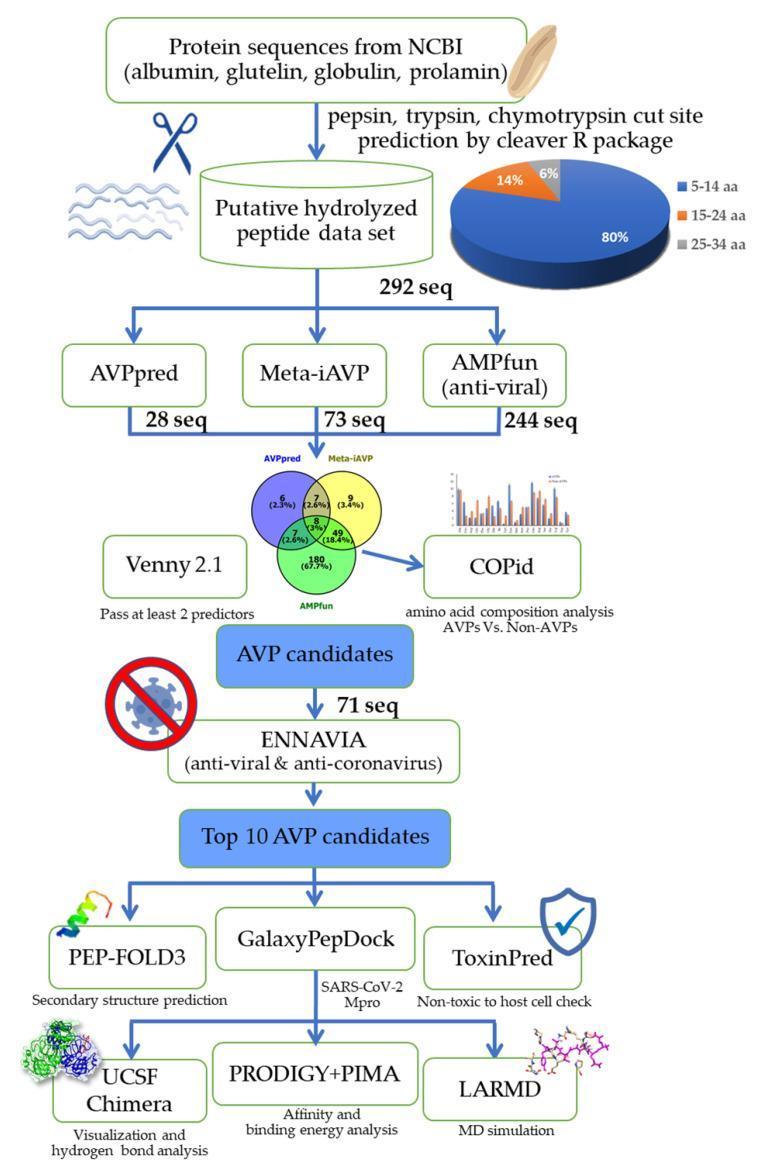
This bioinformatics flowchart details a virtual screening and in silico analysis pipeline for identifying antiviral peptide (AVP) candidates from rice bran proteins targeting SARS-CoV-2. The workflow begins with protein sequences (albumin, glutelin, globulin, prolamin) processed via the 'cleaver' R package for protease cut site prediction (pepsin, trypsin, chymotrypsin). This generates a dataset of 292 sequences, characterized by a pie chart showing peptide lengths (80% at 5-14 aa). The sequences undergo parallel screening using AVPpred, Meta-iAVP, and AMPfun. A Venn diagram illustrates the intersection of these results via Venny 2.1, leading to 71 AVP candidates that passed at least two predictors. COPid performs amino acid composition analysis. Candidates are further filtered through ENNAVIA for anti-coronavirus activity, resulting in the top 10 AVPs. Final characterization includes PEP-FOLD3 for secondary structure, GalaxyPepDock for Mpro interaction, and ToxinPred for toxicity. The pipeline concludes with visualization (UCSF Chimera), affinity analysis (PRODIGY+PIMA), and molecular dynamics simulation (LARMD).
Functions of All Plasma Proteins
Overview: Classification by Electrophoresis
| Fraction | Major Proteins |
|---|---|
| Albumin | Albumin |
| Alpha-1 globulins | alpha-1-antitrypsin, alpha-1-acid glycoprotein (orosomucoid), alpha-fetoprotein |
| Alpha-2 globulins | Haptoglobin, ceruloplasmin, alpha-2-macroglobulin, angiotensinogen |
| Beta globulins | Transferrin, beta-lipoprotein, hemopexin, C3/C4 complement |
| Gamma globulins | Immunoglobulins (IgG, IgA, IgM, IgD, IgE), CRP, SAA |
| Fibrinogen | Fibrinogen |
1. Albumin
- Site of synthesis: Liver exclusively
- Plasma concentration: ~3.5-5.0 g/dL (55-60% of total plasma protein)
- Half-life: ~20 days
- Colloid osmotic (oncotic) pressure - the single most important function; maintains ~25 mmHg oncotic pressure across capillary walls, preventing fluid loss into tissues. Loss of albumin (e.g., nephrotic syndrome, cirrhosis) leads to edema.
- Transport / carrier protein - binds and transports fatty acids, bilirubin, bile salts, hormones (steroids, thyroid hormones), hydrophobic amino acids, vitamins (B12, D), calcium, and numerous drugs (warfarin, aspirin, penicillin).
- Labile protein reserve - macrophages phagocytose plasma proteins via pinocytosis; amino acids are released and redistributed to tissues in starvation or severe illness.
- Buffering - weak acid-base buffer contributing ~15% of blood's total buffering capacity (alongside hemoglobin) via ionizable -COOH and -NH2 groups.
- Nutritional marker - serum albumin correlates with mortality risk across many diseases; falls in malnutrition (though slowly, given its 20-day half-life).
- Guyton and Hall Textbook of Medical Physiology, p. 856; Ganong's Review of Medical Physiology, 26th Ed
2. Fibrinogen
- Synthesis: Liver
- Concentration: 200-450 mg/dL
- MW: 340,000 Da; the largest plasma protein
- Hemostasis - polymerizes into fibrin threads (under thrombin action) to form the blood clot scaffold; essential for sealing vascular damage.
- Wound healing - fibrin matrix serves as substrate for fibroblast migration.
- Acute-phase reactant - levels rise during inflammation, infection, and trauma.
- Junqueira's Basic Histology; Sleisenger & Fordtran's GI and Liver Disease
3. Alpha Globulins
Alpha-1 Fraction
- The major serine protease inhibitor (serpin) of plasma
- Function: Inhibits trypsin, elastase, and other serine proteases released by neutrophils during inflammation, protecting tissues (especially lung alveoli) from proteolytic damage
- Deficiency causes emphysema and liver disease
- Concentration rises during acute-phase response
- Function: Inhibits the proliferating response of peripheral lymphocytes to mitogens; possible role in modulating inflammation. Binds certain drugs (e.g., lidocaine, propranolol).
- Rises sharply during acute-phase response
- Function: Inhibits chymotrypsin-like serine proteinases
- Increases during acute-phase response
- Function: Fetal equivalent of albumin - osmotic regulation and binding/carrier protein in fetal blood; normally very low in adults
- Clinical marker: elevated in hepatocellular carcinoma (HCC) and neural tube defects; mild elevation indicates hepatocellular regeneration
Alpha-2 Fraction
- Concentration: 40-180 mg/dL
- Function: Binds free hemoglobin released during intravascular hemolysis in a 1:1 complex, preventing:
- Renal loss of iron
- Oxidative damage to kidneys from free hemoglobin
- The complex is taken up by the reticuloendothelial system for iron recycling
- Antioxidant role
- Acute-phase protein (rises in inflammation)
- Concentration: 15-60 mg/dL
- Function:
- Transports copper (carries 6 atoms of copper per molecule; transports ~95% of serum copper)
- Has ferroxidase activity - oxidizes Fe2+ to Fe3+, enabling iron loading onto transferrin
- Antioxidant (scavenges free radicals)
- Decreased in Wilson disease; increased in inflammation, cholestasis, pregnancy
- Concentration: 150-420 mg/dL
- Function: Broad-spectrum inhibitor of serum endoproteases (trypsin, chymotrypsin, plasmin, thrombin). Acts as a protease "trap." Important in regulating coagulation and fibrinolysis.
- Function: Precursor to angiotensin I; substrate for renin. The starting point of the renin-angiotensin-aldosterone axis controlling blood pressure and volume.
4. Beta Globulins
- Concentration: 3.0-6.5 mg/dL
- Half-life: ~8-10 days
- Function:
- Transports iron in plasma (binds 2 atoms of Fe3+ per molecule)
- Delivers iron to tissues (especially erythroid precursors for hemoglobin synthesis) via transferrin receptors
- Bacteriostatic effect by sequestering iron away from bacteria
- Increases in iron deficiency, pregnancy; decreases with inflammation (negative acute-phase reactant), liver disease
- Concentration: 50-100 mg/dL
- Function: Binds free heme (porphyrins, especially heme) in a 1:1 complex after haptoglobin is saturated; transports heme to liver for recycling. Prevents heme-mediated oxidative damage.
- Function: Transport cholesterol and triglycerides from liver to peripheral tissues. Apolipoprotein B is their structural scaffold.
- Function: Components of the complement cascade
- C3: central complement component; opsonization, lysis, inflammation
- C4: part of the classical and lectin pathways; contributes to membrane attack complex formation
- Both are acute-phase proteins (increase in inflammation)
- Function: Extracellular matrix adhesion glycoprotein; promotes cell adhesion, wound healing, opsonization of pathogens
5. Gamma Globulins (Immunoglobulins)
| Immunoglobulin | Key Functions |
|---|---|
| IgG (most abundant, 75%) | Secondary immune response; crosses placenta (passive neonatal immunity); opsonization; neutralization of toxins/viruses; complement activation (classical pathway) |
| IgA (serum + secretory) | Primary defense at mucosal surfaces (GI, respiratory, genitourinary tracts); prevents pathogen adherence to mucosa; found in breast milk, saliva, tears |
| IgM (pentamer) | First antibody in primary immune response; very efficient complement activator (classical pathway); agglutination of antigens; ABO blood group antibodies |
| IgD | B cell receptor co-receptor; role in B cell activation and differentiation |
| IgE | Mediates Type I hypersensitivity (allergy, anaphylaxis) by binding mast cells and basophils; defense against helminth parasites |
6. Coagulation Factors (Special Plasma Proteins)
| Factor | Name | Function |
|---|---|---|
| I | Fibrinogen | Clot scaffold (see above) |
| II | Prothrombin | Precursor to thrombin (activates fibrinogen, V, VIII, XIII, platelets) |
| VII | Proconvertin | Initiates extrinsic pathway (with tissue factor) |
| VIII | Antihemophilic factor A | Cofactor in intrinsic pathway (tenase complex) |
| IX | Christmas factor | Intrinsic pathway serine protease |
| X | Stuart-Prower factor | Common pathway; activates prothrombin |
| XI | PTA | Intrinsic pathway |
| XII | Hageman factor | Contact activation; also activates complement and kinins |
| XIII | Fibrin-stabilizing factor | Cross-links fibrin clot |
| vWF | von Willebrand factor | Platelet adhesion; carries/stabilizes factor VIII |
- Antithrombin III - inhibits thrombin and factor Xa; heparin cofactor
- Protein C + Protein S (vitamin K-dependent) - activated protein C inactivates factors Va and VIIIa, limiting clot propagation
- Plasminogen - precursor to plasmin; fibrinolysis
7. Acute-Phase Proteins (Summary)
| Protein | Function in Acute Phase |
|---|---|
| CRP (C-reactive protein) | Binds pathogens/damaged cells; activates complement; opsonization; anti-inflammatory (inhibits superoxide by neutrophils) |
| Serum amyloid A (SAA) | Chemotaxis and adhesion of phagocytes |
| Fibrinogen | Hemostasis; substrate for ESR |
| Haptoglobin | Antioxidant; binds free Hb |
| Ferritin | Iron sequestration (bacteriostatic) |
| Alpha-1-antitrypsin | Protease inhibition |
| Complement C3/C4 | Immune defense |
| Alpha-2-macroglobulin | Protease inhibition |
- Albumin, transferrin, transthyretin (prealbumin), retinol-binding protein
8. Other Notable Plasma Proteins
- Function: Carrier for thyroid hormones (T3/T4) and retinol-binding protein (RBP); also a transport vehicle for retinol
- Short half-life (~2 days); sensitive early marker of hepatic synthetic function and nutritional status
- Function: Carries vitamin A (retinol) from liver to target tissues; circulates bound to transthyretin
- Function: Primary carrier of thyroid hormones (T4 > T3) in blood (~75%)
- Function: Binds and transports testosterone and estradiol; regulates their bioavailability
- Function: Transports cortisol (~75%) and progesterone in plasma
- Function: Mediator of the anabolic effects of growth hormone; promotes cell growth and differentiation
- Function: Antiprotease; inhibits complement activation (C1r, C1s), kallikrein, factor XIIa, and factor XIa. Deficiency causes hereditary angioedema.
Key Points for Exam
- Albumin = #1 by concentration; #1 for oncotic pressure; most versatile carrier
- All plasma proteins except immunoglobulins are synthesized in the liver
- Immunoglobulins are made by plasma cells (derived from B lymphocytes)
- Fibrinogen is the largest plasma protein; absent in serum (consumed in clotting)
- Vitamin K-dependent clotting factors: II, VII, IX, X (and protein C, S)
- Haptoglobin falls in hemolysis (used up); transferrin rises in iron deficiency
- CRP and SAA are the two major acute-phase proteins in humans
- Negative acute-phase reactants: albumin, transferrin, transthyretin, RBP