
Chromosome.
Reading File
The query "Chromosome" is very broad. Let me clarify what you need before diving in.
Asking for Preferences
What aspect of chromosomes are you interested in?
Searching sources...
Finding Sources
Finding Sources
Good hits. Let me read the most comprehensive sections in parallel.
Reading File
Reading File
Reading File
Excellent content. Now let me fetch the key structural diagrams and read the Biochemistry section for more detail.
Loading Image
Loading Image
Reading File
I now have rich textbook content and two excellent diagrams. Here is the full structured answer:
Chromosome: Structure & Organization
Overview
A chromosome is a thread-like structure composed of DNA and associated proteins (chromatin) that carries the genetic information of a cell. Human somatic cells contain 46 chromosomes arranged in 23 pairs (22 pairs of autosomes + 1 pair of sex chromosomes).
If all the DNA in a single human cell were laid end to end, it would stretch roughly 6 feet (~2 meters) - yet it is compacted into a nucleus only ~6 micrometers in diameter. This is achieved through several hierarchical levels of folding.
Levels of DNA Packaging
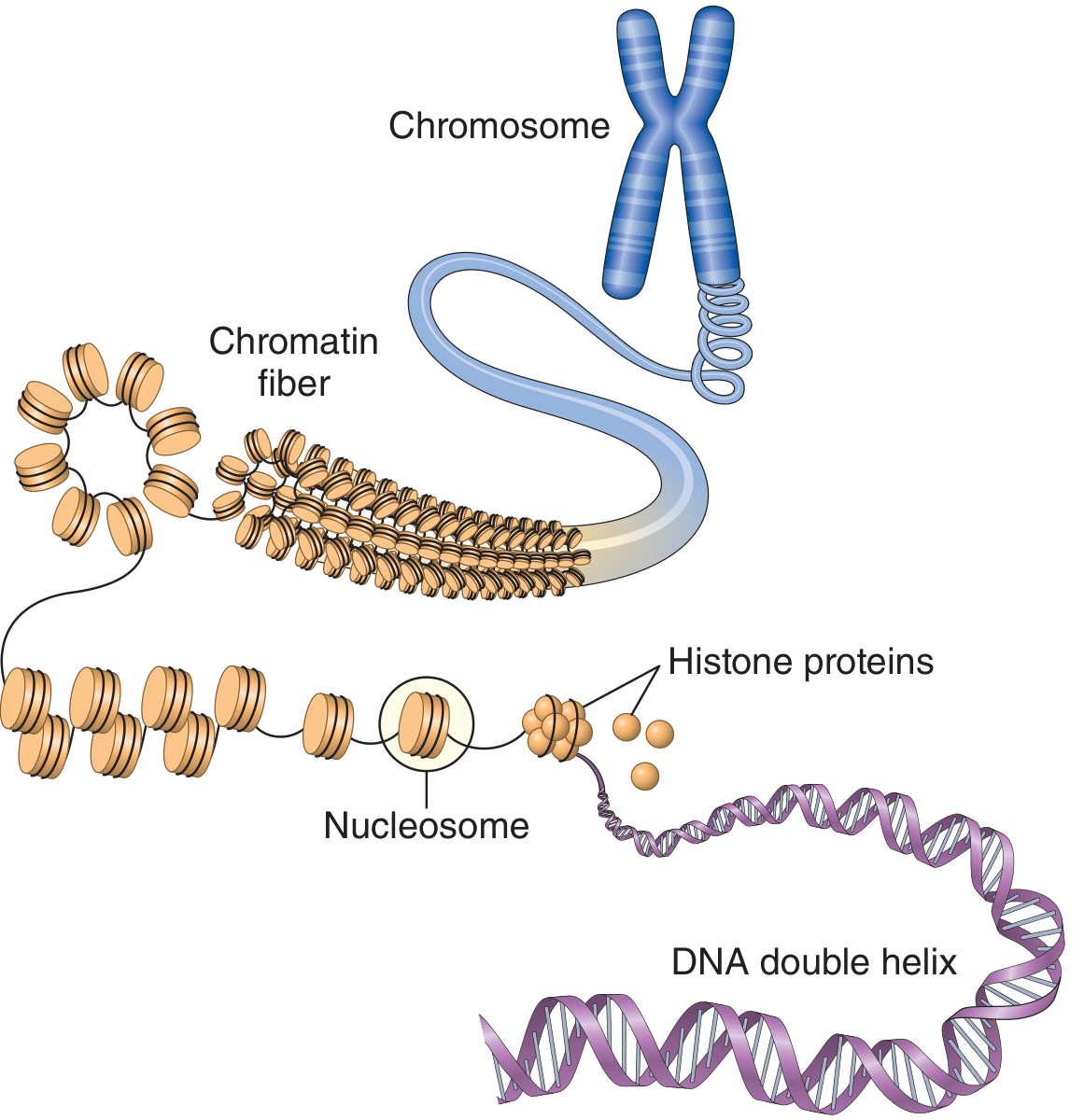
Figure: Organization and packaging of DNA into chromosomes (Guyton & Hall, Textbook of Medical Physiology)
Level 1 - DNA Double Helix (2 nm)
- The fundamental molecule: two antiparallel strands wound in a right-handed helix.
- Each base pair is ~0.34 nm apart; the helix has a diameter of 2 nm.
Level 2 - Nucleosomes ("beads on a string", 11 nm)
- DNA wraps ~1.65 turns (146 nucleotide pairs) around a histone octamer core (2 copies each of H2A, H2B, H3, H4).
- Each nucleosome bead is ~10 nm in diameter.
- Linker DNA (bound by H1 histone) connects adjacent nucleosomes.
- This packing shortens the DNA ~7-fold relative to unfolded DNA.
Level 3 - 30 nm Chromatin Fibril (Solenoid)
- Nucleosomes coil further to form a 30 nm chromatin fibril.
- Six nucleosomes form one turn of this solenoid coil.
- Achieves ~40-fold compaction relative to unfolded DNA.
Level 4 - Loop Domains (~300 nm)
- Long 30 nm fibrils form radial loops (each containing ~15,000-100,000 base pairs) anchored to a central chromosome scaffold of non-histone proteins (topoisomerase II, condensin).
Level 5 - Metaphase Chromosome (~1,400 nm)
- During mitosis, the loop domains condense further into the highly compact metaphase chromosome visible under light microscopy.
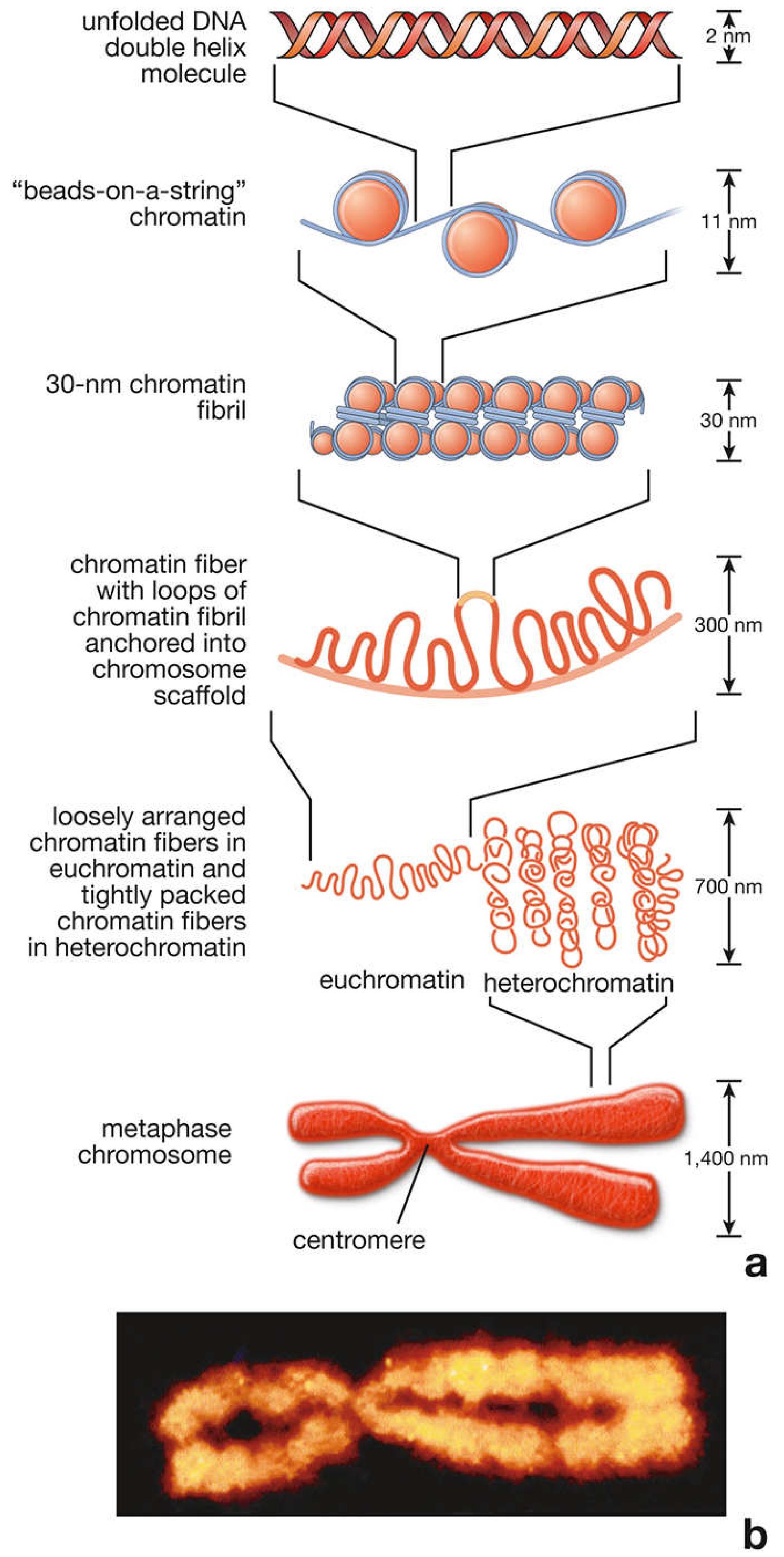
Figure: Sequential steps in chromatin packaging from DNA double helix to metaphase chromosome (Histology - A Text and Atlas, 8th Ed.)
Key Structural Components
Histones
- Small, positively charged (basic) proteins rich in lysine and arginine.
- Their positive charge attracts negatively charged DNA phosphate backbone.
- Five main types: H1, H2A, H2B, H3, H4.
- H2A, H2B, H3, H4 form the octamer core; H1 is the linker histone.
- Posttranslational modifications (acetylation, methylation, phosphorylation) regulate chromatin compaction and gene expression.
Non-Histone Proteins
- Scaffold proteins (structural role).
- Transcription factors, RNA polymerases, DNA repair enzymes (functional roles).
- Regulate gene activation and chromosome architecture.
Centromere and Telomere
Centromere
- A specific DNA region where the two sister chromatids are held together after replication.
- Serves as the attachment point for the kinetochore and spindle microtubules during cell division.
- Chromosome types based on centromere position:
| Type | Centromere Position |
|---|---|
| Metacentric | Middle (equal arms) |
| Submetacentric | Between middle and tip |
| Acrocentric | Near the tip |
| Telocentric | At the very tip (rare in humans) |
Telomeres
- Repetitive DNA sequences (TTAGGG repeats in humans) at the ends of chromosomes.
- Protected by shelterin protein complex.
- Prevent chromosome ends from being recognized as double-strand breaks.
- Shorten with each cell division - a key indicator of cellular aging and replicative lifespan.
- Maintained by the enzyme telomerase (active in stem cells, germ cells, most cancer cells).
Chromatin States: Euchromatin vs. Heterochromatin
| Feature | Euchromatin | Heterochromatin |
|---|---|---|
| Packing | Loosely packed | Tightly packed |
| Transcription | Active - genes can be expressed | Inactive - transcriptionally silent |
| Staining | Lightly staining | Darkly staining |
| Predominant in | Metabolically active cells (neurons, hepatocytes) | Inactive cells (small lymphocytes, sperm) |
- Constitutive heterochromatin: permanently condensed (e.g., centromeric/pericentromeric regions, most of the Y chromosome).
- Facultative heterochromatin: can switch between active/inactive states (e.g., the inactive X chromosome = Barr body).
Chromosome Nomenclature
- The short arm is designated "p" (from French petit).
- The long arm is designated "q".
- Bands are numbered outward from the centromere; e.g., 14q32 = second band, third region of the long arm of chromosome 14.
| Symbol | Meaning |
|---|---|
| del | Deletion |
| dup | Duplication |
| inv | Inversion |
| t | Translocation |
| r | Ring chromosome |
| + / - | Gain / loss of chromosome |
| / | Mosaicism separator |
Human Chromosome Complement
- 46 chromosomes total (diploid = 2n).
- 22 autosomes in pairs (chromosomes 1-22) + XX (female) or XY (male).
- Gene density is unequal: chromosomes 19 and 22 are gene-rich; chromosomes 4 and 18 are relatively gene-poor.
- ~60-70% of the genome is single/low-copy DNA; 30-40% is repetitive (satellite DNA, interspersed sequences like SINEs and LINEs).
Sources:
- Emery's Elements of Medical Genetics and Genomics - Chromosome Structure section
- Guyton & Hall Textbook of Medical Physiology - Chromosomes and Their Replication, p. 59
- Histology: A Text and Atlas - Chromatin, pp. 234-236
- Basic Medical Biochemistry (6e) - Chromosome Structure and Nomenclature, p. 650
This is a shared conversation. Sign in to Orris to start your own chat.