
Pain who criteria
WHO analgesic ladder pain management steps
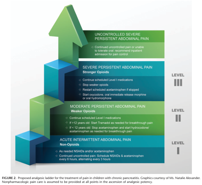
Summary : This figure presents a proposed analgesic ladder for the treatment of pain in children with chronic pancreatitis, illustrating a stepwise approach to pain management based on severity, from non-opioid to stronger opioid interventions. flowchart: # Levels of Pain and Analgesic Intervention : ## Level I – Acute Intermittent Abdominal Pain (Non-Opioids) : • As needed NSAIDs and/or acetaminophen. • For continued uncontrolled pain: Schedule NSAIDs & acetaminophen every 6 hours, alternating every 3 hours. ## Level II – Moderate Persistent Abdominal Pain (Weaker Opioids) : • Continue scheduled Level I medications. • If >12 years old: Start Tramadol as needed for breakthrough pain. • If <12 years old: Stop acetaminophen and start hydrocodone/acetaminophen as needed for breakthrough pain. ## Level III – Severe Persistent Abdominal Pain (Stronger Opioids) : • Continue scheduled Level I medications. • Stop weaker opioids. • Restart scheduled acetaminophen if stopped. • Start oxycodone, oral immediate release morphine, or oral hydromorphone. ## Uncontrolled Severe Persistent Abdominal Pain : • Continued uncontrolled pain or unable to tolerate oral: recommend inpatient admission for pain control. # Layout : • The diagram is structured as a vertical ascending ladder with four steps, each representing increasing pain severity and corresponding escalation in analgesic potency. • Each step is color-coded: blue-green for non-opioids, green for weaker opioids, teal for stronger opioids, and dark green for uncontrolled pain requiring inpatient care. • The steps are labeled as Level I, Level II, and Level III, with the final step above Level III. # Analysis : • The figure visually emphasizes a stepwise, escalating approach to pain management in pediatric chronic pancreatitis, starting with non-opioid medications and progressing to stronger opioids and inpatient care as pain severity increases. • Nonpharmacologic pain care is assumed to be provided at all points in the ladder. • The structure supports careful titration and escalation of analgesic potency, with clear age-based recommendations for opioid use.
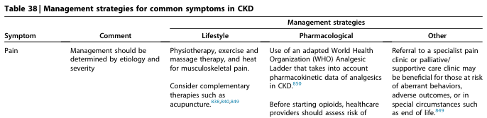
Table 38 | Management strategies for common symptoms in CKD <table><thead><tr><th>Symptom</th><th>Comment</th><th colspan="3">Management strategies</th></tr><tr><th></th><th></th><th>Lifestyle</th><th>Pharmacological</th><th>Other</th></tr></thead><tbody><tr><td>Pain</td><td>Management should be determined by etiology and severity</td><td>Physiotherapy, exercise and massage therapy, and heat for musculoskeletal pain.<br><br>Consider complementary therapies such as acupuncture.<sup>838,840,849</sup></td><td>Use of an adapted World Health Organization (WHO) Analgesic Ladder that takes into account pharmacokinetic data of analgesics in CKD.<sup>850</sup><br><br>Before starting opioids, healthcare providers should assess risk of</td><td>Referral to a specialist pain clinic or palliative/ supportive care clinic may be beneficial for those at risk of aberrant behaviors, adverse outcomes, or in special circumstances such as end of life.<sup>849</sup></td></tr></tbody></table>
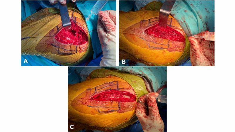
A series of three clinical photographs (A-C) demonstrating the intraoperative steps of peri-articular multimodal drug injection (PMDI) during hip arthroplasty. All images show a lateral approach to the hip with the skin prepped in a sterile yellow antiseptic solution and marked with surgical ink. Figure A illustrates the deep injection into the hip capsule; a metallic retractor reflects the gluteus muscles to expose the joint space while a syringe delivers the analgesic cocktail. Figure B shows the second stage of injection into the gluteus medius muscle and tensor fascia lata after prosthesis insertion and muscle repair, with a retractor maintaining surgical access to the red, fleshy muscle tissue. Figure C depicts the final stage where the solution is injected into the subcutaneous tissue layer before skin closure, with the incision site partially apposed. This sequence serves as an educational guide for orthopedic surgeons on the standardized anatomical layers for local infiltration analgesia to optimize postoperative pain management.
WHO Analgesic Ladder - Pain Criteria & Management
The Three-Step Ladder
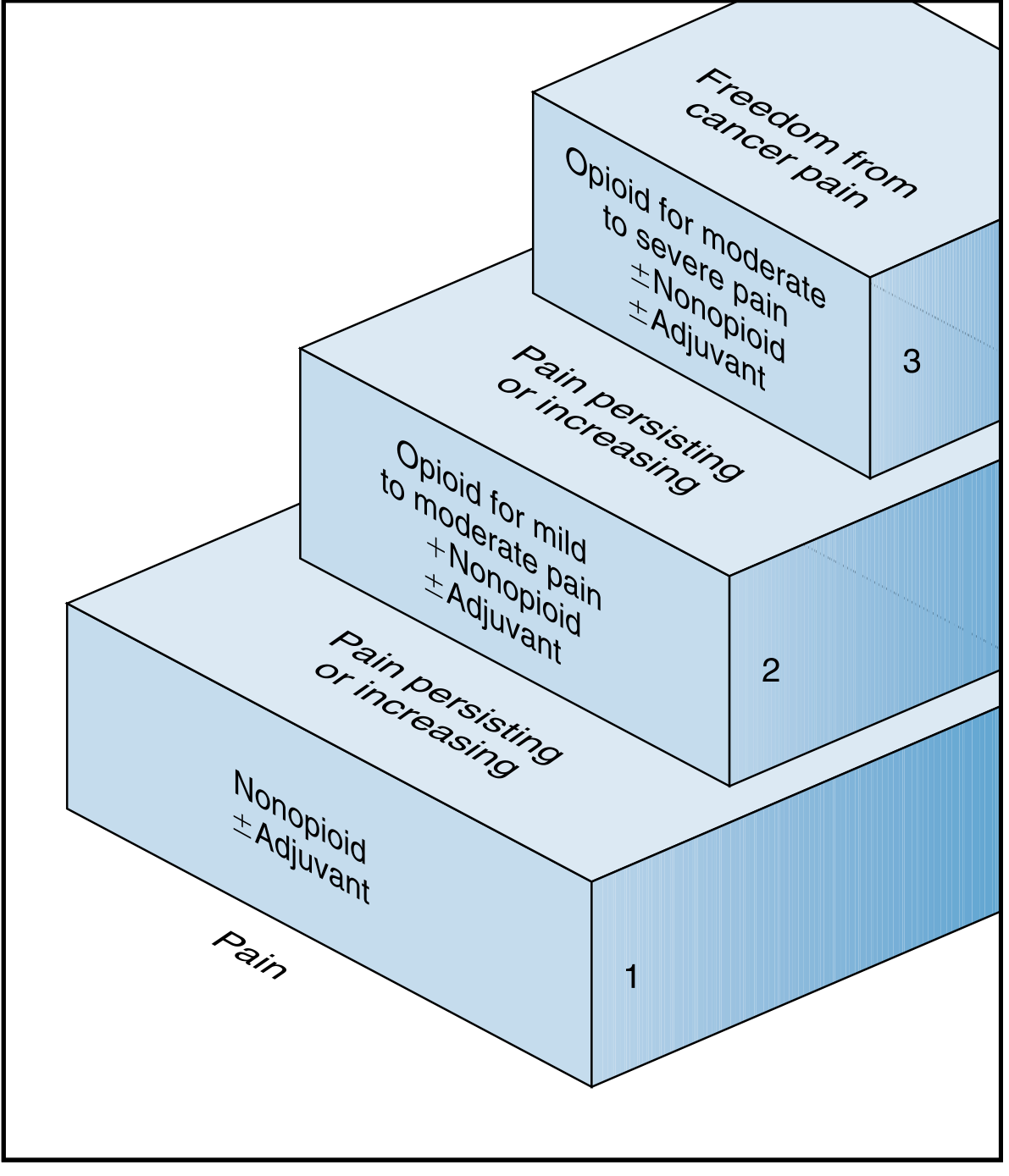
| Step | Pain Level | Drug Class | Examples |
|---|---|---|---|
| Step 1 | Mild pain | Non-opioid ± adjuvant | Paracetamol (acetaminophen), aspirin, NSAIDs |
| Step 2 | Mild-to-moderate pain persisting/increasing | Weak opioid ± non-opioid ± adjuvant | Codeine, tramadol, hydrocodone |
| Step 3 | Moderate-to-severe pain persisting/increasing | Strong opioid ± non-opioid ± adjuvant | Morphine, oxycodone, hydromorphone, fentanyl, methadone |
- ~20-25% of cancer pain patients can be fully controlled with Step 1 agents alone.
- Adjuvants (tricyclic antidepressants, anticonvulsants, corticosteroids) can be added at any step.
WHO's 5 Key Principles ("By mouth, by the clock, by the ladder...")
| Principle | Meaning |
|---|---|
| By mouth | Oral route preferred - effective, inexpensive, easily adjusted |
| By the clock | Give analgesics on a fixed schedule (not PRN), using immediate-release and/or sustained-release formulations to maintain continuous pain relief |
| By the ladder | Pain severity and responsiveness to medication dictates which step to use; step up if pain persists or increases |
| For the individual | Titrate to each patient - there is no single "correct" dose |
| Attention to detail | Vigilantly assess pain relief before and after each new regimen; monitor closely for adverse effects |
Adjuvant Analgesics (Used at Any Step)
- Neuropathic pain: Tricyclic antidepressants (amitriptyline, desipramine), anticonvulsants (gabapentin, pregabalin)
- Bone pain / inflammation: NSAIDs, corticosteroids
- Muscle spasm: Baclofen, muscle relaxants
- Anxiety/depression comorbidity: SNRIs, SSRIs
Key Clinical Notes
- Start at the appropriate step - if pain is already severe, starting directly at Step 3 is acceptable and often necessary.
- "Breakthrough pain" - short-acting opioids should always be available alongside long-acting preparations (e.g., oral morphine every 4 hours for baseline; short-acting morphine for breakthrough).
- Opioid rotation - consider switching opioids if a patient achieves adequate analgesia with one opioid but has intolerable adverse effects.
- Constipation is a persistent opioid side effect - begin laxatives prophylactically.
- Nausea from opioids usually does not persist; treat with antiemetics.
- The ladder is bidirectional - step down if pain improves (e.g., after radiotherapy for bone metastases).
Pain score scale
pain scale visual analogue numeric rating Wong-Baker FACES
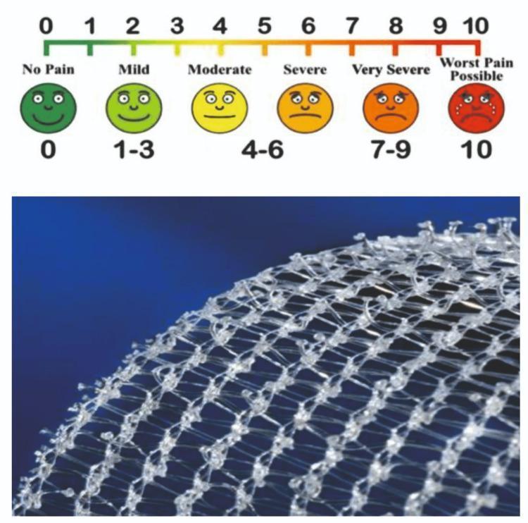
Educational medical graphic combining a pain assessment tool and surgical material. The top section displays a Visual Analog Scale (VAS) used for clinical pain measurement. The scale features a numeric rating from 0 to 10 correlated with descriptive labels and a Wong-Baker style faces scale: 0 (No Pain, dark green smiling face), 1-3 (Mild, light green smiling face), 4-6 (Moderate to Severe, yellow neutral to orange frowning faces), 7-9 (Very Severe, dark orange distressed face), and 10 (Worst Pain Possible, red crying face). Below the scale is a high-resolution photograph of a self-gripping prosthetic mesh used in surgical procedures like inguinal hernia repair. The mesh is composed of a translucent, white synthetic monofilament knit into a diamond-shaped grid. Characteristic micro-grips or bulbous nodes are visible at the strand intersections, designed for sutureless fixation to anatomical tissues. This comparison illustrates tools used for evaluating patient outcomes, specifically postoperative pain levels relative to surgical techniques and materials.
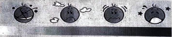
This visual aid is an adaptation of a Faces Pain Rating Scale used for clinical assessment of patient-reported discomfort, often categorized under the Visual Analogue Scale (VAS) methodology. The infographic consists of four progressive facial icons and a corresponding intensity gradient bar below. 1) The first icon features a smiling mouth and 'crossed' eyes, representing no pain or 'very bearable' sensation. 2) The second icon shows a frowning mouth and cloud symbols, indicating mild discomfort or emerging distress. 3) The third icon displays a wavy, distressed mouth and motion lines around the head, signifying moderate to high pain levels. 4) The final icon depicts a wide-open mouth, closed eyes, and star symbols, representing severe or maximum pain. This tool is utilized in clinical settings, such as post-procedural hysteroscopy evaluations, to help patients quantify subjective pain levels when verbal communication may be limited or to supplement numerical ratings.
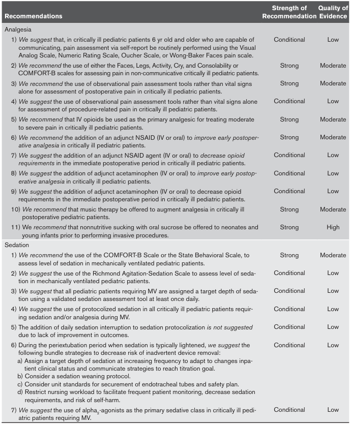
<table><thead><tr><th>Recommendations</th><th>Strength of Recommendation</th><th>Quality of Evidence</th></tr></thead><tbody><tr><td>Analgesia</td><td></td><td></td></tr><tr><td>1) We suggest that, in critically ill pediatric patients 6 yr old and older who are capable of communicating, pain assessment via self-report be routinely performed using the Visual Analog Scale, Numeric Rating Scale, Oucher Scale, or Wong-Baker Faces pain scale.</td><td>Conditional</td><td>Low</td></tr><tr><td>2) We recommend the use of either the Faces, Legs, Activity, Cry, and Consolability or COMFORT-B scales for assessing pain in non-communicative critically ill pediatric patients.</td><td>Strong</td><td>Moderate</td></tr><tr><td>3) We recommend the use of observational pain assessment tools rather than vital signs alone for assessment of postoperative pain in critically ill pediatric patients.</td><td>Strong</td><td>Moderate</td></tr><tr><td>4) We suggest the use of observational pain assessment tools rather than vital signs alone for assessment of procedure-related pain in critically ill pediatric patients.</td><td>Conditional</td><td>Low</td></tr><tr><td>5) We recommend that IV opioids be used as the primary analgesic for treating moderate to severe pain in critically ill pediatric patients.</td><td>Strong</td><td>Moderate</td></tr><tr><td>6) We recommend the addition of an adjunct NSAID (IV or oral) to improve early postoperative analgesia in critically ill pediatric patients.</td><td>Strong</td><td>Moderate</td></tr><tr><td>7) We suggest the addition of an adjunct NSAID agent (IV or oral) to decrease opioid requirements in the immediate postoperative period in critically ill pediatric patients.</td><td>Conditional</td><td>Low</td></tr><tr><td>8) We suggest the addition of adjunct acetaminophen (IV or oral) to improve early postoperative analgesia in critically ill pediatric patients.</td><td>Conditional</td><td>Low</td></tr><tr><td>9) We suggest the addition of adjunct acetaminophen (IV or oral) to decrease opioid requirements in the immediate postoperative period in critically ill pediatric patients.</td><td>Conditional</td><td>Low</td></tr><tr><td>10) We recommend that music therapy be offered to augment analgesia in critically ill postoperative pediatric patients.</td><td>Strong</td><td>Moderate</td></tr><tr><td>11) We recommend that nonnutritive sucking with oral sucrose be offered to neonates and young infants prior to performing invasive procedures.</td><td>Strong</td><td>High</td></tr><tr><td>Sedation</td><td></td><td></td></tr><tr><td>1) We recommend the use of the COMFORT-B Scale or the State Behavioral Scale, to assess level of sedation in mechanically ventilated pediatric patients.</td><td>Strong</td><td>Moderate</td></tr><tr><td>2) We suggest the use of the Richmond Agitation-Sedation Scale to assess level of sedation in mechanically ventilated pediatric patients.</td><td>Conditional</td><td>Low</td></tr><tr><td>3) We suggest that all pediatric patients requiring MV are assigned a target depth of sedation using a validated sedation assessment tool at least once daily.</td><td>Conditional</td><td>Low</td></tr><tr><td>4) We suggest the use of protocolized sedation in all critically ill pediatric patients requiring sedation and/or analgesia during MV.</td><td>Conditional</td><td>Low</td></tr><tr><td>5) The addition of daily sedation interruption to sedation protocolization is not suggested due to lack of improvement in outcomes.</td><td>Conditional</td><td>Low</td></tr><tr><td>6) During the periextubation period when sedation is typically lightened, we suggest the following bundle strategies to decrease risk of inadvertent device removal: a) Assign a target depth of sedation at increasing frequency to adapt to changes inpatient clinical status and communicate strategies to reach titration goal. b) Consider a sedation weaning protocol. c) Consider unit standards for securement of endotracheal tubes and safety plan. d) Restrict nursing workload to facilitate frequent patient monitoring, decrease sedation requirements, and risk of self-harm.</td><td>Conditional</td><td>Low</td></tr><tr><td>7) We suggest the use of alpha2-agonists as the primary sedative class in critically ill pediatric patients requiring MV.</td><td>Conditional</td><td>Low</td></tr></tbody></table>
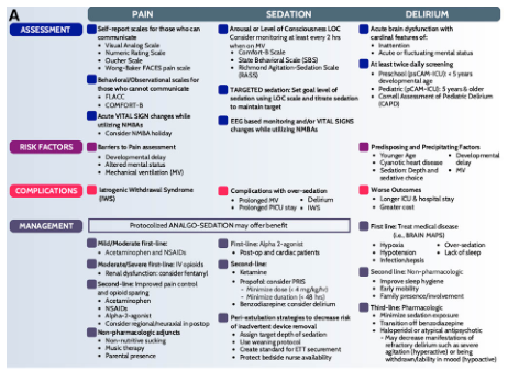
Summary : This figure presents a side-by-side comparison of pain, sedation, and delirium assessment and management in pediatric intensive care, organized into four main categories: Assessment, Risk Factors, Complications, and Management. Each category is subdivided for pain, sedation, and delirium, with specific tools, risk factors, complications, and management strategies listed for each. flowchart/table hybrid: # Overall Structure : • The figure is divided into three vertical columns: Pain, Sedation, and Delirium. • Four horizontal sections: Assessment, Risk Factors, Complications, and Management. • Each cell contains bulleted lists of tools, factors, or strategies. # Pain : ## Assessment : • Self-report scales for communicative children: Visual Analog Scale, Numeric Rating Scale, Oucher Scale, Wong-Baker FACES pain scale. • Behavioral/observational scales for non-communicative children: FLACC, COMFORT-B. • Acute vital sign changes with NMBA use; consider NMBA holiday. ## Risk Factors : • Barriers to pain assessment: Developmental delay, altered mental status, mechanical ventilation (MV). ## Complications : • Iatrogenic Withdrawal Syndrome (IWS). ## Management : • Mild/Moderate: Acetaminophen, NSAIDs. • Moderate/Severe: First-line IV opioids, consider fentanyl for renal dysfunction. • Second-line: Improved pain control and opioid sparing (acetaminophen, NSAIDs, alpha-2 agonist). • Non-pharmacologic: Non-nutritive sucking, music therapy, parental presence. # Sedation : ## Assessment : • Arousal/Level of Consciousness: Monitor every 2 hours when on MV. • Comfort-B scale, State Behavioral Scale (SBS), Richmond Agitation-Sedation Scale (RASS). • Targeted sedation: Set goal, titrate sedation. • EEG-based monitoring and vital sign changes with NMBA use. ## Risk Factors : • Complications with over-sedation: Prolonged MV, prolonged PICU stay, delirium, IWS. ## Complications : • Over-sedation: Prolonged MV, delirium, prolonged PICU stay, IWS. ## Management : • Protocolized analgo-sedation may offer benefit. • First-line: Alpha-2 agonist (post-op, non-cardiac). • Second-line: Ketamine, minimize dose (4 mg/kg/h), minimize duration (<48h), benzodiazepines for delirium. • Peri-extubation strategies: Assign wean target, use extubation protocol, create standard for ETT securement, protect bedside nurse availability. # Delirium : ## Assessment : • Acute brain dysfunction: Inattention, fluctuating mental status. • Acute daily screening: Preschool (psCAM-ICU-5), school-age (pCAM-ICU-5), Cornell Assessment of Pediatric Delirium (CAPD). ## Risk Factors : • Predisposing/precipitating: Young age, developmental delay, congenital heart disease, MV, sedative/analgesic use. ## Complications : • Worse outcomes: Longer ICU/hospital stay, greater cost. ## Management : • First-line: Treat medical disease (labs, brain MAPS). • Hypoactive: Over-sedation, lack of sleep. • Hyperactive: Infections. • Second-line: Non-pharmacologic (sleep hygiene, early mobilization, family presence). • Third-line: Minimize sedation exposure, avoid benzodiazepines, consider alpha-2 agonist, may need dexmedetomidine for withdrawal/mood (hypoactive). # Design Encodings : • Blue, purple, and pink colour coding for Pain, Sedation, and Delirium columns. • Section headers highlighted with background shading. • Bulleted lists for clarity. • Icons for assessment, risk factors, complications, and management. # Analysis : • The figure provides a comprehensive, parallel overview of pain, sedation, and delirium management in pediatric critical care. • Each domain is addressed with specific assessment tools, risk factors, complications, and management strategies. • There is emphasis on protocolized approaches, minimizing complications, and integrating non-pharmacologic interventions. • The structure facilitates rapid comparison and highlights the interconnectedness of pain, sedation, and delirium in clinical practice.
FLACC pain scale table infants behavioral assessment
Summary : This figure presents a side-by-side comparison of pain, sedation, and delirium assessment and management in pediatric intensive care, organized into four main categories: Assessment, Risk Factors, Complications, and Management. Each category is subdivided for pain, sedation, and delirium, with specific tools, risk factors, complications, and management strategies listed for each. flowchart/table hybrid: # Overall Structure : • The figure is divided into three vertical columns: Pain, Sedation, and Delirium. • Four horizontal sections: Assessment, Risk Factors, Complications, and Management. • Each cell contains bulleted lists of tools, factors, or strategies. # Pain : ## Assessment : • Self-report scales for communicative children: Visual Analog Scale, Numeric Rating Scale, Oucher Scale, Wong-Baker FACES pain scale. • Behavioral/observational scales for non-communicative children: FLACC, COMFORT-B. • Acute vital sign changes with NMBA use; consider NMBA holiday. ## Risk Factors : • Barriers to pain assessment: Developmental delay, altered mental status, mechanical ventilation (MV). ## Complications : • Iatrogenic Withdrawal Syndrome (IWS). ## Management : • Mild/Moderate: Acetaminophen, NSAIDs. • Moderate/Severe: First-line IV opioids, consider fentanyl for renal dysfunction. • Second-line: Improved pain control and opioid sparing (acetaminophen, NSAIDs, alpha-2 agonist). • Non-pharmacologic: Non-nutritive sucking, music therapy, parental presence. # Sedation : ## Assessment : • Arousal/Level of Consciousness: Monitor every 2 hours when on MV. • Comfort-B scale, State Behavioral Scale (SBS), Richmond Agitation-Sedation Scale (RASS). • Targeted sedation: Set goal, titrate sedation. • EEG-based monitoring and vital sign changes with NMBA use. ## Risk Factors : • Complications with over-sedation: Prolonged MV, prolonged PICU stay, delirium, IWS. ## Complications : • Over-sedation: Prolonged MV, delirium, prolonged PICU stay, IWS. ## Management : • Protocolized analgo-sedation may offer benefit. • First-line: Alpha-2 agonist (post-op, non-cardiac). • Second-line: Ketamine, minimize dose (4 mg/kg/h), minimize duration (<48h), benzodiazepines for delirium. • Peri-extubation strategies: Assign wean target, use extubation protocol, create standard for ETT securement, protect bedside nurse availability. # Delirium : ## Assessment : • Acute brain dysfunction: Inattention, fluctuating mental status. • Acute daily screening: Preschool (psCAM-ICU-5), school-age (pCAM-ICU-5), Cornell Assessment of Pediatric Delirium (CAPD). ## Risk Factors : • Predisposing/precipitating: Young age, developmental delay, congenital heart disease, MV, sedative/analgesic use. ## Complications : • Worse outcomes: Longer ICU/hospital stay, greater cost. ## Management : • First-line: Treat medical disease (labs, brain MAPS). • Hypoactive: Over-sedation, lack of sleep. • Hyperactive: Infections. • Second-line: Non-pharmacologic (sleep hygiene, early mobilization, family presence). • Third-line: Minimize sedation exposure, avoid benzodiazepines, consider alpha-2 agonist, may need dexmedetomidine for withdrawal/mood (hypoactive). # Design Encodings : • Blue, purple, and pink colour coding for Pain, Sedation, and Delirium columns. • Section headers highlighted with background shading. • Bulleted lists for clarity. • Icons for assessment, risk factors, complications, and management. # Analysis : • The figure provides a comprehensive, parallel overview of pain, sedation, and delirium management in pediatric critical care. • Each domain is addressed with specific assessment tools, risk factors, complications, and management strategies. • There is emphasis on protocolized approaches, minimizing complications, and integrating non-pharmacologic interventions. • The structure facilitates rapid comparison and highlights the interconnectedness of pain, sedation, and delirium in clinical practice.
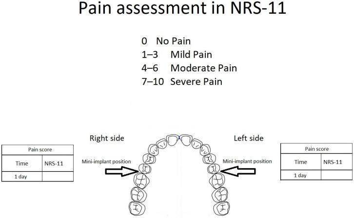
This infographic illustrates the methodology for orthodontic pain assessment using the NRS-11 scale following mini-implant placement. The top section defines the Numeric Rating Scale (NRS-11) categories: 0 (No Pain), 1–3 (Mild Pain), 4–6 (Moderate Pain), and 7–10 (Severe Pain). Central to the figure is an anatomical diagram of the maxillary dental arch. Black arrows indicate the bilateral 'Mini-implant position' in the posterior region, specifically between the second premolars and first molars. Flanking the anatomical diagram are two data entry tables labeled 'Pain score' for the 'Right side' and 'Left side.' Each table contains columns for 'Time' (specified as 1 day post-treatment) and 'NRS-11' score. This layout is designed for a clinical study comparing postoperative pain levels between a treatment group and a control group in orthodontic procedures involving bone-anchored devices.
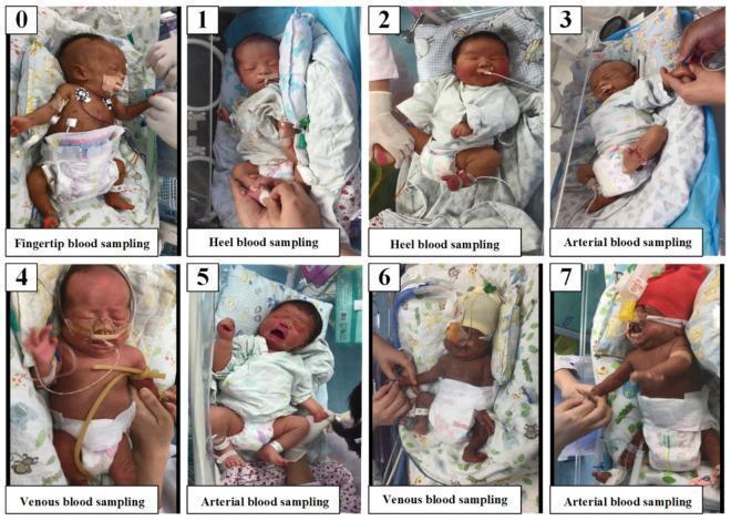
A series of eight clinical photographs (labeled 0-7) documenting various blood sampling procedures performed on neonates in a hospital setting, used to study neonatal pain responses. The images illustrate four distinct sampling modalities: fingertip (0), heel (1, 2), venous (4, 6), and arterial (3, 5, 7) blood collection. The infants are depicted in neonatal intensive care or nursery environments, showing varying degrees of medical support including nasogastric tubes, oxygen cannulae, chest leads for cardiac monitoring, and pulse oximetry sensors. The newborns exhibit a range of behavioral and physiological pain indicators, most notably facial grimacing and crying, particularly evident during arterial sampling in images 5 and 7. Healthcare personnel are shown performing the procedures at different anatomical sites such as the hands, heels, and antecubital fossa. This visual data supports the assessment of the Neonatal Infant Pain Scale (NIPS) and the development of automated pain recognition systems.
Pain Score Scales
1. Self-Report Scales (for communicative patients)
Numeric Rating Scale (NRS) - Most widely used
| Score | Interpretation |
|---|---|
| 0 | No pain |
| 1-3 | Mild pain |
| 4-6 | Moderate pain |
| 7-9 | Severe pain |
| 10 | Worst pain possible |
- Reliable, valid, quick. Horizontal lines preferred over vertical (more normally distributed scores).
- Used in: adolescents, adults, children ≥8 years.
Visual Analogue Scale (VAS)
- Patient marks a point on a 10 cm line anchored at "No pain" and "Worst pain possible."
- Requires intact hand-eye coordination and visual acuity - difficult for elderly, cognitively impaired, or motor-impaired patients.
- Can be converted to a 0-10 numeric score.
Combined VAS + FACES Scale
2. Faces Pain Scales (for children 3-8 years)
Faces Pain Scale - Revised (FPS-R)
- Most commonly recommended for children aged 3-8 years.
- Shows 6 faces ranging from neutral (no pain) to maximum pain expression.
- No tears or smiles - specifically avoids conflating happiness/sadness with pain.
Wong-Baker FACES Scale
- Uses smiley-to-crying faces scored 0, 2, 4, 6, 8, 10.
- Widely used; recommended from age 3 years and older.
- Also used in adults with cognitive or language barriers.
3. Behavioral / Observational Scales (for non-communicative patients)
FLACC Scale - For infants and toddlers (0-3 years) and non-verbal patients of any age
| Category | 0 | 1 | 2 |
|---|---|---|---|
| Face | No expression, smiling | Occasional frown, grimace, withdrawn | Frequent/constant frown, clenched jaw |
| Legs | Normal/relaxed | Uneasy, restless, tense | Kicking or legs drawn up |
| Activity | Lying quietly, normal | Squirming, shifting, tense | Arched, rigid, jerking |
| Cry | No cry | Moans, whimpers | Crying steadily, screams |
| Consolability | Content/relaxed | Reassured by touch/voice | Difficult to console |
- The revised FLACC (r-FLACC) is used for children with cognitive impairment.
CRIES Scale - For neonates (0-6 months)
| Category | Meaning |
|---|---|
| Crying | High-pitched, inconsolable |
| Requires O2 saturation | SpO2 <95% |
| Increased vital signs | HR/BP >baseline |
| Expression | Grimacing |
| Sleeplessness | Awake, not sleeping |
PIPP (Premature Infant Pain Profile)
- Specifically for premature infants.
- Incorporates gestational age, behavioral state, heart rate, oxygen saturation, and facial actions (brow bulge, eye squeeze, nasolabial furrow).
CHEOPS (Children's Hospital of Eastern Ontario Pain Scale)
- For toddlers and young children in post-operative settings.
- Scores 6 behaviors: cry, facial expression, verbal, torso, touch, legs.
- Score range: 4-13 (4 = no pain; 13 = maximum pain).
4. Summary: Scale Selection by Age/Population
| Population | Recommended Scale |
|---|---|
| Premature infants | PIPP |
| Neonates (0-6 months) | CRIES |
| Infants/toddlers (0-3 years) | FLACC |
| Non-verbal / cognitive impairment (any age) | FLACC / r-FLACC |
| Children 3-8 years | FPS-R or Wong-Baker FACES |
| Children 8+ years, adults | NRS (0-10) or VAS |
| Critically ill (pediatric) | FLACC, COMFORT-B |
Key Principle
Adult score
adult pain assessment scale numeric rating verbal NRS VAS
Educational medical graphic combining a pain assessment tool and surgical material. The top section displays a Visual Analog Scale (VAS) used for clinical pain measurement. The scale features a numeric rating from 0 to 10 correlated with descriptive labels and a Wong-Baker style faces scale: 0 (No Pain, dark green smiling face), 1-3 (Mild, light green smiling face), 4-6 (Moderate to Severe, yellow neutral to orange frowning faces), 7-9 (Very Severe, dark orange distressed face), and 10 (Worst Pain Possible, red crying face). Below the scale is a high-resolution photograph of a self-gripping prosthetic mesh used in surgical procedures like inguinal hernia repair. The mesh is composed of a translucent, white synthetic monofilament knit into a diamond-shaped grid. Characteristic micro-grips or bulbous nodes are visible at the strand intersections, designed for sutureless fixation to anatomical tissues. This comparison illustrates tools used for evaluating patient outcomes, specifically postoperative pain levels relative to surgical techniques and materials.
This infographic illustrates the methodology for orthodontic pain assessment using the NRS-11 scale following mini-implant placement. The top section defines the Numeric Rating Scale (NRS-11) categories: 0 (No Pain), 1–3 (Mild Pain), 4–6 (Moderate Pain), and 7–10 (Severe Pain). Central to the figure is an anatomical diagram of the maxillary dental arch. Black arrows indicate the bilateral 'Mini-implant position' in the posterior region, specifically between the second premolars and first molars. Flanking the anatomical diagram are two data entry tables labeled 'Pain score' for the 'Right side' and 'Left side.' Each table contains columns for 'Time' (specified as 1 day post-treatment) and 'NRS-11' score. This layout is designed for a clinical study comparing postoperative pain levels between a treatment group and a control group in orthodontic procedures involving bone-anchored devices.
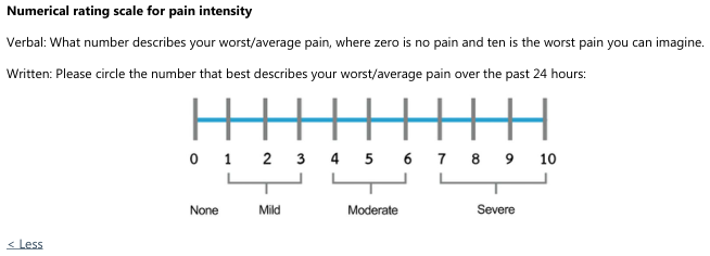
Summary : This figure presents a numerical rating scale for pain intensity, used to assess a patient's worst or average pain over the past 24 hours, with both verbal and written instructions. scale: Title & Axes : • Title: "Numerical rating scale for pain intensity" (implied by heading). • X-axis: Numeric scale from 0 to 10, labeled at each integer. • No Y-axis present. Scale Points & Labels : • 0: None • 1–3: Mild • 4–6: Moderate • 7–10: Severe Instructions : • Verbal: "What number describes your worst/average pain, where zero is no pain and ten is the worst pain you can imagine." • Written: "Please circle the number that best describes your worst/average pain over the past 24 hours." Design Encodings : • Horizontal blue line connecting vertical tick marks at each integer from 0 to 10. • Brackets below the scale group numbers into pain categories (None, Mild, Moderate, Severe). Analysis : • The scale provides a simple, standardized method for patients to quantify pain intensity, facilitating communication and documentation. • Pain is categorized into four levels (None, Mild, Moderate, Severe) based on numeric ranges, allowing for both qualitative and quantitative assessment. • The scale is linear and evenly spaced, supporting consistent interpretation across users.
Adult Pain Score Scales
A. Self-Report Scales (Communicative Adults)
1. Numeric Rating Scale (NRS) - Gold Standard for Adults
| Score | Category |
|---|---|
| 0 | No pain |
| 1-3 | Mild pain |
| 4-6 | Moderate pain |
| 7-10 | Severe pain |
- Patient asked: "What number from 0-10 describes your pain, where 0 = no pain and 10 = worst pain imaginable?"
- Can also be used as a 0-100 scale.
- Horizontal orientation preferred (scores more normally distributed than vertical).
- Quick, reliable, valid; most commonly used in clinical practice and research.
2. Visual Analogue Scale (VAS)
- A 10 cm horizontal line: left end = "No pain", right end = "Worst pain possible."
- Patient marks their pain level; the distance in mm from the left = score (0-100).
- Very sensitive for detecting small changes in pain intensity - useful in research/clinical trials.
- Limitation: Requires adequate hand-eye coordination and visual acuity. Difficult for elderly, post-surgical, or cognitively impaired patients.
3. Verbal Rating Scale (VRS)
| Descriptor | Approximate NRS Equivalent |
|---|---|
| No pain | 0 |
| Mild pain | 1-3 |
| Moderate pain | 4-6 |
| Severe pain | 7-9 |
| Worst possible pain | 10 |
- Simpler than NRS/VAS - good for elderly or those with low health literacy.
- Comparable to NRS in quantifying acute pain.
- Less sensitive to small changes than NRS or VAS.
B. Behavioral/Observational Scales (Non-verbal / ICU / Intubated Adults)
Vital signs (tachycardia, hypertension, tachypnea) may suggest pain but are poor correlates - validated behavioral tools must be used instead.
4. Critical Care Pain Observation Tool (CPOT)
| Indicator | Description | Score |
|---|---|---|
| Facial expression | Relaxed | 0 |
| Tense (frowning, brow lowering) | 1 | |
| Grimacing (eyes tightly closed) | 2 | |
| Body movements | Absent / normal position | 0 |
| Protection (rubbing pain site) | 1 | |
| Restlessness / agitation (pulling tubes, thrashing) | 2 | |
| Muscle tension | Relaxed, no resistance to passive movement | 0 |
| Tense/rigid, resistance to passive movement | 1 | |
| Very tense/rigid, strong resistance | 2 | |
| Compliance with ventilation (intubated) OR Vocalisation (extubated) | Tolerating ventilation / no vocalization | 0 |
| Coughing but tolerating / sighing, moaning | 1 | |
| Fighting ventilator / crying out, sobbing | 2 |
5. Behavioral Pain Scale (BPS)
| Item | Description | Score |
|---|---|---|
| Facial expression | Relaxed | 1 |
| Partially tightened (brow lowering) | 2 | |
| Fully tightened (eyelid closing) | 3 | |
| Grimacing | 4 | |
| Upper limb movements | No movement | 1 |
| Partially bent | 2 | |
| Fully bent with finger flexion | 3 | |
| Permanently retracted | 4 | |
| Compliance with ventilation | Tolerating movement | 1 |
| Coughing but tolerating most of the time | 2 | |
| Fighting ventilator | 3 | |
| Unable to control ventilation | 4 |
6. Wong-Baker FACES Scale (Adults who cannot verbalize)
- Also used in adults with language barriers, cognitive impairment, or post-op confusion.
- 6 faces from smiling (0) to crying (10).
Summary: Which Scale for Which Adult?
| Patient Type | Recommended Scale |
|---|---|
| Communicative adult (ward/ED/post-op) | NRS (0-10) - first line |
| Research / precise measurement needed | VAS |
| Elderly / low literacy / simple communication | VRS (mild/moderate/severe) |
| Language barrier / cognitive impairment | Wong-Baker FACES |
| Intubated / sedated ICU patient | CPOT or BPS |
| Non-verbal ICU but not intubated | CPOT (vocalisation version) |
Different types of pain
types of pain classification nociceptive neuropathic visceral somatic diagram
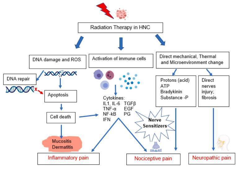
This pathophysiology diagram illustrates the biological mechanisms underlying radiotherapy-induced acute pain in Head and Neck Cancer (HNC). The flowchart categorizes pain into three primary types: Inflammatory, Nociceptive, and Neuropathic. The process begins with Radiation Therapy in HNC, branching into three initial pathways: 1) DNA damage and ROS production, which leads to failed DNA repair, apoptosis, cell death, and clinical manifestations like mucositis and dermatitis, resulting in inflammatory pain. 2) Activation of immune cells, leading to the release of inflammatory cytokines (e.g., IL1, IL-6, TNF-α, NF-κB, IFN, TGFβ, EGF, PG), which serve as nerve sensitizers triggering nociceptive pain. 3) Direct mechanical, thermal, and microenvironmental changes, including the release of protons (acid), ATP, bradykinin, and Substance-P, or direct nerve injury and fibrosis. These mechanical and chemical stimuli converge on nerve sensitizers to induce nociceptive pain, while direct nerve injury and chronic fibrosis specifically facilitate neuropathic pain. The diagram serves as an educational tool for oncology and pain management specialists to understand the multi-modal nature of radiation-induced toxicity.
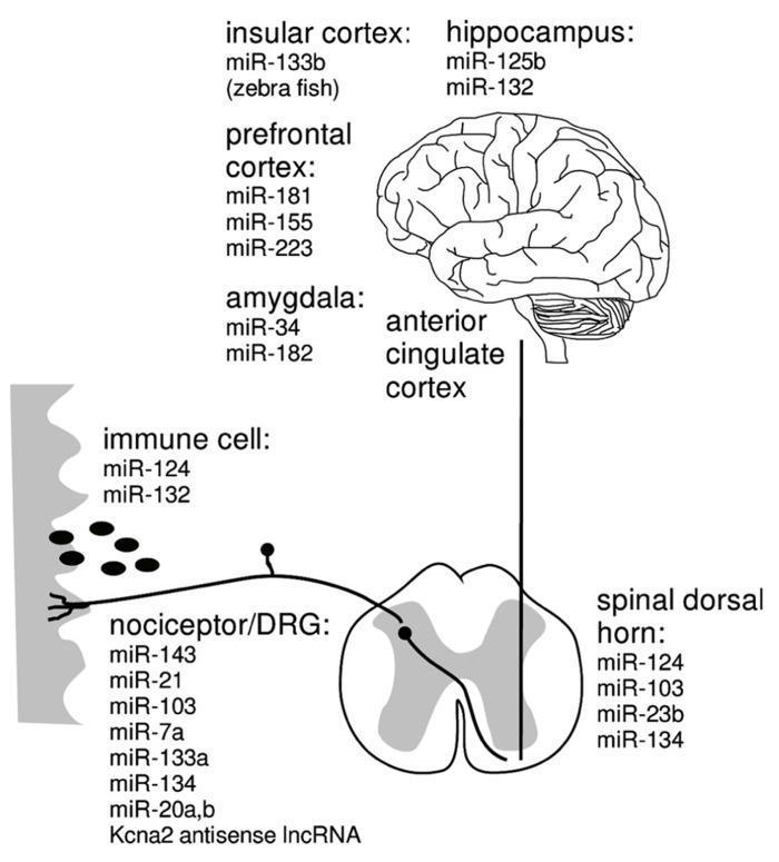
This pathophysiology diagram illustrates the ascending nociceptive pathway and the associated microRNAs (miRNAs) involved in neuropathic pain. The schematic traces the signal flow from the periphery to the central nervous system. It begins with an immune cell and nociceptor/dorsal root ganglion (DRG) at the peripheral level, connecting to the spinal dorsal horn, depicted as a characteristic butterfly-shaped cross-section. The pathway continues cephalad to the anterior cingulate cortex and into various brain regions, including the prefrontal cortex, insular cortex, hippocampus, and amygdala. Each anatomical station is annotated with specific miRNAs (such as miR-124, miR-132, miR-143, and miR-21) that are causally associated with the maintenance of neuropathic pain within those specific tissues. The diagram effectively highlights the complex molecular and cellular landscape of pain chronification across the peripheral and central nervous systems, serving as an educational resource for understanding the epigenetic regulation of chronic pain states.
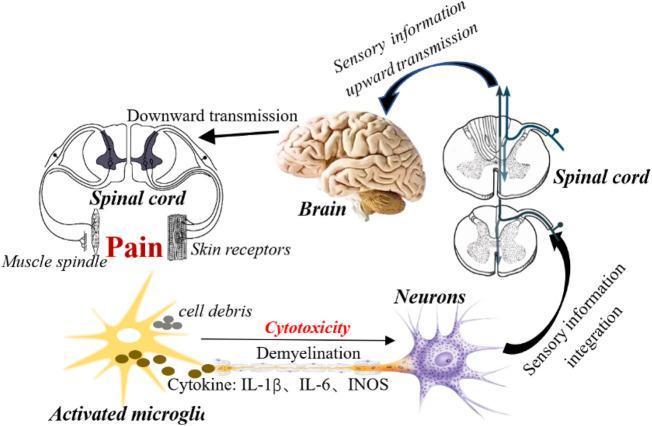
This pathophysiology diagram illustrates the neural pathways and neuroinflammatory mechanisms involved in neuropathic pain (NPP). The schematic follows a feedback loop starting from peripheral receptors, including skin receptors and muscle spindles, which transmit nociceptive signals. These signals undergo 'sensory information integration' within cross-sections of the spinal cord and 'upward transmission' to the brain. The brain subsequently processes this data and sends instructions via 'downward transmission' back through the spinal cord, culminating in the perception of pain. A significant portion of the diagram focuses on the cellular level, depicting an 'Activated microglia' with a star-shaped morphology near 'cell debris.' This microglial cell is shown releasing pro-inflammatory cytokines and factors, specifically IL-1β, IL-6, and iNOS. These substances act upon an adjacent neuron, leading to 'demyelination' and 'cytotoxicity,' which enhances sensory transmission and contributes to the persistence of chronic pain states. This educational visual integrates neuroanatomy, sensory physiology, and neuroimmunology to explain the development of pain following nerve injury.
Types of Pain
Classification 1: By Mechanism
1. Nociceptive Pain
A. Somatic Pain
| Subtype | Source | Character | Localization |
|---|---|---|---|
| Superficial somatic | Skin, mucous membranes, subcutaneous tissue | Sharp, pricking, throbbing, burning | Well-localized |
| Deep somatic | Muscles, tendons, joints, bones | Dull, aching | Less well-localized; radiates with severe/sustained stimuli |
B. Visceral Pain
| Subtype | Character |
|---|---|
| True visceral pain | Dull, diffuse, usually midline; associated with autonomic features (nausea, vomiting, sweating, BP/HR changes) |
| Parietal pain | Sharp, stabbing; localized around organ or referred to distant site |
| Referred visceral/parietal pain | Felt at cutaneous dermatomes due to convergence of visceral + somatic afferents in CNS |
| Organ | Referred Dermatome |
|---|---|
| Heart | T1-T4 (left arm, jaw) |
| Central diaphragm | C4 (shoulder tip) |
| Lungs | T2-T6 |
| Stomach, liver, gallbladder | T6-T9 |
| Pancreas, spleen | T5-T10 |
| Small intestine | T9-T11 |
| Aorta | T1-L2 |
| Kidneys/ureters | T10-L1 |
2. Neuropathic Pain
| Subtype | Source | Examples |
|---|---|---|
| Peripheral neuropathic | Peripheral nerve injury (compression, transection, ischemia, infiltration) | Sciatica, diabetic neuropathy, post-herpetic neuralgia, nerve entrapment, neuromas, trigeminal neuralgia |
| Central neuropathic | Central nervous system lesion | Stroke, Parkinson's disease, brain/spinal cord injury, multiple sclerosis |
- Burning, shooting, stabbing, electric shock-like, "pins and needles", tingling
- Allodynia - pain from normally non-painful stimuli (e.g. light touch)
- Hyperalgesia - exaggerated pain from a normally painful stimulus
- Dysesthesias - unpleasant abnormal sensations
- Often delayed in onset after the original nerve injury
- Difficult for patients to describe
3. Nociplastic Pain (formerly "Dysfunctional Pain")
- Widespread, intense pain
- Associated with fatigue, cognitive difficulties ("brain fog"), insomnia, mood disturbance
- No identifiable structural or neuropathic cause
Classification 2: By Duration
| Type | Duration | Key Features |
|---|---|---|
| Acute pain | Days to weeks; resolves with healing | Almost always nociceptive; proportional to stimulus; associated with neuroendocrine stress response; protective purpose |
| Chronic pain | Persists >3 months beyond expected healing | Maladaptive; persists after original injury/disease resolves; often involves central sensitization; significant psychological component |
When acute pain fails to resolve due to abnormal healing or inadequate treatment, it transitions to chronic pain.
Classification 3: By Temporal Pattern
| Pattern | Description | Example |
|---|---|---|
| Constant/continuous | Present all the time | Cancer pain, neuropathy |
| Intermittent | Episodes with pain-free intervals | Migraine, renal colic |
| Breakthrough pain | Acute flare on background of controlled chronic pain | Cancer pain management |
| Incident pain | Triggered by specific activity/movement | Bone metastasis pain on movement |
Summary Table (Sabiston Textbook of Surgery Classification)
| Classification | Subtype | Description | Examples | Treatment |
|---|---|---|---|---|
| Nociceptive / Inflammatory | Somatic | Well-localized, dull, aching, throbbing | Fracture, incisional pain, arthritis | NSAIDs, muscle relaxants, opioids |
| Visceral | Poorly localized, aching, can be referred | Appendicitis, bowel obstruction, liver mets | Opioids, treat cause | |
| Neuropathic | Peripheral | Shooting, stabbing, burning | Sciatica, diabetic neuropathy | TCAs, anticonvulsants, nerve blocks |
| Central | Variable | Stroke, SCI, Parkinson's | TCAs, anticonvulsants | |
| Nociplastic | - | Widespread; fatigue, insomnia, mood disturbance | Fibromyalgia, CRPS, IBS | SNRIs, TCAs, gabapentinoids |