Brachial plexus

Reading File
Finding Sources
Searching Images

brachial plexus anatomy diagram roots trunks cords

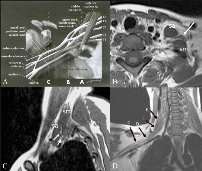
This composite educational graphic illustrates the normal anatomy of the brachial plexus through anatomical diagrams and Magnetic Resonance Imaging (MRI). Panel A is an anatomical diagram depicting the plexus architecture from roots (C5-T1) to terminal branches, labeled with musculoskeletal landmarks like the scalene muscles and coracoid process. Panel B (axial) and Panel C (sagittal) are T1-weighted MRI scans demonstrating the interscalene triangle, defined by the anterior scalene (sa) and middle scalene (sm) muscles. These muscles serve as primary radiological landmarks for identifying the neural roots and trunks (indicated by arrows). Panel D provides an oblique coronal T1-weighted MRI view, tracing the sequential segments of the right brachial plexus: roots (R), trunks (T), divisions (D), and cords (C). This resource is designed for medical education in radiology and neurology to assist in the localization of neural structures and the diagnostic evaluation of conditions like brachial plexopathy, neurogenic tumors, or Pancoast tumors.

This composite educational graphic illustrates the normal anatomy of the brachial plexus through anatomical diagrams and Magnetic Resonance Imaging (MRI). Panel A is an anatomical diagram depicting the plexus architecture from roots (C5-T1) to terminal branches, labeled with musculoskeletal landmarks like the scalene muscles and coracoid process. Panel B (axial) and Panel C (sagittal) are T1-weighted MRI scans demonstrating the interscalene triangle, defined by the anterior scalene (sa) and middle scalene (sm) muscles. These muscles serve as primary radiological landmarks for identifying the neural roots and trunks (indicated by arrows). Panel D provides an oblique coronal T1-weighted MRI view, tracing the sequential segments of the right brachial plexus: roots (R), trunks (T), divisions (D), and cords (C). This resource is designed for medical education in radiology and neurology to assist in the localization of neural structures and the diagnostic evaluation of conditions like brachial plexopathy, neurogenic tumors, or Pancoast tumors.

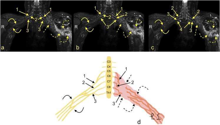
This composite educational resource comprises three Magnetic Resonance Neurography (MRN) coronal views (a, b, c) and a correlating schematic anatomical diagram (d) illustrating a severe traumatic brachial plexus injury. The right side of the images displays normal brachial plexus anatomy, with continuous nerve structures including the upper trunk (1), middle trunk (2), C8 root (3), and Th1 root (4). In contrast, the left side demonstrates a total rupture of the brachial plexus (dashed arrows). This injury is characterized by the complete discontinuity of the neural trunks and cords, accompanied by a large axillary cavity hematoma (curved dashed arrows). The schematic (d) highlights the side-to-side difference, showing the organized right-sided roots (C5-Th1) and trunks versus the disorganized, hemorrhagic area on the left. This material is designed for advanced medical education in radiology and neurosurgery, focusing on diagnostic imaging features of high-grade nerve trauma, specifically Seddon grade 3-5 injuries (neurotmesis) and the identification of postganglionic plexopathy.

This composite educational resource comprises three Magnetic Resonance Neurography (MRN) coronal views (a, b, c) and a correlating schematic anatomical diagram (d) illustrating a severe traumatic brachial plexus injury. The right side of the images displays normal brachial plexus anatomy, with continuous nerve structures including the upper trunk (1), middle trunk (2), C8 root (3), and Th1 root (4). In contrast, the left side demonstrates a total rupture of the brachial plexus (dashed arrows). This injury is characterized by the complete discontinuity of the neural trunks and cords, accompanied by a large axillary cavity hematoma (curved dashed arrows). The schematic (d) highlights the side-to-side difference, showing the organized right-sided roots (C5-Th1) and trunks versus the disorganized, hemorrhagic area on the left. This material is designed for advanced medical education in radiology and neurosurgery, focusing on diagnostic imaging features of high-grade nerve trauma, specifically Seddon grade 3-5 injuries (neurotmesis) and the identification of postganglionic plexopathy.

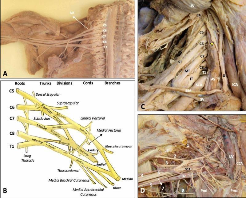
This composite educational graphic details the anatomy of the human brachial plexus (BP) through cadaveric dissections and a schematic diagram. (A) Cadaveric dissection of the cervical spine and axilla with the anterior scalene muscle removed, exposing the C5-T1 nerve roots emerging laterally to the middle scalene muscle (MS). (B) A structured schematic diagram illustrating the hierarchical organization of the BP from roots (C5-T1), trunks (Superior, Middle, Inferior), divisions (Anterior, Posterior), and cords (Lateral, Posterior, Medial) to terminal branches. (C) Dissection showing the roots in situ within the scalene triangle, formed between the anterior (AS) and middle scalene muscles, alongside major vessels including the subclavian artery (SCA) and internal jugular vein (IJV). The phrenic nerve (yellow arrowhead) is seen traversing the anterior scalene. (D) Detailed view of the infraclavicular and axillary regions showing terminal branches (1-8: suprascapular, musculocutaneous, axillary, radial, medial brachial cutaneous, median, ulnar, and intercostobrachial nerves) in relationship to the pectoralis minor (Pmi), pectoralis major (Pma), and subclavian vessels. This content provides essential topographical and clinical anatomy for understanding thoracic outlet syndrome and regional anesthesia.

This composite educational graphic details the anatomy of the human brachial plexus (BP) through cadaveric dissections and a schematic diagram. (A) Cadaveric dissection of the cervical spine and axilla with the anterior scalene muscle removed, exposing the C5-T1 nerve roots emerging laterally to the middle scalene muscle (MS). (B) A structured schematic diagram illustrating the hierarchical organization of the BP from roots (C5-T1), trunks (Superior, Middle, Inferior), divisions (Anterior, Posterior), and cords (Lateral, Posterior, Medial) to terminal branches. (C) Dissection showing the roots in situ within the scalene triangle, formed between the anterior (AS) and middle scalene muscles, alongside major vessels including the subclavian artery (SCA) and internal jugular vein (IJV). The phrenic nerve (yellow arrowhead) is seen traversing the anterior scalene. (D) Detailed view of the infraclavicular and axillary regions showing terminal branches (1-8: suprascapular, musculocutaneous, axillary, radial, medial brachial cutaneous, median, ulnar, and intercostobrachial nerves) in relationship to the pectoralis minor (Pmi), pectoralis major (Pma), and subclavian vessels. This content provides essential topographical and clinical anatomy for understanding thoracic outlet syndrome and regional anesthesia.

Reading File
Reading File
Reading File
Loading Image
Reading File
Loading Image
Reading File
Reading File
Reading File
Finding Sources
Reading File
Reading File
Now I have comprehensive information. Let me compile the full answer.

Brachial Plexus

Overview

The brachial plexus is a somatic nerve plexus formed by the anterior rami of C5 to C8, and most of T1. It originates in the neck, passes laterally and inferiorly over rib I, and enters the axilla. All major nerves innervating the upper limb arise from it. The roots receive gray rami communicantes from the sympathetic trunk, carrying postganglionic sympathetic fibers for peripheral distribution.
(Gray's Anatomy for Students, p. 858)

Anatomy Diagram

Brachial Plexus - Major components in the neck and axilla, showing Roots (C5-T1), Trunks, Divisions, and Cords

Structure: "RTDCB" - Roots, Trunks, Divisions, Cords, Branches

Brachial Plexus schematic showing all branches from roots to terminal nerves, with axillary artery relationships

1. Roots

RootContribution
C5Dorsal scapular nerve; contribution to phrenic nerve
C5-C7Long thoracic nerve
C5-T1Brachial plexus proper
Roots pass between the anterior and middle scalene muscles, lying posterior to the subclavian artery.

2. Trunks

TrunkRoot CompositionPosition
SuperiorC5 + C6Superior position
MiddleC7 onlyMiddle
InferiorC8 + T1Lies on rib I, posterior to subclavian artery
Branches from trunks (from superior trunk only):
  • Suprascapular nerve (C5, C6) - supraspinatus + infraspinatus
  • Nerve to subclavius (C5, C6) - subclavius muscle

3. Divisions

Each trunk splits into anterior and posterior divisions:
  • Anterior divisions - ultimately supply anterior compartments (flexors)
  • Posterior divisions - ultimately supply posterior compartments (extensors)
  • No peripheral nerves arise directly from divisions

4. Cords

Named by position relative to the second part of the axillary artery:
CordOriginRootsPosition
LateralAnterior divisions of superior + middle trunkC5-C7Lateral to axillary artery
MedialAnterior division of inferior trunkC8, T1Medial to axillary artery
PosteriorAll three posterior divisionsC5-T1Posterior to axillary artery

5. Branches (Terminal and Collateral)

From the Roots:

NerveOriginSupplies
Dorsal scapularC5 rootRhomboid major + minor
Long thoracicC5-C7 rootsSerratus anterior

From the Superior Trunk:

NerveSupplies
Suprascapular (C5,C6)Supraspinatus + infraspinatus
Nerve to subclavius (C5,C6)Subclavius

From the Lateral Cord:

NerveSupplies
Lateral pectoral nerve (C5-C7)Pectoralis major (+minor via communication)
Musculocutaneous nerve (C5-C7)Coracobrachialis, biceps, brachialis; skin - lateral forearm
Lateral root of median nerveContributes to median nerve

From the Medial Cord:

NerveSupplies
Medial pectoral nerve (C8, T1)Pectoralis major + minor
Medial cutaneous nerve of arm (C8, T1)Skin - medial distal arm
Medial cutaneous nerve of forearm (C8, T1)Skin - medial forearm to wrist
Medial root of median nerveContributes to median nerve
Ulnar nerve (C8, T1)All intrinsic hand muscles except thenar + 2 lateral lumbricals; flexor carpi ulnaris; medial FDP
Median nerve = lateral root (lateral cord) + medial root (medial cord), formed anterior to the 3rd part of the axillary artery.

From the Posterior Cord:

NerveSupplies
Superior subscapularSubscapularis
Thoracodorsal (C6-C8)Latissimus dorsi
Inferior subscapularSubscapularis + teres major
Axillary nerve (C5, C6)Deltoid + teres minor; skin - lateral shoulder (exits via quadrangular space)
Radial nerve (C5-T1)All posterior compartment muscles of arm + forearm; skin - posterior arm/forearm, lateral hand
Memory aid: The musculocutaneous nerve, lateral root of median, median nerve, medial root of median, and ulnar nerve form an "M" over the third part of the axillary artery.

Relations

  • Proximal (neck): roots and trunks lie posterior to the subclavian artery, between anterior and middle scalene muscles
  • Axilla: cords surround the axillary artery (named by their position relative to the second part)
  • Infraclavicular: cords and terminal branches, related to the pectoralis minor muscle

Clinical: Brachial Plexus Injuries

Erb-Duchenne Palsy (Upper Lesion - C5, C6)

Mechanism: Forcible widening of the angle between head and shoulder (motorcycle accidents, difficult labor)
Muscles affected: Deltoid, biceps, brachialis, brachioradialis, supinator
Posture: Arm internally rotated, elbow extended, forearm pronated - "Waiter's tip" or "Policeman taking a tip" position
Sensory loss: Lateral arm + upper lateral forearm
Prognosis: Best (most common type)

Klumpke's Palsy (Lower Lesion - C8, T1)

Mechanism: Forceful hyperabduction (grabbing a rail while falling, breech delivery with arms above head)
Muscles affected: Intrinsic hand muscles
Features: Claw hand, combined median + ulnar palsy pattern, anaesthesia of medial forearm + hand + inner 1½ fingers
Associated finding: Horner's syndrome (ptosis, enophthalmos, miosis, anhidrosis) - because T1 sympathetic fibers are disrupted
Prognosis: Poor

Complete Plexus Injury

  • Occurs in severe trauma
  • Complete paralysis + anaesthesia of the whole upper limb (except upper medial arm - C3/4/5 territory)
  • Long thoracic nerve or dorsal scapular nerve may occasionally be spared
(S Das A Manual on Clinical Surgery, p. 140; Gray's Anatomy for Students, p. 858-865)

Anesthetic Significance

The brachial plexus can be blocked at multiple levels:
  • Interscalene block - targets roots/trunks between the scalene muscles (best for shoulder)
  • Supraclavicular block - targets trunks/divisions at the clavicle (most complete upper limb coverage)
  • Infraclavicular block - targets cords
  • Axillary block - targets terminal branches around axillary artery
This is a shared conversation. Sign in to Orris to start your own chat.