
Applied of subclavian artery
Reading File
Finding Sources
Finding Sources
Searching Images
subclavian artery anatomy diagram relations
Finding Sources
Finding Sources
Reading File
Applied Anatomy of the Subclavian Artery
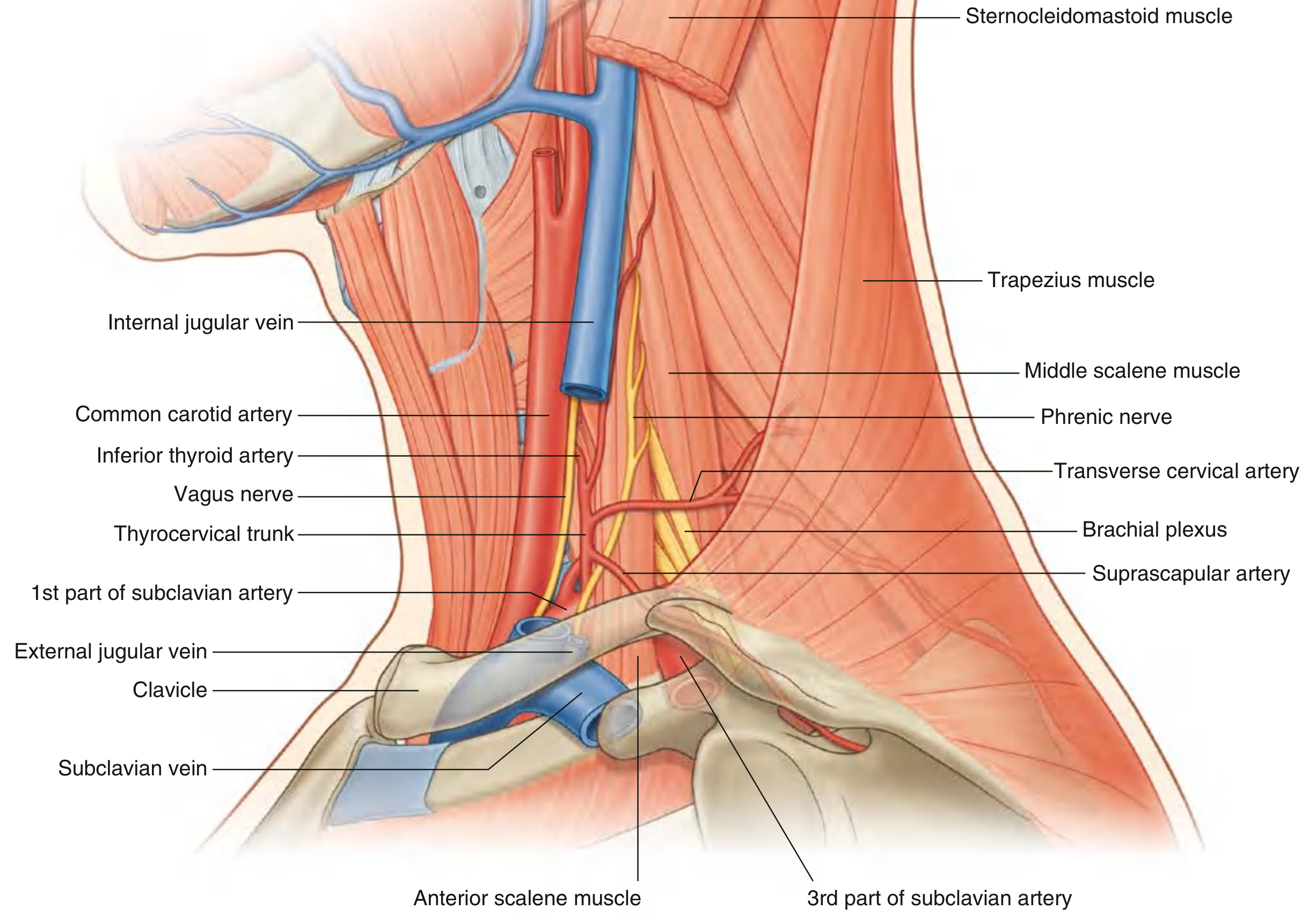
The subclavian artery's course (arising from the brachiocephalic trunk on the right, directly from the aortic arch on the left, arching over the pleural dome, and passing between the anterior and middle scalene muscles) gives rise to several important clinical correlations.
1. Cervical Rib / Thoracic Outlet Syndrome
The artery's third part crosses the base of the posterior triangle of the neck and is closely related to the lower trunk of the brachial plexus as both pass over the first rib. A cervical rib (or a fibrous band from one) narrows the scalene interval and can compress the artery here.
- This produces arterial thoracic outlet syndrome (TOS): compression leads to poststenotic dilatation, mural thrombus, and distal embolization, with reduced pulses/blood pressure and pallor in the affected limb - Harrison's Principles of Internal Medicine, 22E.
- Clinically this overlaps with scalenus anticus syndrome, where the artery and plexus are compressed against the first rib by the anterior scalene, producing identical neurovascular symptoms (paresthesia, weakness, coldness of the hand) - K.J. Lee's Essential Otolaryngology.
- Angulation of the artery over a cervical rib is a classic cause of subclavian artery aneurysm - S. Das, A Manual on Clinical Surgery, 13th ed.
2. Subclavian Steal Syndrome
Stenosis or occlusion of the subclavian artery proximal to the origin of the vertebral artery can cause retrograde ("stolen") flow down the vertebral artery to supply the arm during exercise, at the expense of posterior circulation. This presents with vertigo, dysarthria, diplopia, or syncope precipitated by arm exercise, and is diagnosed by a >40 mmHg difference in blood pressure between the two arms, confirmed on MRA/duplex - Bradley and Daroff's Neurology in Clinical Practice; Rosen's Emergency Medicine.
3. Aberrant Right Subclavian Artery (Dysphagia Lusoria)
When the right subclavian artery arises anomalously from the distal aortic arch, it must cross posterior to the esophagus to reach the arm, producing extrinsic esophageal compression - dysphagia lusoria. This is associated with a non-recurrent right recurrent laryngeal nerve and can be linked with aortic aneurysms - K.J. Lee's Essential Otolaryngology; Yamada's Textbook of Gastroenterology.
4. Aneurysm Formation
Subclavian and axillary artery aneurysms most often arise secondary to TOS-related repetitive compression at the thoracic outlet, causing poststenotic dilation that can become a source of distal emboli. Presentation depends on location - chest/neck/shoulder pain from expansion, hoarseness from recurrent laryngeal nerve compression, or limb ischemia from embolization - Sabiston Textbook of Surgery; Current Surgical Therapy, 14e.
5. Surgical/Interventional Relevance
- Ligation: Because of the rich anastomotic network around the scapula (suprascapular, transverse cervical, and circumflex scapular arteries), the subclavian artery can be ligated with adequate collateral perfusion to the upper limb via the scapular anastomosis.
- Subclavian artery transposition: used to bypass carotid or vertebral lesions, requiring careful dissection proximal to the internal thoracic and vertebral artery origins - Current Surgical Therapy, 14e; Fischer's Mastery of Surgery.
- It is a donor vessel in reconstructive cardiac procedures (e.g., classic Blalock-Taussig shunt) and the internal thoracic artery, a branch of its first part, is the primary graft vessel in coronary artery bypass grafting.
- Coarctation of the aorta: if the coarctation lies proximal to the left subclavian artery origin, blood pressure and pulses are reduced in the left arm relative to the right, an important bedside clue.
6. Landmark Value
The third part of the subclavian artery, palpable in the supraclavicular fossa, serves as a landmark for supraclavicular brachial plexus block and for locating the artery in trauma or vascular access procedures. Its close relationship to the phrenic nerve, vagus nerve, and thoracic duct (on the left) means iatrogenic injury during central line placement or surgery can cause phrenic nerve palsy, recurrent laryngeal nerve injury, or chylothorax.
If you want, I can go deeper into any one of these (e.g., detailed TOS management, or the surgical steps for ligation and collateral circulation).
This is a shared conversation. Sign in to Orris to start your own chat.