
Miosis occurs in which pathological condition explain refer ophthal Khurana
miosis pupil constriction causes ophthalmology
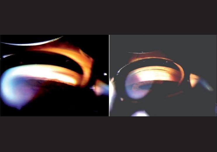
This diagnostic image consists of two side-by-side clinical photographs showing gonioscopy of the anterior chamber angle of the eye under different lighting conditions. The left panel demonstrates the angle with bright illumination and a long slit beam. This lighting causes miosis (pupil constriction), which visually pulls the iris away from the trabecular meshwork, creating the appearance of an open angle. A prominent bright white and orange light reflex is visible across the iris and cornea. The right panel demonstrates the same anterior chamber angle under dim illumination. In these appropriate testing conditions, the lack of light stimulus prevents pupillary constriction, revealing the true state of the angle, which appears closed or significantly narrowed as the iris root approximates the corneal endothelium. The comparison illustrates the clinical importance of controlling ambient light during gonioscopy to avoid false-negative results in angle-closure glaucoma screening.
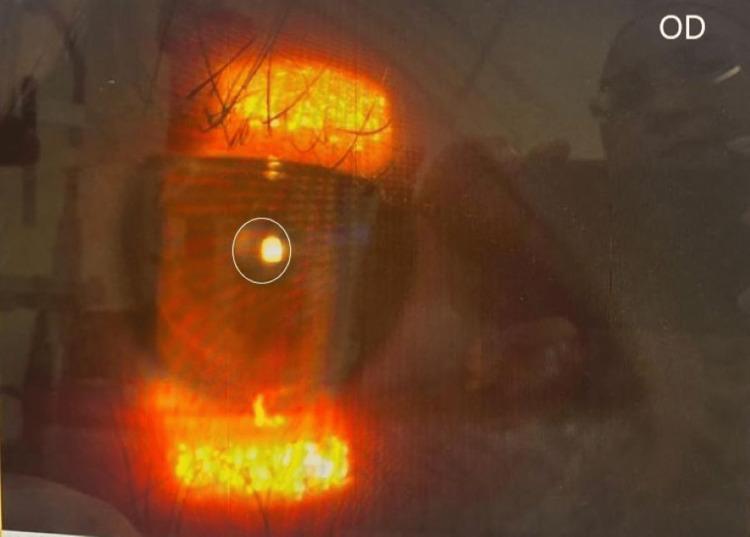
A close-up clinical photograph of the right eye (oculus dexter, OD) under examination, demonstrating miosis. The image highlights a constricted pupil, approximately 2 mm in diameter, which is circular and displays a sharp corneal light reflex from the slit lamp or illumination source. The iris is visible but partially obscured by reflection. The image is used to document clinical findings in Horner's syndrome, specifically pupillary asymmetry (anisocoria) where the affected eye exhibits miosis. The photograph provides a clear visual of the anterior segment focusing on pupillary size and reactivity for diagnostic purposes in neuro-ophthalmology. Key educational concepts include the assessment of the oculosympathetic pathway and the identification of post-surgical Horner's syndrome manifestations.
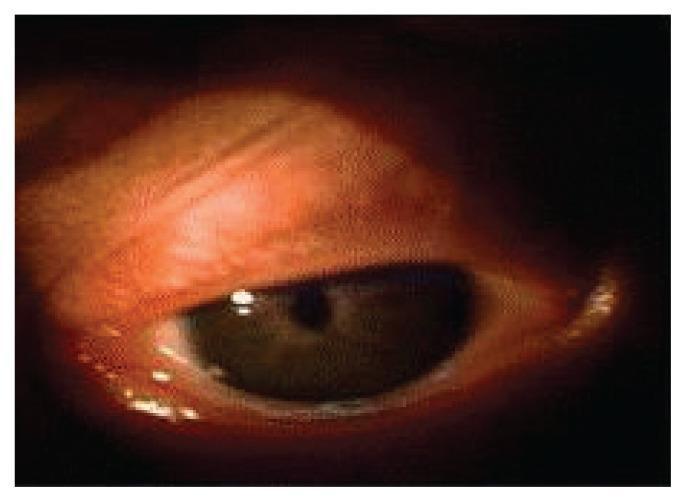
A clinical photograph focusing on the left eye of an infant, demonstrating microcoria. The primary finding is a pathologically small, pinpoint pupil (miosis) that is centrally positioned within the iris. The iris appears dark brown and occupies most of the visible corneal area due to the extreme pupillary constriction. The sclera shows some mild periorbital erythema along the eyelid margins, while the palpebral fissure appears narrowed. This visual finding is a hallmark ocular manifestation of Pierson syndrome (congenital nephrotic syndrome with ocular abnormalities), representing a fixed, non-reactive pupil. The image serves as an educational example of congenital ocular pathology and its association with systemic multi-organ syndromes in pediatric patients.
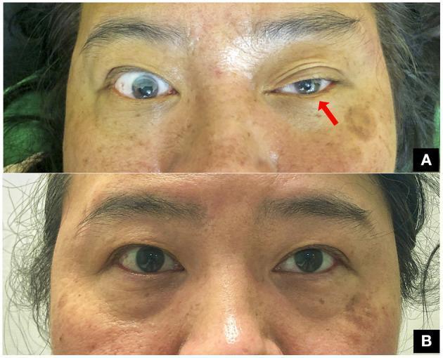
This clinical comparison photograph shows a 47-year-old woman presenting with classic oculofacial features of Horner’s syndrome following a clavicle fracture. Image A demonstrates the acute presentation of the oculosympathetic defect in the left eye, characterized by significant blepharoptosis (drooping of the upper eyelid) and ipsilateral miosis (constriction of the pupil) compared to the normal right eye. A red arrow points to the affected left eye to emphasize these findings. Image B shows the same patient at a three-month follow-up after surgical fixation of the fracture. It illustrates complete resolution of the syndrome, with symmetric eyelid positions and equal pupillary diameters (isocoria) in both eyes. This visual progression highlights the classic clinical triad (ptosis and miosis, with anhidrosis typically associated) and the potential for recovery following the management of the underlying traumatic compression of the preganglionic sympathetic pathway. The image is a valuable educational resource for neurology, ophthalmology, and trauma surgery.
Miosis - Pathological Conditions
Mechanism of Pupillary Control (Basis for Miosis)
| Pathway | Effect |
|---|---|
| Parasympathetic (CN III → ciliary ganglion → sphincter pupillae) | Constricts pupil (miosis) |
| Sympathetic (hypothalamus → ciliospinal centre of Budge C8-T2 → superior cervical ganglion → dilator pupillae) | Dilates pupil (mydriasis) |
Pathological Conditions Causing Miosis
1. Horner Syndrome (Oculosympathetic Palsy) - MOST IMPORTANT
- Miosis - due to unopposed sphincter pupillae action (dilator paralysed)
- Ptosis (1-2 mm) - weakness of Muller's muscle
- Anhidrosis - only if lesion is below the superior cervical ganglion
| Level | Causes |
|---|---|
| 1st order (central) | Brainstem stroke (lateral medullary infarction/Wallenberg), brainstem tumour, demyelination, syringomyelia, cervical spinal cord lesion, diabetic autonomic neuropathy |
| 2nd order (preganglionic) | Pancoast tumour (apical bronchogenic carcinoma), carotid/aortic aneurysm and dissection, thoracic cord lesion, neck lesions (thyroid tumour, lymphadenopathy, trauma, post-surgical) |
| 3rd order (postganglionic) | Internal carotid artery dissection, nasopharyngeal tumour, cavernous sinus mass, otitis media, cluster headache |
2. Iridocyclitis (Anterior Uveitis)
- Irritative miosis - due to reflex spasm of the sphincter pupillae in response to inflammatory stimulation
- The miosis is typically accompanied by photophobia, ciliary flush, aqueous flare/cells
- In lepromatous uveitis specifically: miosis and iris atrophy result from impaired dilator pupillae innervation (Kanski's, p. 12.59)
- Risk: miosis predisposes to posterior synechiae formation if untreated
3. Spasm of the Near Reflex
- A functional or organic disorder where the near triad (accommodation, convergence, miosis) goes into spasm
- Signs: Esotropia, pseudomyopia (excess accommodation), and miosis
- Observation of miosis is the key to diagnosis (distinguishes it from true convergence excess esotropia)
- Refraction under cycloplegia confirms pseudomyopia
-
- Kanski's Clinical Ophthalmology, Spasm of near reflex section (block 5, line 2431)
4. Pontine Hemorrhage / Pontine Lesions
- Bilateral pinpoint pupils (extreme miosis) - classic sign
- Mechanism: interruption of descending pupillodilator fibres (sympathetic) bilaterally
- Pupils remain reactive to light (though magnification may be needed to confirm)
- Associated: coma, quadriplegia, hyperthermia, irregular breathing
5. Argyll Robertson Pupil (Neurosyphilis)
- Small, irregular, bilateral miosis
- Pupils do NOT react to light, but DO react to near (accommodation-reflex dissociation)
- "Prostitute's pupil" - accommodates but does not react
- Caused by destruction of pretectal nuclei by syphilitic endarteritis
- The reflex arc for near is preserved via a separate pathway
6. Pharmacological / Toxicological Miosis
| Cause | Mechanism |
|---|---|
| Opioid overdose (morphine, heroin, fentanyl) | Central inhibition of sympathetic + enhanced parasympathetic; classic triad: miosis + respiratory depression + coma |
| Organophosphate / anticholinesterase poisoning | Excess acetylcholine → prolonged sphincter stimulation (DUMBELS mnemonic includes miosis) |
| Pilocarpine (glaucoma drops) | Direct muscarinic agonist |
| Echothiophate, physostigmine | Anticholinesterase - miosis + accommodative spasm |
| Clonidine overdose | Central alpha-2 agonist - reduces sympathetic tone |
7. Acute Angle Closure Glaucoma
- A mid-dilated (4-6 mm) NON-reactive pupil is classic during an acute attack, BUT:
- Corneal edema + iris ischemia can cause a fixed slightly constricted pupil post-attack
- Instillation of pilocarpine deliberately induces miosis to break the attack by pulling iris away from the angle
8. Horner Syndrome in Specific Ophthalmological Contexts
- Corneal foreign body/ulcer: Mild secondary uveitis causes irritative miosis with photophobia (Kanski's, p. 3299)
- Lepromatous leprosy: Chronic anterior uveitis → miosis + iris atrophy + iris pearls (pathognomonic)
- Media opacities (cataract): Effect on visual field testing is exaggerated by miosis (Kanski's, p. 683)
9. Other Neurological Causes
| Condition | Notes |
|---|---|
| Lateral medullary (Wallenberg) syndrome | Ipsilateral Horner (central 1st-order neurone lesion) |
| Tabes dorsalis (neurosyphilis) | Argyll Robertson pupils |
| Parinaud syndrome (dorsal midbrain) | Convergence-retraction nystagmus + light-near dissociation |
| Cerebellar tonsillar herniation | Pinpoint pupils + flaccid paralysis + sudden death risk |
| Upward transtentorial herniation | Conjugate downward gaze + pinpoint pupils |
| Old age (senile miosis) | Dilator pupillae atrophy - not truly pathological |
Summary Table: Miosis at a Glance
| Condition | Laterality | Reaction to light | Reaction to near | Key associated features |
|---|---|---|---|---|
| Horner syndrome | Unilateral (usually) | Normal | Normal | Ptosis, anhidrosis, anisocoria worse in dark |
| Iridocyclitis | Unilateral | May be sluggish | - | Photophobia, keratic precipitates, flare |
| Argyll Robertson | Bilateral | Absent | Present | Irregular pupils, neurosyphilis |
| Pontine hemorrhage | Bilateral pinpoint | Preserved (barely) | - | Coma, quadriplegia |
| Opioid overdose | Bilateral | Preserved | - | Respiratory depression, coma |
| Spasm of near reflex | Bilateral | Normal | Excessive | Pseudomyopia, esotropia |
| Organophosphate | Bilateral | Normal | - | DUMBELS toxidrome |