
Explain everything about stress cardiomyopathy
"Takotsubo Cardiomyopathy"[MeSH Terms]
Takotsubo cardiomyopathy left ventriculogram apical ballooning
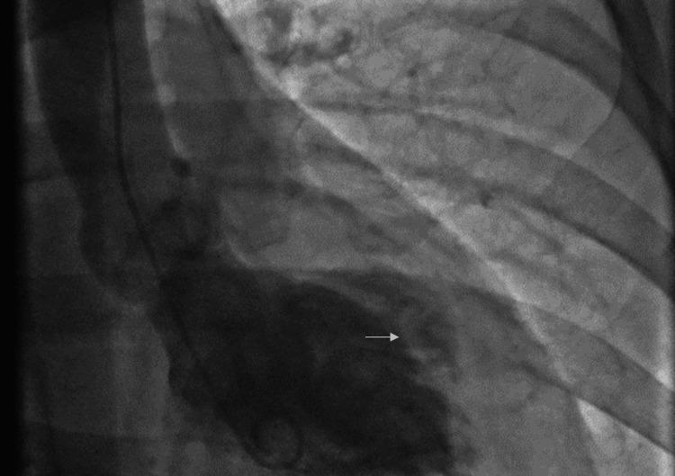
This diagnostic image is a left ventriculogram obtained during cardiac catheterization, displayed via fluoroscopic X-ray radiography. The image demonstrates a characteristic wall motion abnormality consistent with the reverse (inverted) subtype of Takotsubo Cardiomyopathy (TCM). Unlike the classic apical ballooning pattern, this visual shows preserved apical and basal contraction with prominent ballooning and akinesis localized to the mid-ventricular region. Specifically, the ventriculogram identifies akinesis of the diaphragmatic segment and dyskinesis of the high anterolateral wall. Contrast opacification reveals the distorted morphology of the left ventricular chamber, highlighting the regional transition from hyperdynamic basal contraction to the stunned mid-ventricular zone. This imaging is clinically significant for differentiating atypical variants of stress-induced cardiomyopathy from acute coronary syndromes, particularly when coronary arteries are found to be angiographically normal.
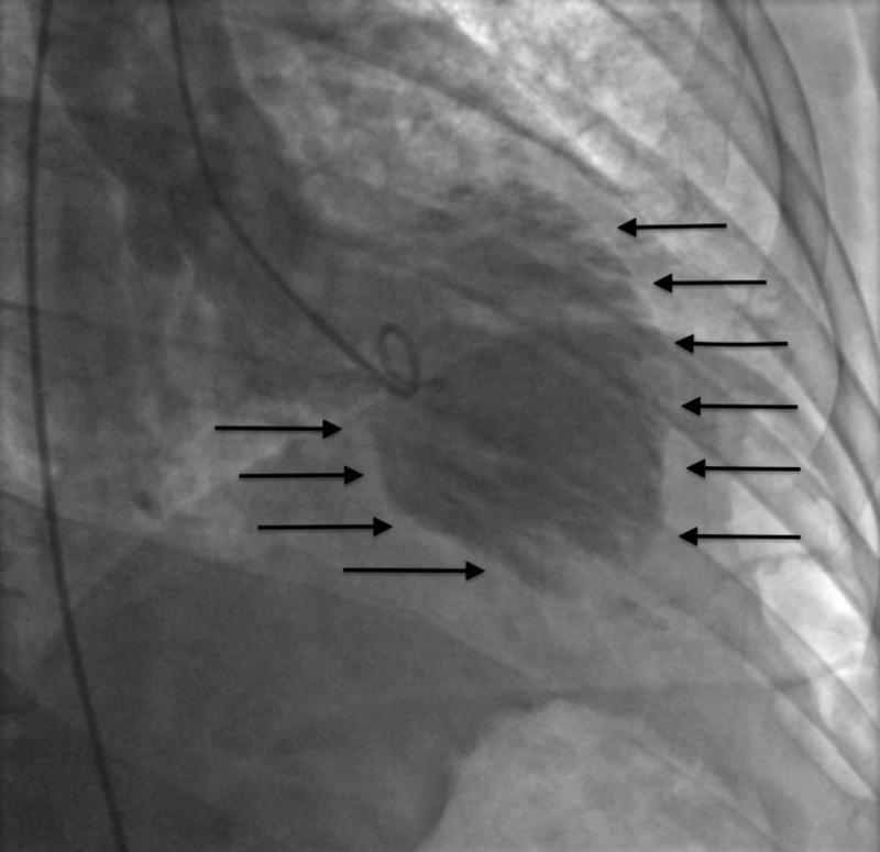
This diagnostic image is a left ventriculogram obtained via cardiac catheterization during ventricular systole. The radiograph displays the left ventricle opacified with contrast medium, revealing a characteristic 'apical ballooning' morphology. Black arrows highlight significant wall motion abnormalities, specifically akinesis in the mid-to-apical anterolateral and mid-to-apical inferior walls. In contrast, the cardiac base demonstrates relatively preserved contractility. This visual pattern is a hallmark finding of Takotsubo cardiomyopathy (stress-induced cardiomyopathy), often mimicking the presentation of an acute myocardial infarction despite the absence of obstructive coronary artery disease. The image is intended for medical education in cardiology and radiology to demonstrate classic diagnostic features of apical ballooning syndromes and the assessment of regional wall motion abnormalities using invasive ventriculography.
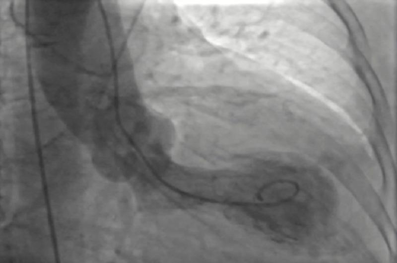
This diagnostic image is a coronary angiogram (specifically a left ventriculogram) illustrating the classic morphology of Takotsubo cardiomyopathy. The image depicts the left ventricle in a state of systolic dysfunction, characterized by prominent apical ballooning. There is a visible contrast opacification showing a dilated, rounded apical segment and hypokinetic/dyskinetic wall motion in the apical and mid-ventricular regions, contrasting with the relatively preserved or hypercontractile basal segments. A pigtail catheter is positioned within the left ventricular cavity to deliver the radiopaque contrast medium. The appearance is reminiscent of a 'takotsubo' (a Japanese octopus trap), which is the hallmark of stress-induced cardiomyopathy. The image also captures portions of the coronary vasculature, which typically show an absence of obstructive coronary artery disease in this clinical context. This visual is a key educational tool for cardiology to differentiate stress-induced cardiomyopathy from acute myocardial infarction caused by arterial occlusion.
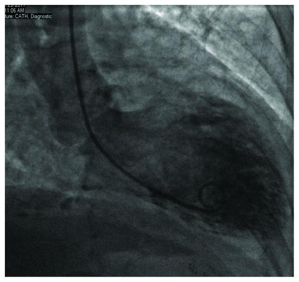
A diagnostic left ventriculogram image obtained during cardiac catheterization, shown in a standard fluoroscopic view. The image captures the left ventricle in diastole, filled with radiopaque contrast medium. A pigtail catheter is clearly visible, entering from the base and positioned within the ventricular cavity. The primary pathological finding is a distinct apical ballooning, characterized by a rounded, dilated appearance of the apex, while the basal regions demonstrate relatively preserved morphology. This visual pattern is a classic diagnostic feature of Takotsubo cardiomyopathy (stress-induced cardiomyopathy), resembling a 'tako-tsubo' (Japanese octopus trap). The image serves as a clinical teaching tool for identifying wall motion abnormalities and the specific morphology of transient apical ballooning syndrome in the context of acute coronary syndrome-like presentations with non-obstructive coronary arteries.
Takotsubo syndrome management recommendations InterTAK diagnostic criteria 2018 ESC 2023 consensus
Stress (Takotsubo) cardiomyopathy
1. Definition and names
2. Epidemiology
- TTS is found in about 0.5% to 3% of all ACS presentations. It is found in 5% to 6% of women with ACS, and 5.9% to 7.5% of postmenopausal women in Braunwald. - Fuster and Hurst's The Heart, 15th Ed; Braunwald
- About 90% of patients are women, and about 80% are 50 or older. Mean age is 65 to 70.
- Recorded incidence has risen over time, probably because of better recognition. It is somewhat higher in summer.
- During COVID-19, a two-centre Ohio study found stress cardiomyopathy in 7.8% of cath lab presentations, against 1.5% to 1.8% before the pandemic. - Fuster and Hurst's The Heart
3. Pathogenesis
- Sympathetic surge and catecholamine excess. Epinephrine is released from the adrenal medulla. Norepinephrine is released locally from cardiac nerve terminals, with reduced reuptake (shown on MIBG scans). Pheochromocytoma and catecholamine infusions can produce the same picture.
- Direct catecholamine toxicity. Biopsies show contraction band changes without necrosis.
- Adrenoceptor signalling switch. High epinephrine may switch beta-2 coupling from stimulatory to inhibitory G-protein, causing myocardial stunning. This may be protective, and it favours the apex, which has a high beta-receptor density.
- Microvascular and epicardial vasomotor dysfunction, and diffuse microvascular spasm. It is unclear whether this is a cause or a consequence.
- Brain-heart axis. Many patients have abnormal structure and activity in limbic and autonomic brain regions. Reduced parasympathetic tone (lower heart rate variability) is described. - Goldman-Cecil Medicine
- Susceptibility factors. These include postmenopausal estrogen loss, possible adrenoceptor polymorphisms (inconsistent data), familial clustering, and LV or mitral geometry resembling mild hypertrophic cardiomyopathy.
- Inflammation. Myocardial inflammation occurs in the acute phase and may persist. - Goldman-Cecil Medicine
- Emotional: grief, fear, anger, conflict, or even joy ("happy heart").
- Physical: surgery, sepsis, asthma or COPD exacerbation, pain, trauma.
- Neurologic: subarachnoid hemorrhage, stroke, seizures.
- Endocrine and drugs: pheochromocytoma, and catecholamine, epinephrine or other sympathomimetic drugs.
4. Clinical presentation
- Symptoms: chest pain (76%), dyspnea (47%), syncope (8%). Others present with pulmonary edema, hypotension, or shock.
- ECG:
- ST elevation in 44%, T-wave inversion in 41%, QTc prolongation in 48%, and ST depression in 8%.
- Rare cases have no ECG change.
- T-wave inversion with a long QT often evolves over days.
- Biomarkers:
- Troponin is elevated in nearly all patients, but modestly relative to the extent of dysfunction. In InterTAK the median peak was about 13 times the upper limit of normal with a median LVEF of 40%.
- BNP is often markedly raised, more so than in MI.
5. Wall motion patterns (InterTAK registry)
- Apical (classic), 82%.
- Midventricular, 15%.
- Basal (inverted), 2%.
- Focal, 1.5%.
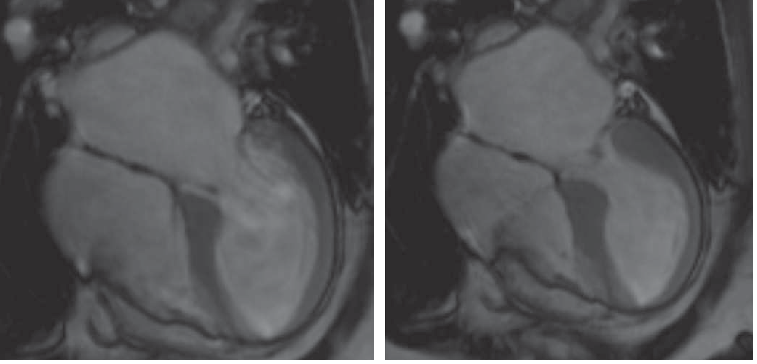
6. Diagnosis
- Transient LV dysfunction (apical, midventricular, basal or focal), usually beyond one coronary territory.
- An emotional or physical trigger may precede it but is not required.
- Neurologic disorders and pheochromocytoma can be triggers.
- New ECG abnormalities (rare cases have none).
- Moderately elevated troponin and CK, with commonly raised BNP.
- Significant CAD does not exclude TTS.
- Myocarditis is excluded. (I did not see this item in the passage I read, but it is part of the published criteria.)
- Female sex: 25.
- Emotional trigger: 24.
- Physical trigger: 13.
- No ST depression (except aVR): 12.
- Psychiatric disorder: 11.
- Neurologic disorder: 9.
- QTc prolongation: 6.
- Coronary angiography with ventriculography should not be deferred on the basis of ECG or wall motion pattern alone. Stress also precipitates plaque rupture, and an apical pattern can come from a proximal or wraparound LAD occlusion. - Fuster and Hurst's The Heart
- Obstructive CAD can coexist as a bystander. Spontaneous coronary artery dissection (SCAD) of the LAD can mimic TTS, so review the angiogram carefully.
- No culprit lesion: the differential is MINOCA, myocarditis, or TTS. Cardiac MRI helps to separate them:
- TTS shows circumferential edema in the dysfunctional segments, with no dense late gadolinium enhancement (LGE).
- MI shows subendocardial or transmural LGE.
- Myocarditis shows non-ischemic LGE.
- Echo shows the wall motion pattern and screens for complications (LVOT obstruction, mitral regurgitation, thrombus, effusion). Follow-up imaging showing recovery confirms the diagnosis retrospectively.
- Noninvasive angiography should be reserved for patients with very high likelihood of TTS who have no ST elevation or hemodynamic compromise.
7. Complications
- Cardiogenic shock in 5% to 10%, and pulmonary edema in about 5%.
- LVOT obstruction. Basal hypercontractility with a poorly contracting apex can cause it, sometimes with systolic anterior motion of the mitral valve and mitral regurgitation.
- Arrhythmias. These include AF, VT/VF, and torsades from QT prolongation. Bradyarrhythmias and asystole after conversion are also described.
- LV thrombus in 1% to 3%, with a risk of embolic stroke. It is linked to the apical pattern and larger troponin rise.
- Ventricular wall rupture is rare (0.2%). Pericarditis or effusion is also uncommon. - Braunwald; Fuster and Hurst's The Heart
8. Management
- Treat the patient as ACS until angiography excludes an occlusion.
- Treat the trigger (pain, sepsis, bronchospasm) and avoid sympathomimetics where possible.
- Use diuretics and nitrates, provided there is no LVOT obstruction.
- Harrison's suggests combined alpha and beta blockers rather than selective beta blockade if the patient is hemodynamically stable.
- Pump failure alone: vasoactive support; levosimendan is preferred where available because catecholamines are implicated.
- LVOT obstruction:
- Avoid inotropes.
- Give fluids or elevate the legs.
- Use a beta-blocker as a negative inotrope.
- If a vasopressor is needed, use a pure alpha agonist such as phenylephrine.
- Mechanical support: consider it if these measures fail. An intra-aortic balloon pump is used only if there is no LVOT obstruction (Harrison's). VA-ECMO is an option for refractory shock.
- Give magnesium, correct electrolytes, and avoid QT-prolonging drugs.
- Give it if LV thrombus is found.
- Many centres give it for a large akinetic apex. Evidence is limited, and I did not find a firm recommendation in the passages I read.
- ACE inhibitors are associated with better 1-year survival in observational data, but the duration is unknown.
- Beta-blockers did not prevent the index event or recurrence in registry data. - Braunwald
- Treat comorbid psychiatric illness and offer stress-management support. Cardiac rehabilitation may help.
- Repeat echo at about 4 to 8 weeks to confirm recovery.
9. Prognosis
- In-hospital mortality is 1% to 5% (4.1% in InterTAK), between NSTEMI and STEMI.
- Long-term mortality is 2% to 6% per year (InterTAK 5.6% long term), similar to MI. Non-cardiovascular causes, including cancer, outnumber cardiovascular ones.
- Worse outcomes are linked to physical triggers or no trigger, a neurologic trigger (particularly adverse), male sex, older age, diabetes, low LVEF, and shock. Emotional triggers carry the best prognosis.
- Recurrence is about 2% per year, or 5% to 10% overall.
- Thirty-day readmission is 12%, and heart failure is the most common cardiac cause.
- LVEF normalizes in most patients, but strain imaging and MRI extracellular volume can stay abnormal. Some patients have persistent symptoms. - Fuster and Hurst's The Heart; Braunwald
10. Recent literature check
- PMID 41173762 (2026): review of advances in pathophysiology, biomarkers, genetics, multisystem involvement, and treatment.
- PMID 41159266 (2026): meta-analysis of predictors of major adverse cardiovascular events.
- PMID 38897052 (2024): systematic review of the prognostic role of cardiac MRI.