
I needa ospe question on the names of foramenof norma basalis
OSPE Question: Foramina of Norma Basalis
Station Setup
Questions & Model Answers
| Foramen | Bone | Contents |
|---|---|---|
| Foramen magnum | Occipital bone | Medulla oblongata, meninges, vertebral arteries, meningeal branches of vertebral arteries, spinal roots of CN XI |
| Foramen ovale | Greater wing of sphenoid | CN V3 (mandibular nerve), accessory meningeal artery, lesser petrosal nerve (occasionally) |
| Foramen spinosum | Greater wing of sphenoid | Middle meningeal artery and vein, meningeal branch of mandibular nerve |
| Foramen lacerum | Between sphenoid, temporal, and occipital bones | Filled with fibrocartilage in life; internal carotid artery crosses its upper part; greater petrosal nerve and deep petrosal nerve traverse it |
| Jugular foramen | Between temporal and occipital bones | CN IX (glossopharyngeal), CN X (vagus), CN XI (accessory), inferior petrosal sinus, sigmoid sinus, posterior meningeal artery |
| Hypoglossal canal | Occipital bone | CN XII (hypoglossal nerve), meningeal branch of ascending pharyngeal artery |
| Carotid canal | Petrous temporal bone | Internal carotid artery, sympathetic plexus |
| Stylomastoid foramen | Between styloid and mastoid processes of temporal bone | CN VII (facial nerve), stylomastoid artery |
| Greater palatine foramen | Greater wing of palatine bone | Greater palatine nerve and vessels |
| Lesser palatine foramina | Palatine bone | Lesser palatine nerves and vessels |
| Incisive fossa / foramen | Maxilla (anterior palate) | Nasopalatine nerve, sphenopalatine artery branch |
| Condylar canal (inconstant) | Occipital bone | Emissary vein, meningeal branch of ascending pharyngeal artery |
| Mastoid foramen (inconstant) | Temporal bone | Emissary vein (branch of occipital artery) |
| Foramen | Cranial Nerve(s) |
|---|---|
| Foramen ovale | CN V3 (mandibular) |
| Jugular foramen | CN IX, CN X, CN XI |
| Hypoglossal canal | CN XII |
| Stylomastoid foramen | CN VII |
| Foramen magnum | Spinal roots of CN XI |
- Condylar canal - transmits an emissary vein
- Mastoid foramen - transmits an emissary vein (branch of the occipital artery)
Visual Reference
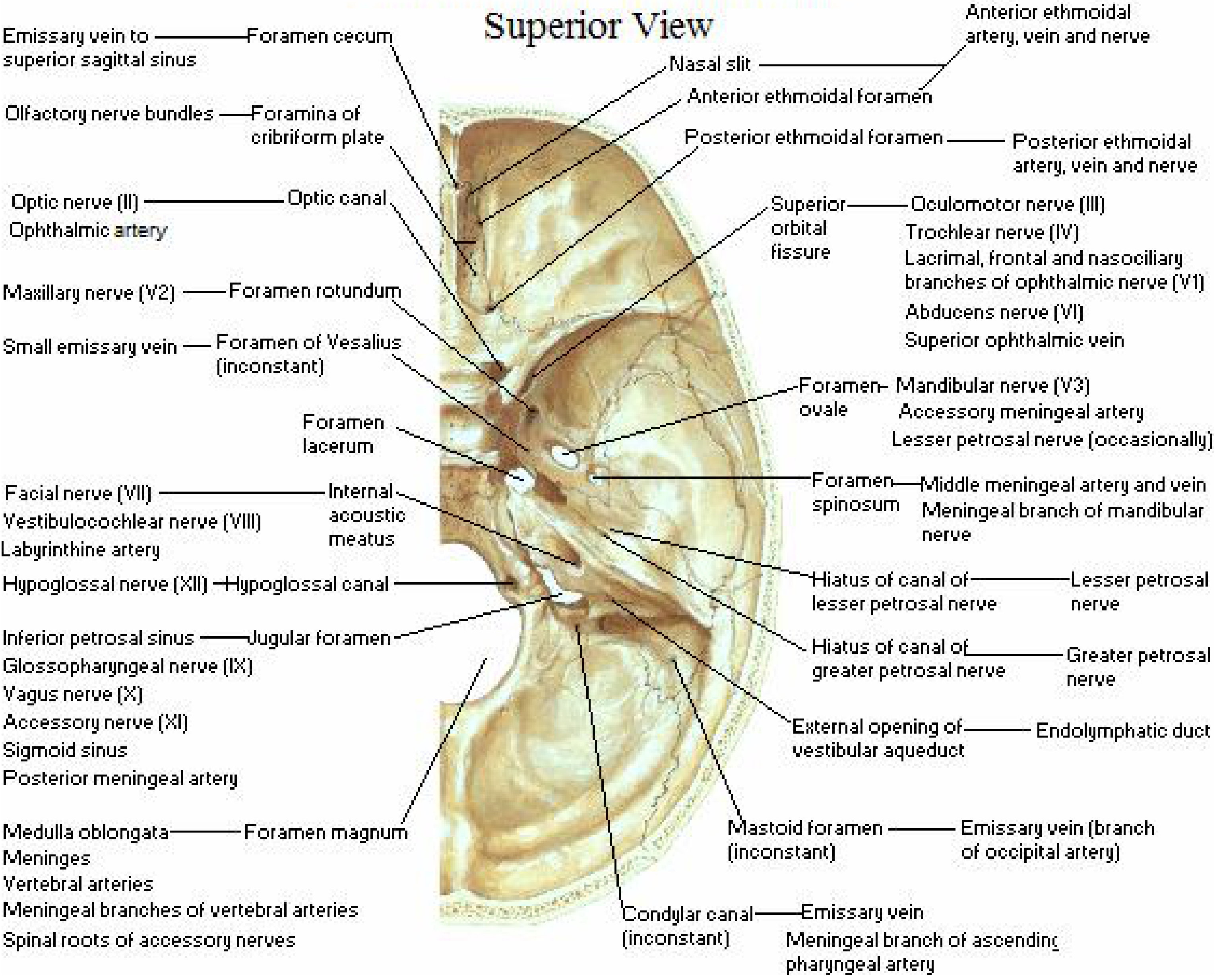
Quick Memory Aid (for OSPE)
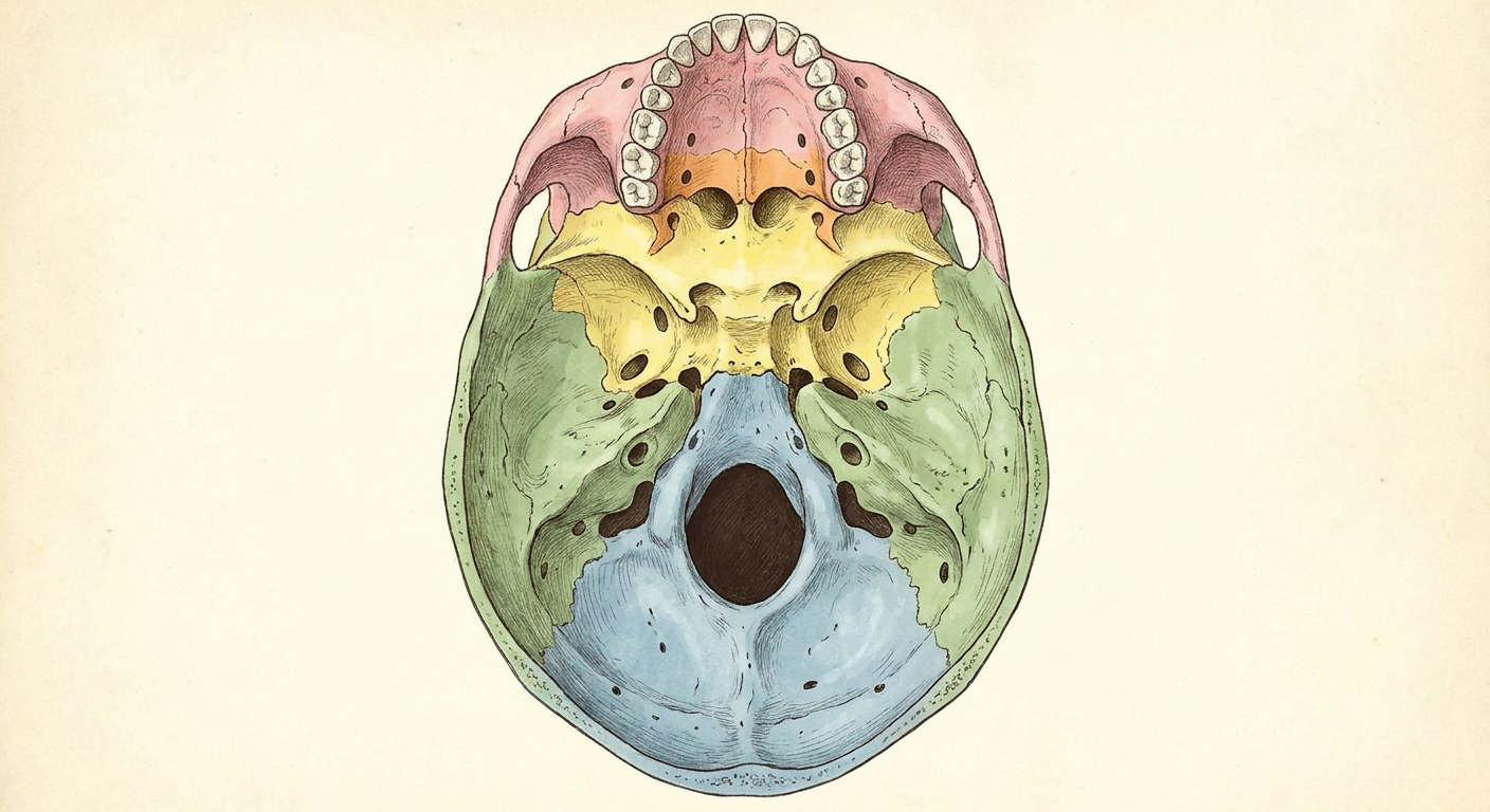
Images of base of sull with foramens visible but unlabelled
base of skull inferior view foramina unlabelled anatomy image
generate_image tool to create a clean, unlabelled diagram of the base of the skull showing foramina, suitable for OSPE spotting.Anatomical diagram of the base of the skull, inferior view (norma basalis), showing all major foramina clearly visible as holes and canals. The foramina to show include: foramen magnum (large central oval hole), jugular foramen (bilateral irregular holes posterolaterally), carotid canal openings (bilateral circular holes), foramen ovale (bilateral oval holes in greater wing of sphenoid), foramen spinosum (small bilateral holes), foramen lacerum (irregular bilateral holes), stylomastoid foramen (bilateral small holes), hypoglossal canal (bilateral holes near foramen magnum), greater palatine foramen (bilateral in palatine bone), lesser palatine foramina (small bilateral holes), incisive fossa (anterior midline). The skull bones are color-coded by bone (maxilla pink, sphenoid yellow, temporal bone green, occipital bone blue, palatine bone orange). All foramina are clearly visible as dark holes. NO text labels, NO arrows, NO numbers, NO annotations of any kind. Clean anatomical illustration, high detail, medical textbook style.
Base of Skull (Norma Basalis) - OSPE Images
Unlabelled - for OSPE Practice
Labelled Reference - Gray's Anatomy (Fig. 8.23)
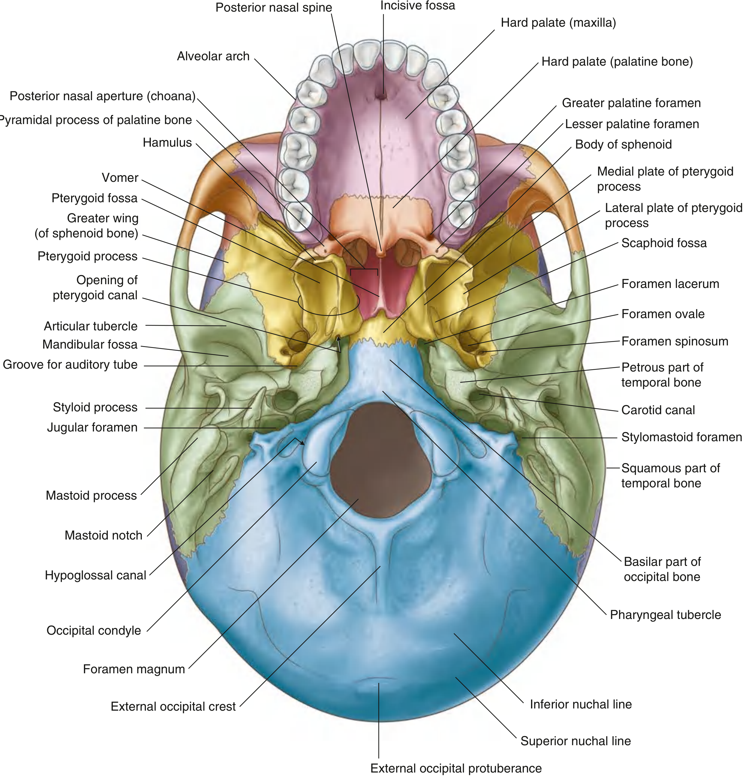
What to Identify (OSPE Spotting List)
| Region | Foramen to Identify |
|---|---|
| Anterior (palate area) | Incisive fossa, Greater palatine foramen, Lesser palatine foramina |
| Middle (sphenoid area) | Foramen ovale, Foramen spinosum, Foramen lacerum, Carotid canal, Opening of pterygoid canal |
| Posterior (temporal area) | Jugular foramen, Stylomastoid foramen, External acoustic meatus |
| Midline posterior | Foramen magnum, Hypoglossal canal, Condylar canal |