
Approach to aub in reproductive age group
"abnormal uterine bleeding"[MeSH Terms] AND management
PALM-COEIN classification abnormal uterine bleeding FIGO diagram
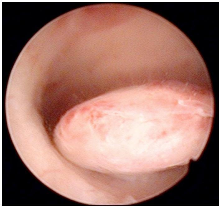
This diagnostic image is an intraoperative hysteroscopic photograph of the uterine cavity. The visual focus is a well-defined, oval-shaped pedunculated endometrial polyp protruding from the posterior uterine wall. The polyp demonstrates a smooth, glistening surface with a pale pink to white color, interspersed with focal areas of erythema and fine vertical striations across its body. Its morphology is slightly lobulated, and it is attached to the uterine wall by a distinct stalk. The surrounding endometrial tissue appears healthy, smooth, and pink, although there is a slight darkening near the base of the pedicle, likely representing minor blood accumulation. This clinical photograph illustrates a common structural cause of abnormal uterine bleeding (AUB), frequently categorized under the 'Polyp' component of the FIGO PALM-COEIN classification system. Such findings are critical in the diagnostic workup of perimenopausal and postmenopausal women to differentiate benign focal abnormalities from endometrial hyperplasia or malignancy.
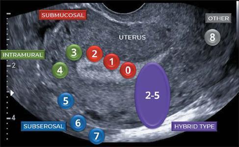
This diagnostic ultrasound image demonstrates the FIGO (International Federation of Gynecology and Obstetrics) classification system for uterine fibroids (leiomyomas). The image shows a grayscale sagittal view of a uterus with heterogeneous, hypoechoic texture. Numbered circular overlays are used to map fibroid locations relative to the uterine wall layers. Red markers 0, 1, and 2 identify 'Submucosal' fibroids (ranging from pedunculated intracavitary to ≥ 50% intramural). Green markers 3 and 4 identify 'Intramural' fibroids. Blue markers 5, 6, and 7 represent 'Subserosal' positions (ranging from ≥ 50% intramural to pedunculated subserosal). A large purple oval labeled '2-5' indicates the 'Hybrid Type' (contacting both the endometrium and serosa, but < 50% of each), and a gray marker '8' indicates 'Other' locations such as cervical or parasitic fibroids. This anatomical diagram serves as an educational guide for clinicians to determine surgical approaches, such as hysteroscopic versus laparoscopic myomectomy, based on lesion classification.
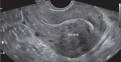
This diagnostic image is a transvaginal ultrasound (TVUS) showing a longitudinal view of a retroverted uterus. A prominent uterine leiomyoma (fibroid) is clearly visible and labeled within the anterior wall. The lesion is characterized as a solid, round, and well-defined mass. Visually, it presents as hypoechoic relative to the surrounding myometrium with a heterogeneous internal echo texture. According to the FIGO classification system for uterine fibroids, this is a type 2 submucosal fibroid, meaning it is partially intramural but has a significant component (at least 50%) projecting into the endometrial cavity, resulting in the displacement of the endometrial line. This imaging is clinically significant for evaluating causes of abnormal uterine bleeding and infertility, and for preoperative planning of hysteroscopic myomectomy.
Approach to Abnormal Uterine Bleeding (AUB) in the Reproductive Age Group
Definition and Normal Parameters
- Frequency: every 24-38 days
- Duration: 4-8 days of flow
- Volume: not interfering with quality of life
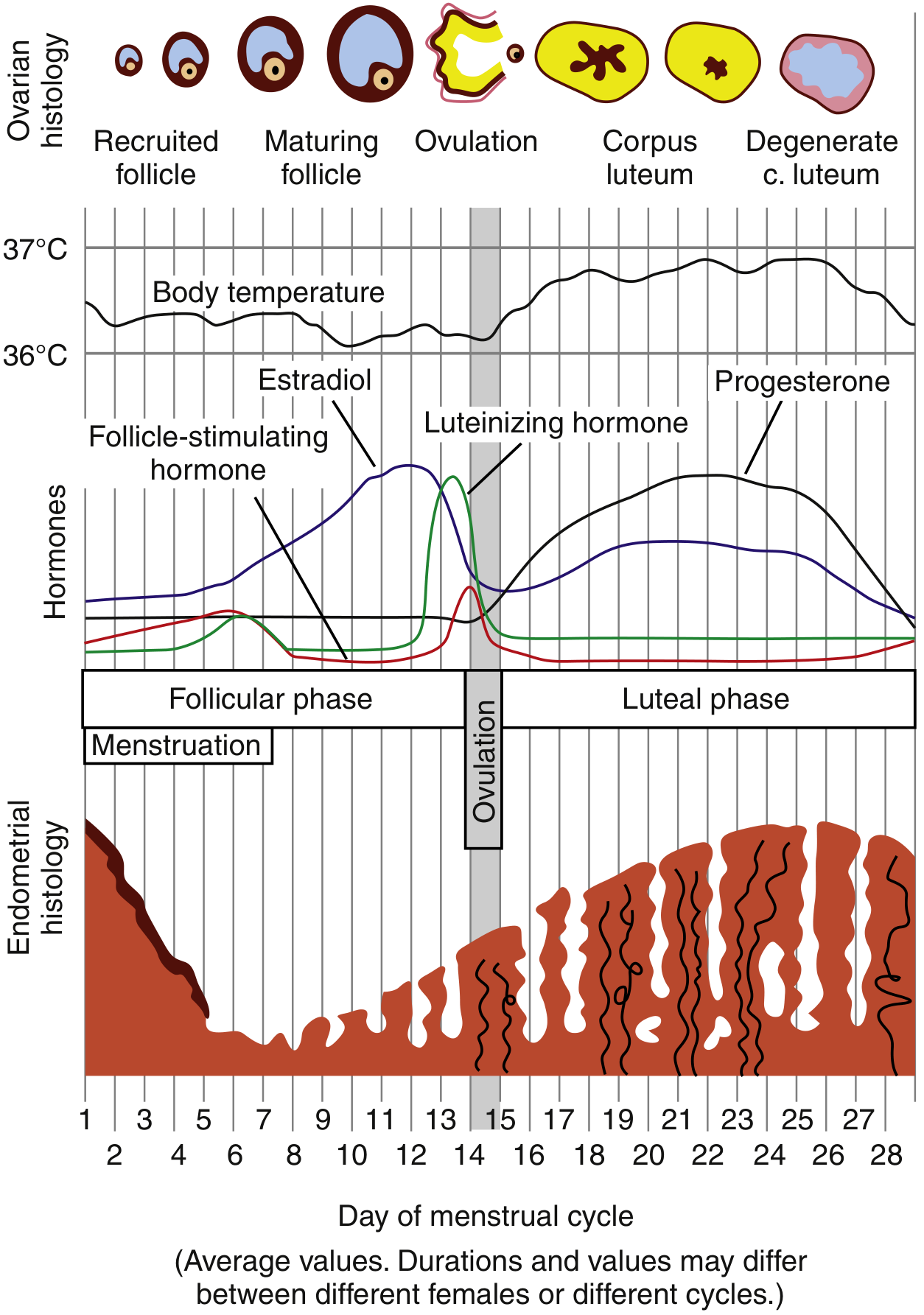
The Normal Menstrual Cycle (Context)
FIGO PALM-COEIN Classification (2011)
PALM - Structural Causes
| Category | Notes |
|---|---|
| AUB-P - Polyp | Endometrial or endocervical polyps; diagnosed on ultrasound/hysteroscopy |
| AUB-A - Adenomyosis | Historically a histologic diagnosis; now diagnosable on TVUS/MRI before hysterectomy |
| AUB-L - Leiomyoma | Present in up to 70-80% of women; submucosal leiomyomas most likely to cause bleeding. Subclassified as AUB-LSM (submucosal) and AUB-LO (other) |
| AUB-M - Malignancy & Hyperplasia | Endometrial hyperplasia (with or without atypia), endometrial carcinoma, cervical carcinoma |
COEIN - Non-structural Causes
| Category | Notes |
|---|---|
| AUB-C - Coagulopathy | Von Willebrand disease (most common, up to 13% of AUB cases), ITP, liver disease, anticoagulant use. Up to 20% of women with heavy menstrual bleeding have an underlying coagulopathy |
| AUB-O - Ovulatory dysfunction | Most common category in reproductive age. Includes PCOS, hyperprolactinemia, thyroid disorders, hypothalamic dysfunction (eating disorders, excessive exercise), perimenopause |
| AUB-E - Endometrial | Primary disorder of endometrial hemostasis (e.g., deficient local prostaglandin production, altered fibrinolysis) - a diagnosis of exclusion |
| AUB-I - Iatrogenic | Exogenous hormones (OCP breakthrough bleeding), anticoagulants, antipsychotics causing hyperprolactinemia, IUDs |
| AUB-N - Not yet classified | Rare entities not fitting other categories (e.g., arteriovenous malformation, myometrial hypertrophy) |
Causes Specific to the Reproductive Age Group
- Complications of pregnancy - threatened/incomplete/missed abortion, ectopic pregnancy, gestational trophoblastic disease (ALWAYS exclude first)
- Functional/anovulatory - PCOS (most common cause), hypothalamic dysfunction, hyperprolactinemia, thyroid disease
- Structural lesions - polyps, leiomyomas (especially submucosal), adenomyosis
- Coagulation disorders - Von Willebrand disease, ITP
- Endocrine disorders - diabetes mellitus, hypothyroidism/hyperthyroidism, Cushing syndrome, Addison disease
- Medications - OCP use, anticoagulants, antipsychotics
- Iatrogenic/Trauma - post-cesarean scar defect (niche), foreign body, coital trauma
- Malignancy - endometrial hyperplasia/carcinoma (less common in this age group but must be excluded in high-risk patients)
Clinical Evaluation
Step 1: History
- Characterize the bleeding: frequency, duration, volume (number of pads/tampons, presence of clots, flooding)
- Menstrual calendar - prospective charting may be required
- Relationship of bleeding to the cycle (intermenstrual, postcoital, or heavy regular menses)
- Obstetric history - last menstrual period, sexual activity, contraception
- Symptoms of pregnancy - any possibility must be excluded immediately
- Symptoms of anovulation - irregular cycles, no predictable PMS symptoms
- Symptoms of systemic disease - fatigue, weight change, heat/cold intolerance, galactorrhea
- Bleeding tendency - bruising, epistaxis, family history of bleeding disorder
- Medications - especially anticoagulants, hormonal therapy, antipsychotics
- Risk factors for endometrial cancer - obesity, PCOS, nulliparity, diabetes, family history (Lynch syndrome)
Step 2: Physical Examination
- General: signs of anemia (pallor, tachycardia), signs of hypovolemia in acute bleeding
- Signs of PCOS: obesity, acne, hirsutism, acanthosis nigricans
- Thyroid: nodules, enlargement
- Skin: petechiae, ecchymosis (coagulopathy)
- Abdomen: uterine or adnexal mass
- Pelvic exam: vaginal/cervical lesions, discharge, uterine size and tenderness, bimanual - fibroid uterus palpable
- Speculum: source of bleeding (cervical vs. uterine), cervical lesion requiring biopsy
Investigations
Essential (All Cases)
| Investigation | Purpose |
|---|---|
| Urine/serum beta-hCG | Exclude pregnancy - MANDATORY first test |
| Complete blood count | Assess anemia, thrombocytopenia |
| Coagulation screen (PT, aPTT, bleeding time) + von Willebrand factor panel | Exclude coagulopathy - especially if heavy bleeding since menarche |
| Thyroid function tests (TSH) | Exclude thyroid disease |
| Prolactin | Exclude hyperprolactinemia |
| Fasting blood glucose | Endocrine disorder |
| Pap smear / cervical screening | If not up to date |
| STI screen (Chlamydia, Gonorrhoea) | If clinically indicated |
Imaging
- Pelvic ultrasound (TVUS): First-line imaging. Evaluates endometrial thickness, polyps, fibroids (with FIGO subclassification), adenomyosis, ovarian pathology
- Saline infusion sonohysterography (SIS): Better visualization of intracavitary lesions (polyps, submucosal fibroids)
- MRI: For adenomyosis, complex leiomyoma mapping, or pre-operative planning
Endometrial Sampling - When Indicated
- Age ≥45 years with AUB (including intermenstrual bleeding)
- Age <45 years with risk factors:
- Unopposed estrogen exposure (obesity, PCOS)
- Persistent AUB or AUB refractory to medical management
- Elevated familial cancer risk (Lynch syndrome)
- Prolonged anovulatory bleeding
Management
Step 1: Resuscitate if Hemodynamically Unstable
Step 2: Medical Management (First-Line)
For Acute Heavy Anovulatory Bleeding (Hemodynamically Stable)
- Combined OCP: One pill every 6 hours for 5-7 days (off-label). Bleeding should cease within 24 hours. Warn the patient to expect heavy withdrawal bleed 2-4 days after stopping.
- Conjugated estrogens IV: 25 mg every 4 hours for up to 3 doses until bleeding ceases, then simultaneously start progestin (medroxyprogesterone acetate 5-10 mg/day for 10 days). Follow with cyclic OCP for at least 3 cycles.
- Progestin alone: Medroxyprogesterone acetate 5-10 mg/day, or norethindrone 5 mg/day, for 10 days - for mild-moderate anovulatory bleeding or where estrogen is contraindicated.
For Chronic/Ongoing Heavy Menstrual Bleeding
| Agent | Mechanism | Notes |
|---|---|---|
| Tranexamic acid 1 g QID for 4 days | Antifibrinolytic | FDA-approved for HMB; most effective non-hormonal option |
| NSAIDs (mefenamic acid, ibuprofen) | Reduce prostaglandin-mediated bleeding | Effective in ovulatory HMB |
| Combined OCP (cyclic) | Suppress endometrium | First-line if contraception also desired |
| Progestins (oral or injectable) | Oppose estrogen | Cyclic or continuous; useful in anovulatory AUB |
| Levonorgestrel IUD (LNG-IUS) | Local progestin - endometrial suppression | Highly effective for ovulatory AUB and leiomyoma-related bleeding; also provides contraception |
| GnRH agonists (leuprolide) | Medical menopause | Short-term use (max 6 months); for preoperative fibroid reduction; causes hypoestrogenism |
| GnRH antagonists (relugolix 40 mg/day, elagolix 300 mg BD) | Rapid medical menopause | Newer agents for fibroid-related AUB |
Step 3: Cause-Specific Treatment
| Cause | Specific Treatment |
|---|---|
| PCOS (AUB-O) | OCP to regulate cycles; metformin if insulin resistant; ovulation induction if fertility desired |
| Thyroid/Prolactin disorder | Treat the underlying endocrinopathy |
| Von Willebrand disease (AUB-C) | Tranexamic acid, DDAVP, VWF concentrates; avoid ASA/NSAIDs |
| Endometrial polyp (AUB-P) | Hysteroscopic polypectomy |
| Submucosal fibroid (AUB-LSM) | Hysteroscopic myomectomy; UAE; GnRH agonist pre-operatively |
| Intramural/subserosal fibroids | Laparoscopic/open myomectomy if symptomatic; UAE |
| Adenomyosis (AUB-A) | LNG-IUS, GnRH agonist; hysterectomy definitive |
| Endometrial hyperplasia without atypia | Progestin therapy (oral or LNG-IUS) |
| Endometrial hyperplasia with atypia | Hysterectomy (high risk of progression to carcinoma) |
| Iatrogenic (breakthrough bleeding on OCP) | Change formulation; ensure adherence |
Step 4: Surgical Management (Second-Line or Refractory Cases)
- Endometrial ablation: Effective in up to 70-80% of cases; not appropriate for women desiring future fertility. Note: 29% undergo eventual hysterectomy by 5 years.
- Uterine artery embolization (UAE): For fibroid-related AUB in women wishing to avoid surgery; 31% ultimately undergo hysterectomy.
- Hysterectomy: Definitive treatment; reserved for patients who fail or cannot tolerate medical therapy, have completed family, or have malignancy. Endometrial sampling should always precede hysterectomy.
Special Considerations in the Reproductive Age Group
- Always exclude pregnancy first - ectopic pregnancy is a life-threatening emergency that mimics AUB.
- Coagulopathy screening is indicated when: heavy bleeding since menarche, family history of bleeding disorder, signs of systemic bleeding, or on medications associated with AUB. Up to 20% of women with heavy periods have an undiagnosed coagulopathy.
- Fertility preservation guides choice of treatment - medical management and uterine-preserving procedures are preferred over hysterectomy in women who desire future pregnancy.
- PCOS is the most common cause of anovulatory AUB in this age group. Unopposed estrogen in PCOS leads to endometrial proliferation and irregular shedding, and increases long-term risk of endometrial hyperplasia/cancer.
- Endometrial sampling before any surgical treatment is non-negotiable - D&C alone does not adequately treat AUB and is not required before hysterectomy, but a sample must be obtained.
Summary Algorithm
AUB in Reproductive Age
        |
        v
Exclude PREGNANCY (beta-hCG) ← ALWAYS first
        |
        v
Hemodynamic assessment
    Unstable → Resuscitate → IV estrogens / urgent D&C
    Stable ↓
        |
        v
History + Examination + Labs (CBC, TSH, Prolactin, Coags, VWF)
        |
        v
Pelvic Ultrasound (TVUS)
        |
   Structural (PALM)?           Nonstructural (COEIN)?
   Polyp → Hysteroscopy         Anovulatory (PCOS, thyroid) → Medical Rx
   Fibroid → Myomectomy/UAE     Coagulopathy → Haematology referral
   Adenomyosis → LNG-IUS/Hyst  Iatrogenic → Modify medication
   Hyperplasia/Ca → Biopsy      Endometrial cause → Tranexamic acid / NSAIDs
        |
        v
Endometrial biopsy if age ≥45 or risk factors
        |
        v
Medical treatment FIRST (OCP / Progestins / LNG-IUS / Tranexamic acid)
        |
        v
Failure → Surgical (Ablation / Myomectomy / UAE / Hysterectomy)