Explain trails sign in trachea

Reading File
Finding Sources
Searching Images

trails sign trachea radiology

This diagnostic image is a focused frontal view of a chest radiograph (X-ray) centered on the mediastinum and right hilar region. The primary finding is an 'air crescent sign' located in the right hilum, indicated by three black arrows. The sign consists of a curvilinear, crescent-shaped lucency (airspace) that partially surrounds a rounded soft-tissue opacity within a cavitary lesion. This radiographic phenomenon typically represents air trapped between a central mass and the wall of a cavity. While commonly associated with invasive pulmonary aspergillosis (Monod sign), the clinical context here demonstrates the sign in the right hilar vessels, characteristic of pneumomediastinum or pulmonary interstitial emphysema. The image highlights key anatomical landmarks including the trachea, vertebral column, and cardiac silhouette, serving as an educational example of identifying subtle gas patterns in thoracic radiology.

This diagnostic image is a focused frontal view of a chest radiograph (X-ray) centered on the mediastinum and right hilar region. The primary finding is an 'air crescent sign' located in the right hilum, indicated by three black arrows. The sign consists of a curvilinear, crescent-shaped lucency (airspace) that partially surrounds a rounded soft-tissue opacity within a cavitary lesion. This radiographic phenomenon typically represents air trapped between a central mass and the wall of a cavity. While commonly associated with invasive pulmonary aspergillosis (Monod sign), the clinical context here demonstrates the sign in the right hilar vessels, characteristic of pneumomediastinum or pulmonary interstitial emphysema. The image highlights key anatomical landmarks including the trachea, vertebral column, and cardiac silhouette, serving as an educational example of identifying subtle gas patterns in thoracic radiology.

This diagnostic image is a posteroanterior (PA) view chest X-ray illustrating a significant abnormality of the cardiac silhouette. A distinct, rounded prominence is visible along the left cardiac border, suggestive of an enlarged pulmonary trunk or a localized mass. A key radiographic feature is the 'crescent sign'—a thin, radiolucent (dark) rim located at the superior aspect of the prominence, indicating air or low-density interface between the lesion and the adjacent lung or vascular structures. The trachea appears midline, and the right lung field shows no gross consolidations, although the right hilar region appears prominent. This visual is characteristic of pulmonary artery dilation or specific cystic lesions (such as a hydatid cyst) and serves as an educational example of the crescent sign in thoracic radiology for medical students and clinicians specializing in pulmonology and radiology.
This diagnostic image is a posteroanterior (PA) view chest X-ray illustrating a significant abnormality of the cardiac silhouette. A distinct, rounded prominence is visible along the left cardiac border, suggestive of an enlarged pulmonary trunk or a localized mass. A key radiographic feature is the 'crescent sign'—a thin, radiolucent (dark) rim located at the superior aspect of the prominence, indicating air or low-density interface between the lesion and the adjacent lung or vascular structures. The trachea appears midline, and the right lung field shows no gross consolidations, although the right hilar region appears prominent. This visual is characteristic of pulmonary artery dilation or specific cystic lesions (such as a hydatid cyst) and serves as an educational example of the crescent sign in thoracic radiology for medical students and clinicians specializing in pulmonology and radiology.

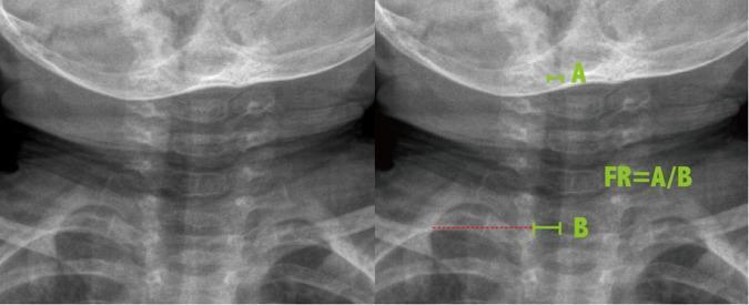
Antero-posterior (AP) soft tissue neck radiograph of a pediatric patient, presented as side-by-side images (unannotated and annotated) to demonstrate the methodology for calculating the Frontal Ratio (FR) of tracheal narrowing. The radiograph displays the cervical spine, base of the skull, clavicles, and the air-filled tracheal column. The annotated image on the right illustrates the measurement technique for subglottic stenosis: label 'A' marks the narrowest diameter of the subglottic trachea, while label 'B' indicates the reference tracheal width measured at the level of the clavicles. The mathematical relationship is provided as FR = A/B. This diagnostic imaging technique is typically used in the evaluation of upper airway obstruction, such as croup (laryngotracheobronchitis), where the 'steeple sign'—the characteristic tapering of the subglottic airway—is a hallmark finding. This material is designed for radiology and pediatric educational purposes to quantify the degree of airway constriction.
Antero-posterior (AP) soft tissue neck radiograph of a pediatric patient, presented as side-by-side images (unannotated and annotated) to demonstrate the methodology for calculating the Frontal Ratio (FR) of tracheal narrowing. The radiograph displays the cervical spine, base of the skull, clavicles, and the air-filled tracheal column. The annotated image on the right illustrates the measurement technique for subglottic stenosis: label 'A' marks the narrowest diameter of the subglottic trachea, while label 'B' indicates the reference tracheal width measured at the level of the clavicles. The mathematical relationship is provided as FR = A/B. This diagnostic imaging technique is typically used in the evaluation of upper airway obstruction, such as croup (laryngotracheobronchitis), where the 'steeple sign'—the characteristic tapering of the subglottic airway—is a hallmark finding. This material is designed for radiology and pediatric educational purposes to quantify the degree of airway constriction.

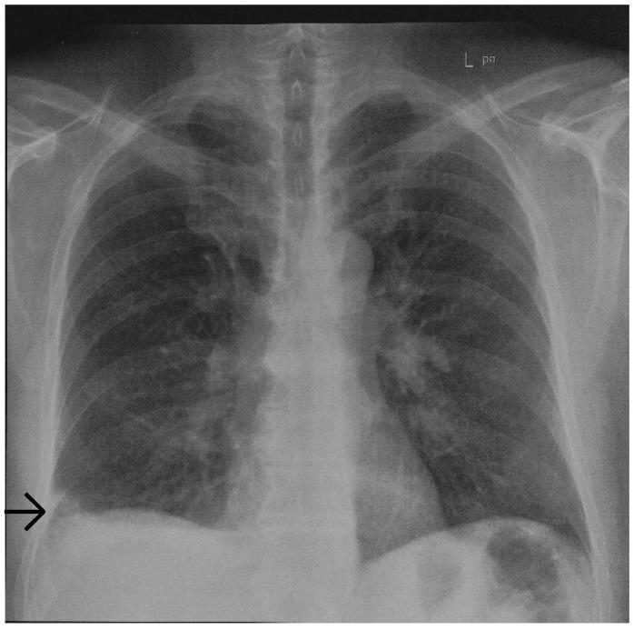
This diagnostic image is a posterior-anterior (PA) chest radiograph. The primary finding is a focal, wedge-shaped area of increased opacity located in the periphery of the right lower lung field, situated just above the diaphragm and abutting the pleura. This radiographic sign, known as a Hampton’s hump, is characterized by its rounded apex pointing toward the hilum and its broad base against the pleural surface, classically indicating pulmonary infarction secondary to pulmonary embolism. The rest of the lung fields appear largely clear with normal lucency and branching pulmonary vasculature. The cardiac silhouette is centrally located and of normal size. Bony structures, including the ribs and clavicles, are intact, and the costophrenic angles are well-visualized. The trachea is midline, and the diaphragmatic contours are sharp, except where the right-sided consolidation overlaps the lateral basal segment. This image serves as a key educational example of a specific radiographic sign associated with vascular pulmonary pathology and is suitable for teaching diagnostic radiology and emergency medicine triage.

This diagnostic image is a posterior-anterior (PA) chest radiograph. The primary finding is a focal, wedge-shaped area of increased opacity located in the periphery of the right lower lung field, situated just above the diaphragm and abutting the pleura. This radiographic sign, known as a Hampton’s hump, is characterized by its rounded apex pointing toward the hilum and its broad base against the pleural surface, classically indicating pulmonary infarction secondary to pulmonary embolism. The rest of the lung fields appear largely clear with normal lucency and branching pulmonary vasculature. The cardiac silhouette is centrally located and of normal size. Bony structures, including the ribs and clavicles, are intact, and the costophrenic angles are well-visualized. The trachea is midline, and the diaphragmatic contours are sharp, except where the right-sided consolidation overlaps the lateral basal segment. This image serves as a key educational example of a specific radiographic sign associated with vascular pulmonary pathology and is suitable for teaching diagnostic radiology and emergency medicine triage.

Searching the Web

trails sign trachea radiology finding explanation

Finding Sources
Searching the Web

"trails sign" trachea thyroid goiter radiology CT

Finding Sources
Searching the Web

"trails sign" OR "trail sign" trachea ENT surgery neck

Finding Sources
Finding Sources
Searching Images

tracheal deviation sternocleidomastoid muscle neck anatomy
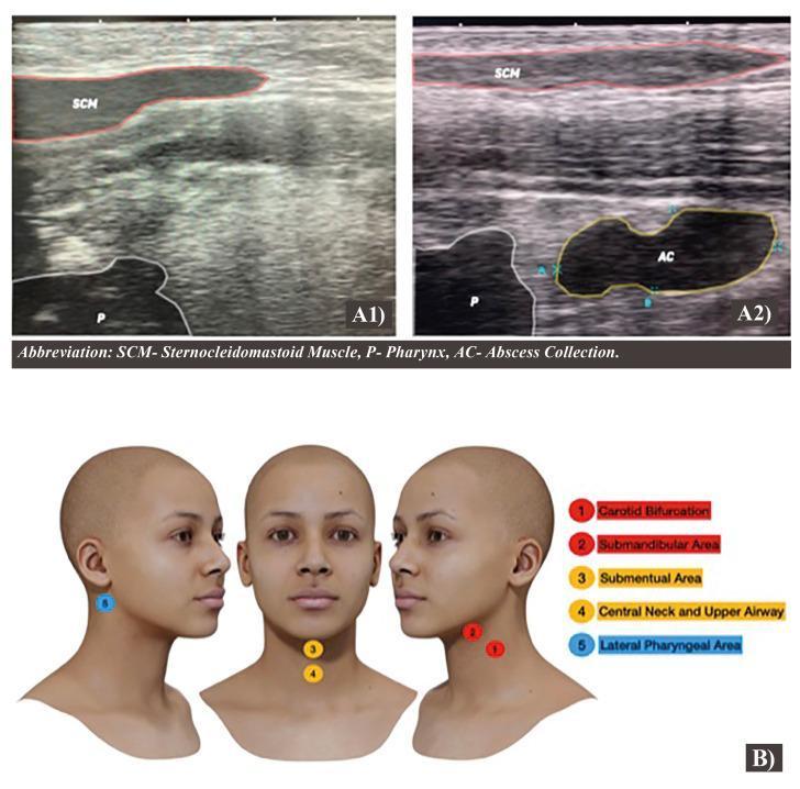
This composite educational graphic combines sagittal ultrasonography images and a 3D anatomical reproduction to illustrate a diagnostic protocol for the lateral pharyngeal area. Section A presents a side-by-side comparison of normal (A1) and pathological (A2) anatomy. In the normal scan, the sternocleidomastoid muscle (SCM) is visible superficially, with the pharynx (P) identified via the airway column shadow. In the affected scan (A2), a distinct, hypoechoic abscess collection (AC) is visualized, causing anatomical distortion and deviation of the pharyngeal structures. Section B provides a 3D anatomical model of the head and neck, mapping five key examination zones: (1) carotid bifurcation, (2) submandibular area, (3) submental area, (4) central neck and upper airway, and (5) lateral pharyngeal area. This resource is designed for clinical imaging instruction, demonstrating how point-of-care ultrasound can detect deep neck space infections and localized collections while outlining a systematic scanning protocol for the cervical region.
This composite educational graphic combines sagittal ultrasonography images and a 3D anatomical reproduction to illustrate a diagnostic protocol for the lateral pharyngeal area. Section A presents a side-by-side comparison of normal (A1) and pathological (A2) anatomy. In the normal scan, the sternocleidomastoid muscle (SCM) is visible superficially, with the pharynx (P) identified via the airway column shadow. In the affected scan (A2), a distinct, hypoechoic abscess collection (AC) is visualized, causing anatomical distortion and deviation of the pharyngeal structures. Section B provides a 3D anatomical model of the head and neck, mapping five key examination zones: (1) carotid bifurcation, (2) submandibular area, (3) submental area, (4) central neck and upper airway, and (5) lateral pharyngeal area. This resource is designed for clinical imaging instruction, demonstrating how point-of-care ultrasound can detect deep neck space infections and localized collections while outlining a systematic scanning protocol for the cervical region.

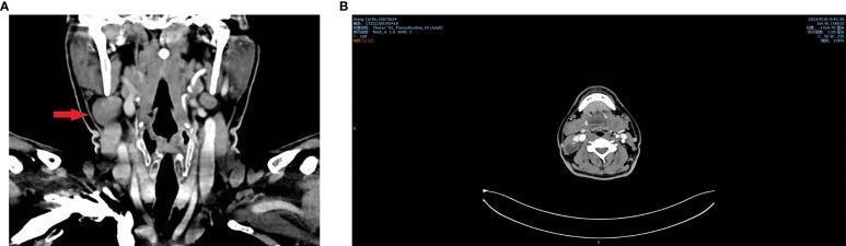
Contrast-enhanced CT scan of the neck presented in coronal (A) and horizontal/axial (B) views. The images demonstrate a well-defined, heterogeneous solid-cystic mass located in the right lateral neck (Level II/III). In the coronal view, the lesion is indicated by a red arrow, positioned lateral to the carotid sheath and posterior-lateral to the thyroid gland. The axial view confirms the mass's position lateral to the trachea and deep to the sternocleidomastoid muscle. Internal features include areas of low attenuation representing cystic components and hyperdense foci consistent with calcification. There is no evidence of significant tracheal deviation or invasion of the adjacent major cervical vasculature. Bilateral small cervical lymph nodes are also visible. This imaging is characteristic of secondary pathologies such as branchial cleft cysts or metastatic papillary thyroid carcinoma with cystic changes. The educational focus is on identifying regional neck masses, their anatomical relationships to the carotid sheath, and recognizing heterogeneous cystic-solid internal morphology on diagnostic radiology.
Contrast-enhanced CT scan of the neck presented in coronal (A) and horizontal/axial (B) views. The images demonstrate a well-defined, heterogeneous solid-cystic mass located in the right lateral neck (Level II/III). In the coronal view, the lesion is indicated by a red arrow, positioned lateral to the carotid sheath and posterior-lateral to the thyroid gland. The axial view confirms the mass's position lateral to the trachea and deep to the sternocleidomastoid muscle. Internal features include areas of low attenuation representing cystic components and hyperdense foci consistent with calcification. There is no evidence of significant tracheal deviation or invasion of the adjacent major cervical vasculature. Bilateral small cervical lymph nodes are also visible. This imaging is characteristic of secondary pathologies such as branchial cleft cysts or metastatic papillary thyroid carcinoma with cystic changes. The educational focus is on identifying regional neck masses, their anatomical relationships to the carotid sheath, and recognizing heterogeneous cystic-solid internal morphology on diagnostic radiology.

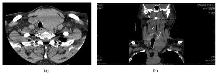
Non-contrast computed tomography (CT) of the neck and superior mediastinum presented in axial (a) and coronal (b) views. The images demonstrate a large, well-circumscribed, heterogeneous soft-tissue mass arising from the left thyroid lobe. In the axial view, the mass causes significant mass effect, resulting in marked rightward tracheal deviation and mild luminal compression. The coronal reconstruction highlights the vertical extent of the lesion, approximately 7.2 cm in sagittal height, showing early substernal extension into the superior mediastinum. The mass is positioned anterior to the vertebral column and displaces the left sternocleidomastoid muscle laterally. The imaging is consistent with a large thyroid goiter or neoplasm, such as Hürthle cell carcinoma, requiring assessment for tracheal patency and potential surgical planning. The surrounding vascular structures and cervical musculature are visible, serving as anatomical landmarks for the extent of the pathology.

Non-contrast computed tomography (CT) of the neck and superior mediastinum presented in axial (a) and coronal (b) views. The images demonstrate a large, well-circumscribed, heterogeneous soft-tissue mass arising from the left thyroid lobe. In the axial view, the mass causes significant mass effect, resulting in marked rightward tracheal deviation and mild luminal compression. The coronal reconstruction highlights the vertical extent of the lesion, approximately 7.2 cm in sagittal height, showing early substernal extension into the superior mediastinum. The mass is positioned anterior to the vertebral column and displaces the left sternocleidomastoid muscle laterally. The imaging is consistent with a large thyroid goiter or neoplasm, such as Hürthle cell carcinoma, requiring assessment for tracheal patency and potential surgical planning. The surrounding vascular structures and cervical musculature are visible, serving as anatomical landmarks for the extent of the pathology.

Loading Image
Loading Image
I now have everything needed for a thorough answer. Here is the full explanation:

Trail's Sign (Trachea)

Definition

Trail's sign is a clinical sign in which shift of the trachea produces prominence of the sternal (or clavicular) head of the sternocleidomastoid (SCM) muscle on the side toward which the trachea has deviated.
It is a bedside physical examination finding used to detect and confirm tracheal deviation.

Anatomical Basis

The key to understanding this sign lies in the arrangement of the deep cervical fascia:
  • The middle (pretracheal/visceral) layer of the deep cervical fascia encloses the visceral structures of the neck - the trachea, esophagus, and thyroid gland. It also wraps around the clavicular (sternal) heads of both sternocleidomastoid muscles on either side.
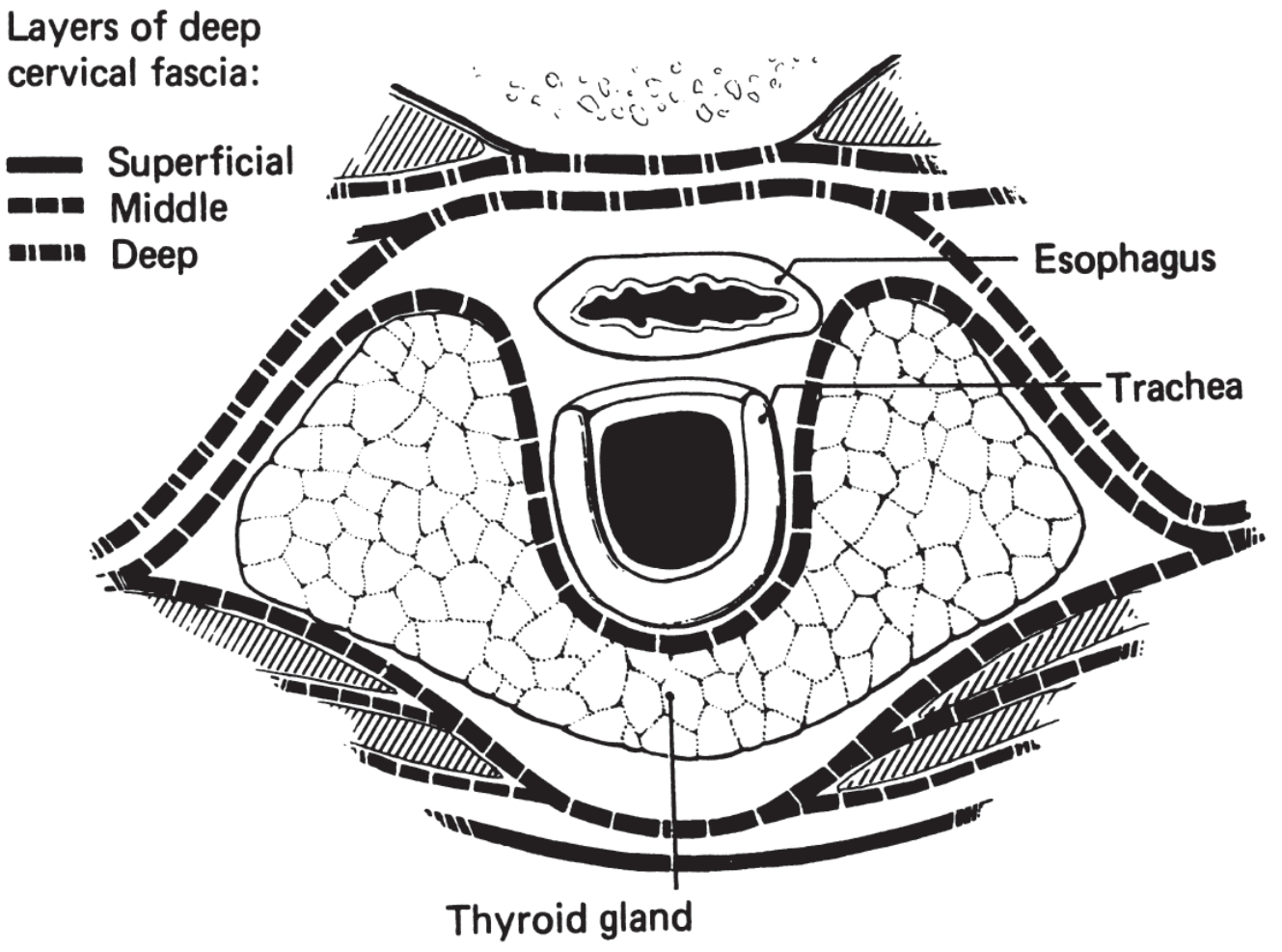
Layers of deep cervical fascia - cross-section showing the pretracheal fascia enclosing the trachea, esophagus, and thyroid gland bilaterally
(K J Lee's Essential Otolaryngology - cross-section of cervical fascia)
When the trachea is in its normal midline position, the pretracheal fascia exerts equal bilateral tension on both SCM clavicular heads, holding them relatively flat against the neck.

Mechanism

When a pathological process displaces the trachea to one side (e.g., to the right):
  1. The pretracheal fascia is pulled to the right along with the trachea.
  2. This causes the fascia on the right side to become slack/relaxed.
  3. The right SCM clavicular head, now no longer held taut against the neck by the fascia, bulges forward and becomes visibly prominent.
  4. On the opposite (left) side, the fascia is put under tension, and the SCM head appears flattened.
In summary: The sternal/clavicular head of the SCM is more prominent on the same side to which the trachea is deviated.

How to Elicit the Sign

  • Ask the patient to sit upright, with the neck slightly extended.
  • Inspect and palpate the lower neck just above the clavicles.
  • Palpate the trachea for deviation.
  • Look for a prominent, forward-bulging sternal/clavicular head of the SCM on the side of tracheal shift.
  • Compare both sides.

Clinical Significance

Trail's sign is useful when tracheal deviation is subtle and may not be immediately obvious on palpation alone. The bulging SCM head serves as an additional visual clue pointing to the direction of shift.
Causes of tracheal deviation (and therefore Trail's sign):
Pushed Away (Contralateral)Pulled Toward (Ipsilateral)
Tension pneumothoraxLung collapse / atelectasis
Large pleural effusionPulmonary fibrosis
Large goiter / thyroid massPneumonectomy
Mediastinal tumorPleural adhesions
Aortic aneurysm
The CT image below shows a large thyroid mass causing rightward tracheal deviation - the clinical correlate would be a prominent right SCM clavicular head (positive Trail's sign on the right):
CT neck showing large left thyroid mass with marked rightward tracheal deviation and lateral displacement of left sternocleidomastoid

Key Point to Remember

Trail's sign = Prominent SCM clavicular head on the SAME side as tracheal deviation
This is the opposite of what one might intuitively expect (you might think the SCM would be stretched taut and more prominent on the opposite side). The sign works because the relaxation of the pretracheal fascia ipsilaterally allows the muscle head to fall forward, not because of active muscle contraction.

Sources: Medicowesome - Trail's Sign | K J Lee's Essential Otolaryngology (pretracheal fascia anatomy) | Cummings Otolaryngology Head and Neck Surgery (middle layer of deep cervical fascia)
This is a shared conversation. Sign in to Orris to start your own chat.