
Part of corpus callosum
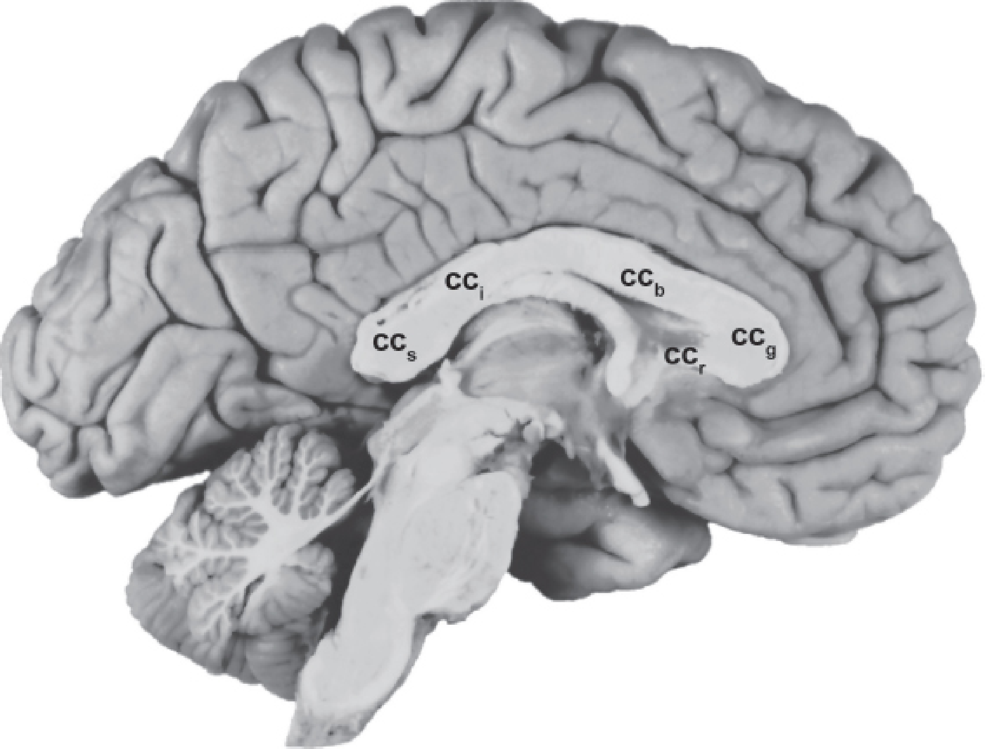
The corpus callosum has 4 main parts (from anterior to posterior):
- Rostrum - the most anterior, inferior part
- Genu - the "knee," the anterior bend
- Body (Trunk) - the large middle section
- Splenium - the posterior, rounded end
Some sources also describe the isthmus as a narrowed segment between the body and splenium.
Searching sources...
Reading File
Finding Sources
Searching Images
corpus callosum parts diagram anatomy genu splenium rostrum body
Finding Sources
Reading File
Reading File
Reading File
Loading Image
Here is a complete, textbook-sourced breakdown of the parts of the corpus callosum:
Parts of the Corpus Callosum
The corpus callosum is the largest fiber bundle in the brain (~300 million axons), connecting the two cerebral hemispheres. It is divided into 5 parts (from anterior/inferior to posterior), best seen on a midsagittal section:
| # | Part | Location | Key Features |
|---|---|---|---|
| 1 | Rostrum | Anteroinferior | The most inferior anterior part; thin, curves backward toward the lamina terminalis |
| 2 | Genu | Anterior "knee" | Curves anteriorly and dorsally; fibers form the forceps minor connecting the two frontal lobes |
| 3 | Body (Trunk) | Long middle section | The largest part; runs horizontally; fibers form the tapetum laterally |
| 4 | Isthmus | Narrow junction | The constriction between the body and splenium; sometimes counted separately |
| 5 | Splenium | Posterior, enlarged rounded end | The thickest part; fibers form the forceps major connecting the two occipital/temporal lobes |
Fiber Bundles by Region
- Genu & splenium - carry higher-order cognitive, sensory information from prefrontal, temporal, and parietal cortices
- Body & isthmus - carry visual, auditory, and somatosensory information
Development Note
The corpus callosum develops from posterior genu -> body -> splenium -> anterior genu -> rostrum (all present by ~20 weeks' gestation, but continues thickening postnatally). This is clinically important: if the splenium and rostrum are present but the body/genu are absent, it suggests destruction rather than failure of development.
Mnemonic
"Really Good Bodies In Sports" = Rostrum, Genu, Body, Isthmus, Splenium (anterior to posterior)
Sources: Kaplan & Sadock's Comprehensive Textbook of Psychiatry; Grainger & Allison's Diagnostic Radiology; Gray's Anatomy for Students
Supranuclear facial palsy
In supranuclear (UMN) facial palsy, the upper face is spared and only the lower face is weak. This is because the upper facial nucleus (forehead/eye) receives bilateral cortical input, while the lower facial nucleus receives only contralateral input. It is caused by lesions above the facial nerve nucleus - typically stroke, tumor, or MS affecting the corticobulbar tract.
Searching sources...
Finding Sources
Searching Images
supranuclear facial palsy upper lower motor neuron facial nerve anatomy
Reading File
Reading File
Reading File
Loading Image
Loading Image
Finding Sources
Here is a complete account of supranuclear facial palsy, drawn from multiple authoritative textbooks:
Supranuclear (UMN) Facial Palsy
Anatomy - Why the Upper Face Is Spared
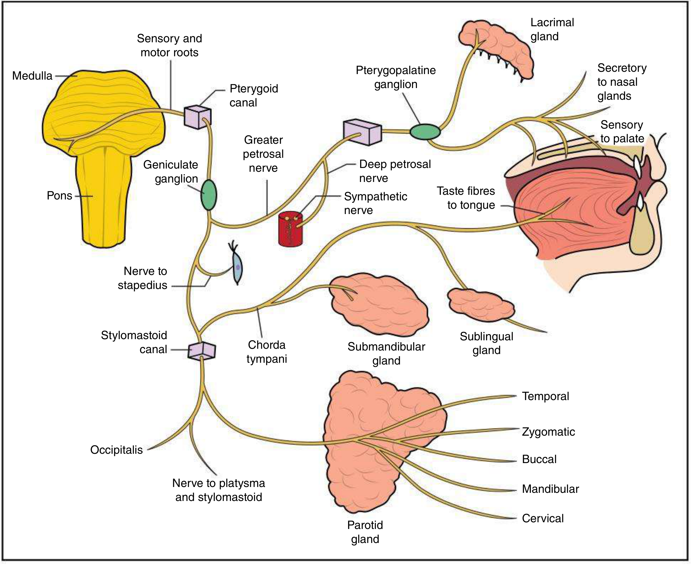
The key is dual cortical representation of the upper face:
- The upper facial nucleus (supplies frontalis, orbicularis oculi) receives corticobulbar input from both hemispheres (bilateral)
- The lower facial nucleus (supplies lower face muscles) receives corticobulbar input only from the contralateral hemisphere
So when one hemisphere or its descending corticobulbar tract is damaged, the lower face loses all input but the upper face still has input from the intact opposite side.
Clinical Features
| Feature | Supranuclear (UMN) | Nuclear/Peripheral (LMN) |
|---|---|---|
| Side affected | Contralateral to lesion | Ipsilateral to lesion |
| Upper face (forehead, eye) | Spared (bilateral cortical input) | Paralyzed |
| Lower face (mouth, cheek) | Paralyzed | Paralyzed |
| Eye closure | Intact | Incomplete (lagophthalmos) |
| Forehead wrinkling | Intact | Lost |
| Taste, lacrimation, hyperacusis | Absent (pure motor lesion) | Present if lesion is proximal |
| Emotional vs voluntary dissociation | Present (see below) | Absent |
| Corneal reflex | Intact or hyperactive | May be reduced |
| Associated signs | Hemiplegia, aphasia | Depends on level |
Emotional-Voluntary Dissociation
A hallmark of supranuclear facial palsy - as noted in Harrison's:
"In supranuclear lesions, there may be a dissociation of emotional and voluntary facial movements."
- Volitional pathway (from motor cortex via corticobulbar tract) - damaged in UMN lesion → patient cannot voluntarily smile on the affected side
- Emotional/involuntary pathway (from limbic system via extrapyramidal connections) - often intact → spontaneous emotional smile may be symmetric
The reverse also exists: in some basal ganglia or subcortical lesions, emotional smile is lost but voluntary smile is preserved ("mimic palsy").
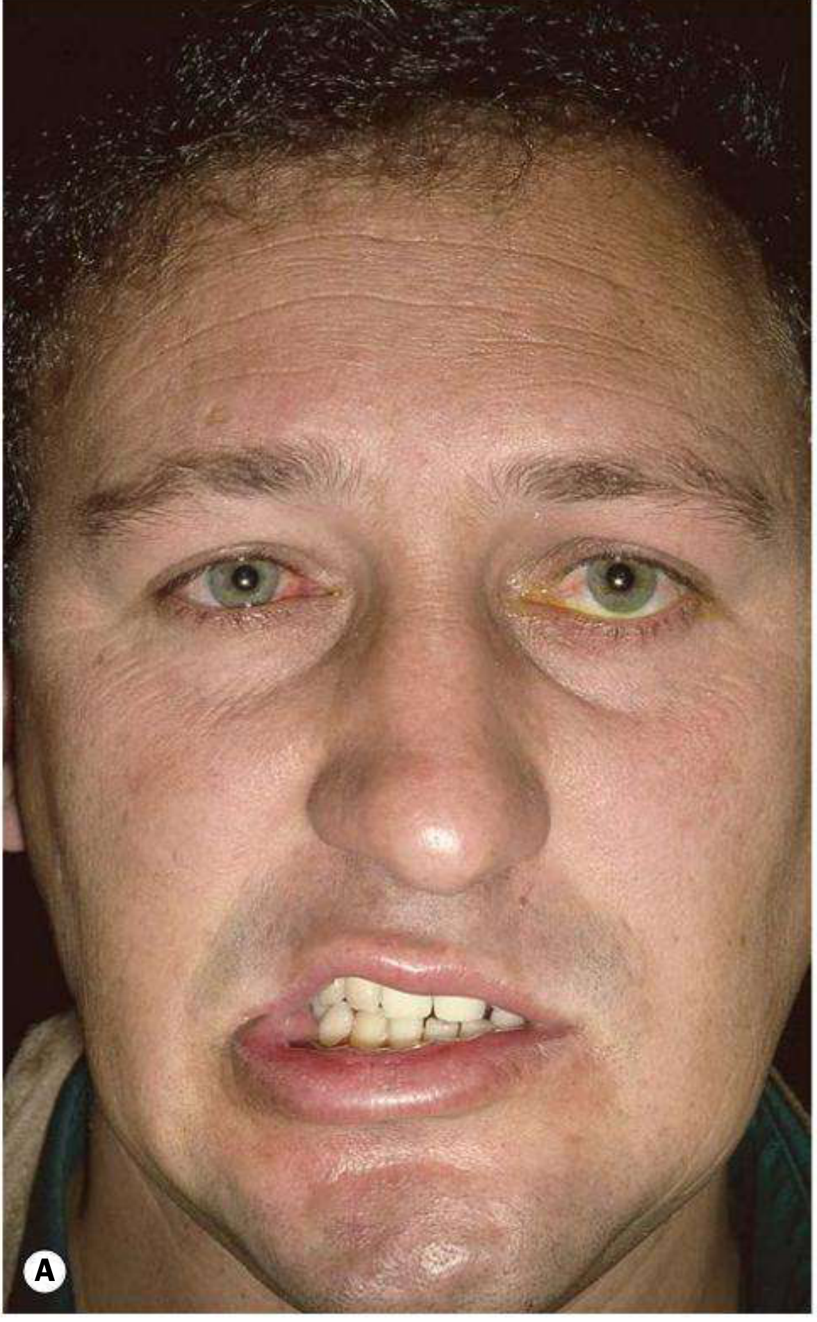
Clinical photo - Left UMN (Supranuclear) Facial Palsy
The image below (A) shows a left upper motor neurone palsy secondary to cerebral trauma. Note the forehead is symmetric (upper face spared) but there is drooping of the left lower face:
Localization of Lesion
The lesion is above the facial nerve nucleus (in the pons). Common sites and associated signs:
| Lesion Level | Signs |
|---|---|
| Motor cortex / internal capsule | Contralateral lower face palsy + hemiplegia ± aphasia (dominant side) |
| Pons (above nucleus) | Contralateral lower face palsy + ipsilateral CN VI, PPRF gaze palsy |
| Below the nucleus (LMN) | Complete ipsilateral face palsy (upper + lower) |
Causes of Supranuclear Facial Palsy
- Stroke (most common) - MCA territory infarct or hemorrhage in internal capsule
- Brain tumor - compressing motor cortex or corticobulbar fibers
- Multiple sclerosis - demyelinating plaque in corticobulbar tract
- Traumatic brain injury
Key Distinguishing Point (Exam Favorite)
"Forehead sparing = UMN (supranuclear) lesion" "Forehead involved = LMN (nuclear or peripheral) lesion"
This is because the forehead (frontalis) has bilateral cortical supply - the intact opposite hemisphere keeps it working even when one side is damaged.
Sources: Harrison's Principles of Internal Medicine 22E; Kanski's Clinical Ophthalmology 10th ed.; Plum and Posner's Diagnosis and Treatment of Stupor and Coma
This is a shared conversation. Sign in to Orris to start your own chat.