

What is this diagnose treatment
tinea barbae annular pustular ringworm face neck
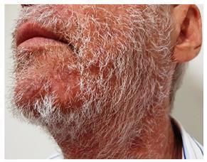
This clinical photograph displays the lower face and neck of an elderly male patient with a white beard, showcasing the resolution phase of tinea barbae. The skin in the perioral and submental regions exhibits mild residual erythema and post-inflammatory changes, but lacks the active, suppurative lesions typical of acute sycosiform tinea barbae, such as yellow papules, pustules, or nodular-cystic structures. The beard hair appears clean without significant crusting or matting, indicating a positive response to antifungal therapy (terbinafine). The anatomical focus is on the hair-bearing areas of the face and the upper neck. This image serves as an educational example of the clinical progression and healing of a deep fungal folliculitis after appropriate dermatological treatment, emphasizing the reduction in inflammatory infiltrate and the restoration of normal skin texture in the beard area.
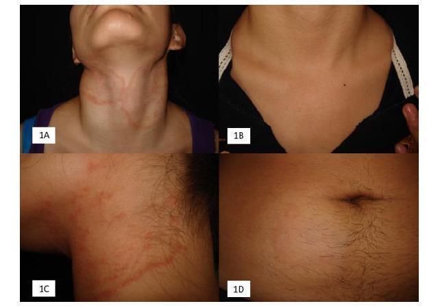
This composite of clinical photographs illustrates the presentation and treatment response of Tinea gladiatorum (tinea corporis), a fungal infection common in contact sports. Panel 1A shows extensive, erythematous, annular plaques with raised borders on the neck and upper chest, demonstrating the characteristic ‘ringworm’ morphology. Panel 1B presents the same patient post-treatment, showing near-complete clinical resolution with minimal residual hyperpigmentation. Panel 1C provides a close-up view of the inflammatory lesions, highlighting the irregular, serpiginous, and papular nature of the raised border. Panel 1D displays a different clinical manifestation on the abdomen: a subtle, hypochromic plaque with fine peripheral desquamation (scaling), representing a localized fungal infection. These images are educational for dermatology and sports medicine, emphasizing the varied clinical appearances of Trichophyton tonsurans infections—from highly inflammatory disseminated lesions to more subtle, scaly plaques—and the efficacy of antifungal therapy.
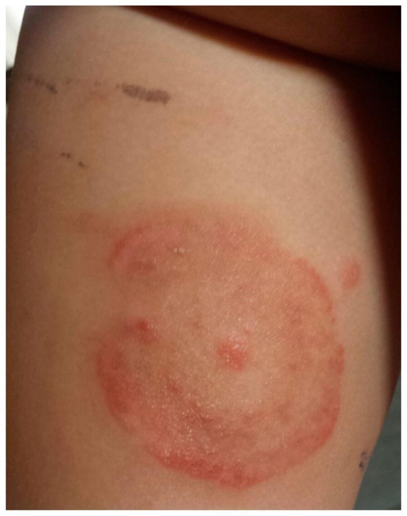
This clinical photograph displays a dermatological presentation of tinea corporis (ringworm) in a pediatric patient. The primary feature is an annular, erythematous plaque located on the trunk or proximal limb. The lesion is characterized by a well-demarcated, raised red border with evidence of mild scaling or vesiculation, indicating active peripheral growth. The center of the plaque shows partial clearing with a lighter pink hue, though it remains occupied by a few scattered inflammatory papules. Above the primary lesion, incidental linear hyperpigmented marks are visible, likely representing excoriations or minor superficial trauma. The image serves as a classic educational example of a fungal skin infection, specifically demonstrating the centrifugal spread and central clearing characteristic of Microsporum canis dermatophytosis.
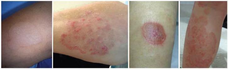
This composite of clinical photographs illustrates various clinical presentations of tinea corporis, a common dermatophyte fungal infection. The images depict the characteristic morphology of 'ringworm' across different skin areas. Key diagnostic features visible include well-demarcated, erythematous, annular (ring-shaped) patches and plaques. Several lesions demonstrate classic central clearing, where the center of the ring appears less inflamed than the periphery. The advancing borders are notably active, exhibiting varying degrees of elevation and fine scaling. The presentation varies from discrete, solitary circular lesions to larger, confluent, and irregularly shaped polycyclic patterns. The color intensity ranges from faint pink to vivid red, representing different stages of inflammatory response. These visual characteristics are essential for distinguishing tinea corporis from other annular dermatoses such as nummular eczema, granuloma annulare, or subacute cutaneous lupus erythematosus. The collection serves as an educational tool for identifying the hallmark features of cutaneous fungal infections in a clinical setting.
Most Likely Diagnosis: Tinea Barbae (Superficial Type) / Inflammatory Dermatophytosis
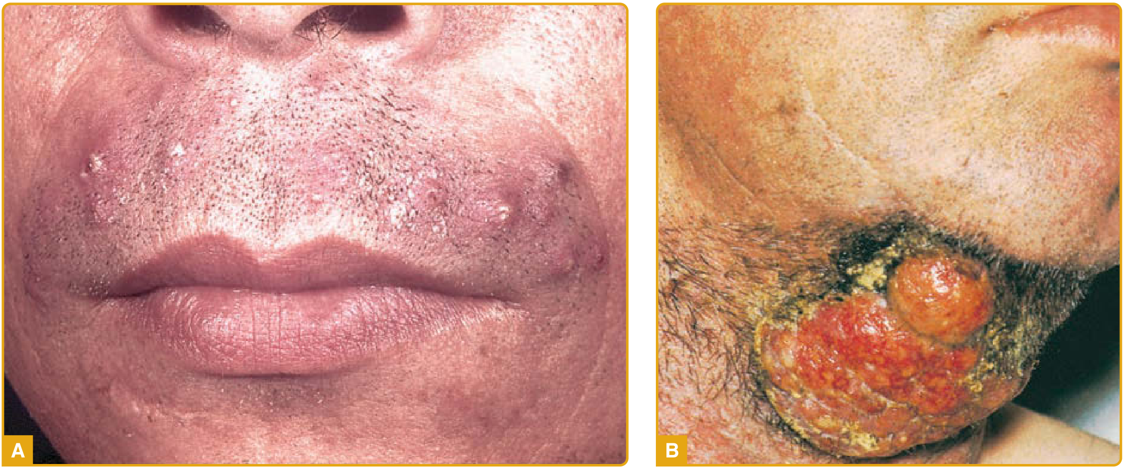
What Is It?
- Use of topical steroids (can worsen/mask the infection - so do NOT apply steroid creams)
- Contact with infected animals (cattle, horses, dogs)
- Diabetes mellitus
- Poor hygiene or contaminated shaving equipment
Differential Diagnoses to Consider
| Condition | How to Distinguish |
|---|---|
| Tinea barbae (most likely) | Annular, pustular border, fungal hyphae on KOH prep |
| Sycosis barbae (bacterial folliculitis) | S. aureus, hairs NOT loosened, no true ring shape |
| Annular impetigo | Honey-colored crust; bacterial culture positive |
| Majocchi granuloma | Deeper follicular involvement, often after steroid use |
Diagnosis
- KOH (potassium hydroxide) preparation - scrape from the active border; look for fungal hyphae (confirms fungal infection)
- Fungal culture - for species identification
- Bacterial swab/culture - if bacterial folliculitis (sycosis) cannot be excluded
Treatment
For Tinea Barbae (Fungal - Requires Oral Antifungals)
| Drug | Dose | Duration |
|---|---|---|
| Terbinafine (preferred) | 250 mg orally once daily | 2 to 4 weeks |
| Griseofulvin (microsize) | 500 mg orally once daily | 6 weeks |
| Griseofulvin (ultramicrosize) | 375 mg orally once daily | 6 weeks |
| Itraconazole (alternative) | 200 mg orally once daily | 2 to 4 weeks |
Note: Both griseofulvin and terbinafine should be avoided in patients with hepatic dysfunction. Check liver function if prolonged treatment is anticipated.
Supportive Care
- Shaving or depilating the beard hair in the affected area
- Warm compresses to remove crust and soften the lesions
- Avoid all topical steroids - they will worsen fungal infection and alter its appearance
If Bacterial Folliculitis (Sycosis Barbae) is Confirmed Instead
- Mild: warm compresses + mupirocin ointment topically
- Moderate/severe or chronic: systemic antibiotics with S. aureus coverage (e.g., dicloxacillin, cephalexin, or doxycycline if MRSA risk)
Important Warning
What would be the cause of etiology
Etiology of Tinea Barbae
1. The Causative Organisms
Zoophilic Species (from animals - most common cause)
| Organism | Animal Source | Clinical Pattern |
|---|---|---|
| T. interdigitale (formerly T. mentagrophytes var. mentagrophytes) | Small mammals (rabbits, guinea pigs, mice) | Highly inflammatory; pustular, kerion-like plaques |
| T. verrucosum | Cattle, horses | May mimic bacterial furunculosis; severe inflammation |
| M. canis | Cats and dogs | Moderately inflammatory; associated with pet contact |
Anthropophilic Species (human-to-human - less common, less inflammatory)
| Organism | Notes |
|---|---|
| T. violaceum | Endemic areas; causes superficial type resembling bacterial folliculitis |
| T. schoenleinii | Rare; endemic in certain regions |
| T. rubrum | Most common dermatophyte overall; harbored in hair follicles; can cause concentric rings |
A newer concern: T. indotineae (formerly T. mentagrophytes var. VIII) is an emerging terbinafine-resistant strain causing widespread annular lesions, particularly in South Asia. - Dermatology 2-Volume Set 5e
2. Why These Fungi Cause Infection - The Pathogenic Mechanism
"Dermatophytoses are fungal infections caused by three genera of fungi that have the unique ability to invade and multiply within keratinized tissue (hair, skin, and nails)." - Dermatology 5e
- Adherence - Fungal spores (arthroconidia) adhere to the keratin of the stratum corneum, beard hair shaft, or follicular epithelium
- Invasion - The fungus produces keratinases and proteases that break down keratin, allowing it to penetrate deeper into the hair follicle
- Multiplication - Hyphae grow centrifugally (outward in a ring pattern), causing the characteristic annular lesion with an active advancing border
- Host inflammatory response - The immune system recognizes fungal antigens; the degree of inflammation depends on whether the species is zoophilic (provokes intense reaction) or anthropophilic (milder reaction)
- Pustule formation - In the beard area, fungal hyphae invade follicles and trigger a neutrophilic response, forming perifollicular pustules - exactly what is visible in the image
3. Predisposing / Risk Factors
| Risk Factor | How It Contributes |
|---|---|
| Animal contact (cattle, dogs, cats, rodents) | Direct inoculation from zoophilic species |
| Topical corticosteroid use | Suppresses local immune defence; causes "tinea incognito" - masked, more aggressive infection |
| Diabetes mellitus | Impaired neutrophil function and poor skin barrier |
| Contaminated shaving equipment | Direct mechanical inoculation into follicles; historically the "barber's itch" route |
| Immunosuppression (HIV, steroids, chemotherapy) | Loss of cell-mediated immunity against dermatophytes |
| Overcrowding / poor hygiene | Increased exposure and transmission |
| Skin trauma / microabrasions | Disrupts keratin barrier, allowing spore entry |
| Warm, humid environment | Fungi thrive in moist conditions |
4. Why the Beard Area Is Specifically Vulnerable
- Cannot be treated with topical antifungals alone (the drug cannot penetrate deep enough)
- Produces follicular pustules rather than just surface scaling
- Can cause permanent scarring alopecia if untreated, because the hair follicle bulb gets destroyed
5. Classification Summary
Tinea Barbae
├── Zoophilic (most common, most inflammatory)
│   ├── T. interdigitale → from small mammals
│   ├── T. verrucosum → from cattle/horses
│   └── M. canis → from cats/dogs
└── Anthropophilic (less common, less inflammatory)
    ├── T. violaceum → human-to-human
    ├── T. rubrum → human-to-human
    └── T. schoenleinii → endemic areas
Does insect bite cause pus