
Can u give answer for bams also
I'd love to help with BAMS (Bachelor of Ayurvedic Medicine and Surgery) content! Could you clarify what you're looking for?
Asking for Preferences
What topic or subject do you need answers for? · What kind of help do you need?
Samita related
Asking for Preferences
Which Samhita are you referring to? · What kind of help do you need?
Can u explain femoral triangle
The femoral triangle is a triangular hollow in the upper inner thigh, just below the inguinal ligament. Its boundaries are: the inguinal ligament (base/top), the medial border of sartorius (lateral side), and the medial border of adductor longus (medial side). The floor is formed by iliopsoas and pectineus muscles. Contents from lateral to medial: femoral nerve, femoral artery, femoral vein, and lymphatics (NAVY - Nerve, Artery, Vein, Y-fronts/lymphatics).
Searching sources...
Reading File
Finding Sources
Searching the Web
femoral triangle anatomy boundaries contents floor BAMS
Reading File
Loading Image
Loading Image
Loading Image
Here is a complete explanation of the Femoral Triangle for your BAMS anatomy:
Femoral Triangle
The femoral triangle is a wedge-shaped (triangular) depression in the upper anterior thigh, at the junction between the anterior abdominal wall and the lower limb.
Boundaries
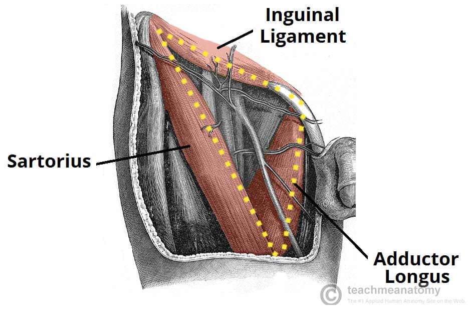
| Boundary | Structure |
|---|---|
| Base (Superior) | Inguinal ligament |
| Lateral border | Medial margin of Sartorius muscle |
| Medial border | Medial margin of Adductor Longus muscle |
| Apex | Where sartorius crosses adductor longus (points inferiorly) |
Floor (from lateral to medial)
The floor is formed by 3 muscles:
- Iliopsoas (lateral part)
- Pectineus (middle)
- Adductor Longus (medial part)
Mnemonic: "I Pee Always" - Iliopsoas, Pectineus, Adductor longus
Roof
- Fascia lata (deep fascia of the thigh)
- Skin and subcutaneous tissue
Contents (lateral to medial)
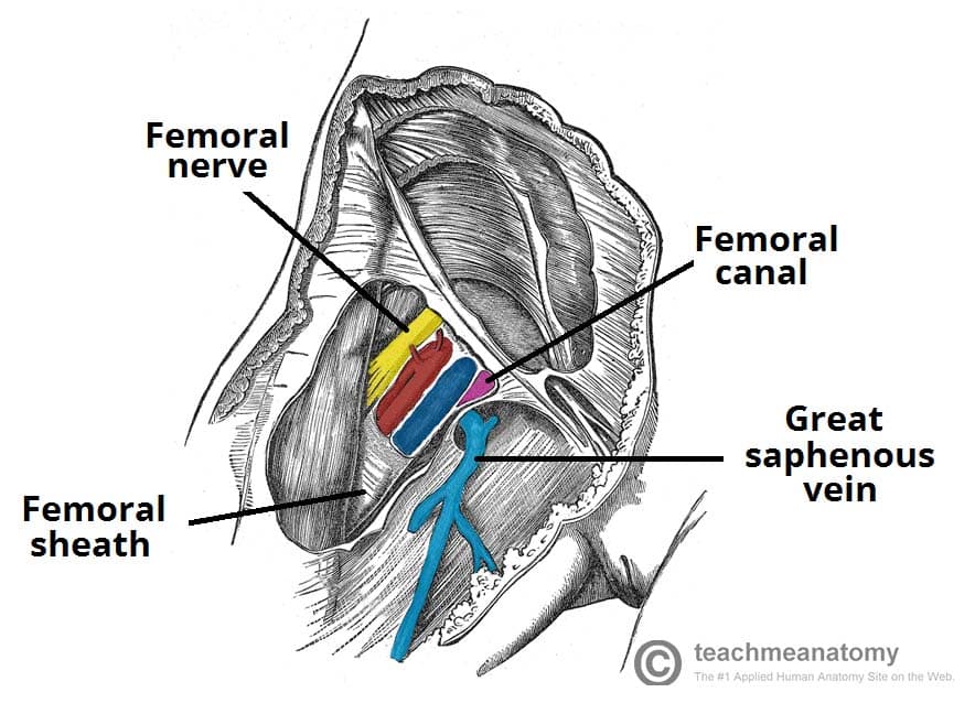
Remember with the mnemonic NAVY (lateral to medial):
| Letter | Structure |
|---|---|
| N | Femoral Nerve |
| A | Femoral Artery |
| V | Femoral Vein |
| Y | Y-fronts (Lymphatics / Femoral canal) |
Important: The femoral nerve is outside the femoral sheath. The artery, vein, and lymphatics are enclosed within the femoral sheath.
Femoral Sheath
The femoral sheath is a funnel-shaped fascial sleeve enclosing the femoral artery, vein, and lymphatics in 3 compartments:
- Lateral - Femoral artery
- Middle - Femoral vein
- Medial - Femoral canal (lymphatics) -- this is the site of femoral hernias
Continuation
The apex of the femoral triangle is continuous with the Adductor Canal (Hunter's canal), which carries the femoral vessels down the thigh. The femoral artery and vein pass through the adductor hiatus in adductor magnus to become the popliteal vessels behind the knee.
Clinical Importance
- Femoral pulse can be palpated just below the midpoint of the inguinal ligament (midway between ASIS and pubic symphysis) - this point is called the mid-inguinal point
- Site for femoral artery cannulation and angiography
- Femoral hernia occurs through the femoral canal (medial compartment of femoral sheath) - more common in females
- Femoral nerve block can be performed here for lower limb analgesia
Source: Gray's Anatomy for Students, p. 675
This is a shared conversation. Sign in to Orris to start your own chat.