
Placenta barrier 5 mark answer
placental barrier layers diagram syncytiotrophoblast fetal maternal
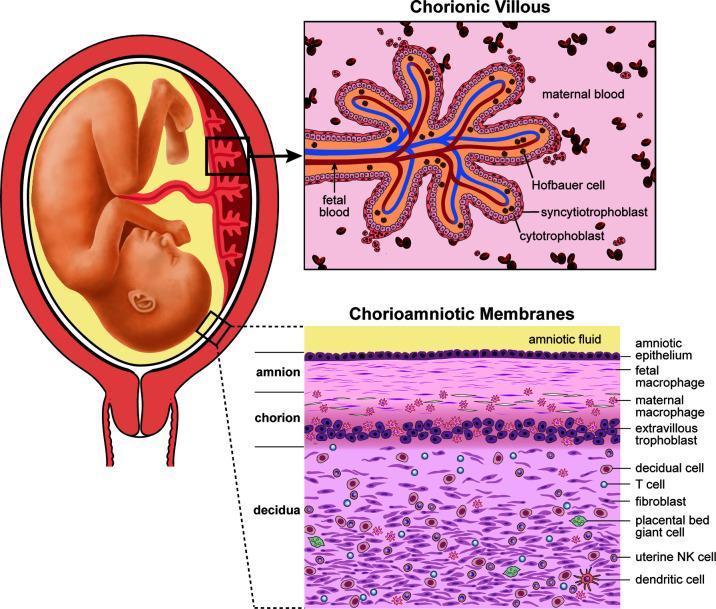
This medical illustration details the human placental anatomy and maternal-fetal interface across three views. On the left, a macro-level diagram shows a fetus in utero with the umbilical cord and placental attachment. Two inset panels provide microscopic structural detail. The upper right panel displays a cross-section of a Chorionic Villous, illustrating the exchange interface where fetal blood vessels are separated from the maternal blood space by three distinct layers: the syncytiotrophoblast, cytotrophoblast, and underlying stroma containing Hofbauer cells (placental macrophages). The lower right panel illustrates the layered Chorioamniotic Membranes. From the amniotic fluid inward, the layers are identified as the amniotic epithelium, the amnion (containing fetal macrophages), the chorion (containing maternal macrophages and extravillous trophoblasts), and the maternal decidua. The decidual layer is shown to be rich in diverse immune and stromal cells, including T cells, fibroblasts, placental bed giant cells, uterine Natural Killer (NK) cells, and dendritic cells. The diagram serves as an educational tool for understanding placental barrier composition and immune cell distribution at the maternal-fetal interface.
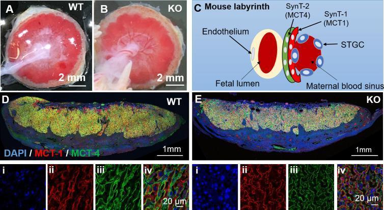
This composite image evaluates the placental morphology and trophoblast syncytialization in wild-type (WT) and TRPV4 knockout (KO) mouse models at embryonic day E18.5. Panels A and B present gross clinical photographs of WT and KO placentas, respectively, showing comparable circular cross-sections and red-pink coloration with a 2mm scale. Panel C is an anatomical diagram of the mouse labyrinth, illustrating the spatial relationship between the fetal lumen (endothelium), SynT-2 layer (MCT4 positive), SynT-1 layer (MCT1 positive), and maternal blood sinuses. Panels D and E show low-magnification immunofluorescence sections of WT and KO placentas, stained for DAPI (blue, nuclei), MCT1 (red), and MCT4 (green). High-magnification subsets (i-iv) confirm the cellular organization of the syncytiotrophoblast layers: MCT1 specifically labels the SynT-1 layer facing maternal blood, while MCT4 labels the SynT-2 layer surrounding fetal vessels. The visual evidence demonstrates that TRPV4 deficiency does not cause gross anatomical defects or disrupt the integrity of the trophoblast layers in the mouse labyrinth, highlighting species-specific differences in placental TRPV4 function.
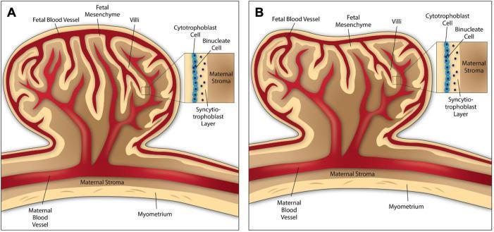
This comparative anatomical diagram illustrates the structural morphology of ruminant placentomes, serving as an animal model for studying placental development and human pregnancy complications. Diagram A depicts the cow placentome, characterized by a convex structure where the fetal cotyledon and maternal caruncle interface in an outward-bulging arrangement. Diagram B shows the sheep placentome, which exhibits a concave morphology with an inward-curving indentation at the fetal-maternal interface. Both diagrams detail the internal architecture, including fetal blood vessels, fetal mesenchyme, and villi. High-magnification callouts highlight the cellular layers forming the synepitheliochorial placenta: a layer of mononuclear cytotrophoblast cells adjacent to binucleate cells, and the syncytiotrophoblast layer which facilitates direct contact with the maternal stroma. The diagrams also delineate the maternal environment, showing the maternal blood vessels, stroma, and the underlying myometrium. This visual resource is used to teach concepts of comparative placentation, trophoblast differentiation, and the spatial relationships within the maternal-fetal unit.
The Placental Barrier
Definition
Layers of the Placental Barrier
- Syncytiotrophoblast - the outermost continuous layer covering the villi, with numerous microvilli projecting into maternal blood; contains transport vesicles, mitochondria, rER, and sER
- Cytotrophoblast (inner layer) - becomes thin and discontinuous after the 4th month (discontinuous after ~20 weeks)
- Trophoblast basal lamina
- Connective (mesenchymal) tissue of the villus - contains Hofbauer cells (placental macrophages, analogous to alveolar macrophages)
- Endothelial basal lamina
- Endothelium of the fetal capillary (within the tertiary villus)
What Crosses the Placental Barrier?
- O₂, water, electrolytes, glucose, amino acids, lipids
- Vitamins and trace elements
- IgG antibodies (passive immunity against diphtheria, measles, smallpox)
- Hormones (steroidal hormones readily cross; thyroxine crosses slowly)
- Drugs, toxins (e.g., tetracyclines, heroin, cocaine)
- Viruses: rubella, CMV, coxsackie, varicella, poliovirus
- CO₂, water, electrolytes
- Urea, uric acid, bilirubin, creatinine, hormones
Comparison to Air-Blood Barrier
Clinical Significance
- Viruses (rubella, CMV) cross easily and cause congenital infections and birth defects
- Drugs: Most drugs and their metabolites cross freely (e.g., tetracyclines accumulate in fetal bones and teeth; heroin/cocaine cause fetal habituation)
- Synthetic hormones: DES (diethylstilbestrol) crosses and causes clear-cell carcinoma of vagina in females and testicular abnormalities in males; synthetic progestins can masculinize female fetuses
- Maternal cells can cross into fetal circulation (microchimerism)
- Mother and fetus can have different blood groups because the barrier normally prevents mixing