
Vien visible in palm and hand and in leg is normal or not
visible veins in hands palms legs normal causes when to worry
Are Visible Veins in the Hands, Palms, and Legs Normal?
Why Veins Become Visible - Normal Reasons
| Cause | Explanation |
|---|---|
| Low body fat | Less fat under the skin means veins sit closer to the surface |
| Fair/light skin tone | The color of veins shows through more easily |
| Exercise | Blood flow increases and muscles press on veins, making them bulge |
| Heat | Warm temperatures cause veins to dilate and become more prominent |
| Age | Skin becomes thinner and loses elasticity over time |
| Genetics | Family history plays a role in vein prominence |
| Dehydration | Can sometimes make veins more visible |
| Standing or sitting for long periods | Gravity causes blood to pool in leg veins |
| Hormonal changes | Puberty, pregnancy, and menopause can all make veins more visible |
The CEAP Classification (Medical Standard)
- C0 - No visible or palpable signs of venous disease (fully normal)
- C1 - Telangiectasias (spider veins) or reticular veins - generally benign, cosmetic
- C2 - Varicose veins - twisted, enlarged veins, may need evaluation
- C3 - Edema (swelling)
- C4 - Skin changes (pigmentation, eczema, lipodermatosclerosis)
- C5/C6 - Healed or active venous ulcer
When to See a Doctor
- Pain, aching, or heaviness in the legs
- Swelling in the legs or ankles
- Itching or burning over the veins
- Skin color changes (redness, brownish discoloration) around the vein
- Hardness or tenderness along a vein (could indicate blood clot / thrombophlebitis)
- Sudden appearance of many new veins without an obvious reason
- Varicose veins - twisted, rope-like bulging veins, especially in the legs
Summary
Search for images of normal vs abnormal visible veins
normal visible veins hands palms
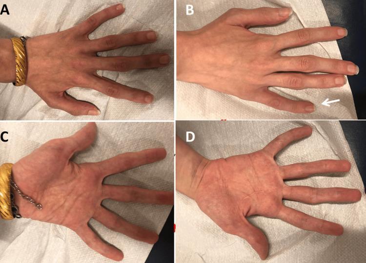
Clinical photograph of a patient's hands in dorsal (A, B) and palmar (C, D) views, demonstrating characteristic features of a connective tissue disorder. The image shows significant arachnodactyly, characterized by abnormally long and slender fingers. In panel B, a white arrow indicates clinodactyly (radial curvature) of the fifth digit. The palmar views (C, D) reveal thin palms with visible superficial veins and elongated phalanges. The fingers also exhibit signs of joint hypermobility/hyperextensibility at the metacarpophalangeal and interphalangeal joints. These musculoskeletal findings are classically associated with conditions such as Marfan syndrome or related genetic connective tissue diseases. The skin appears of normal texture without edema, though the hands show evidence of diffuse muscle wasting consistent with distal weakness.
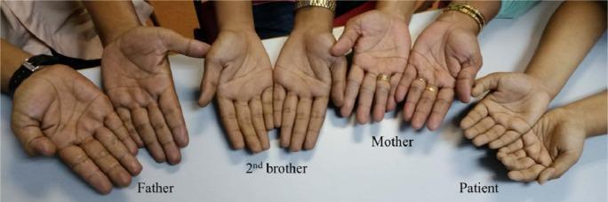
A clinical photograph displaying the palms of four family members side-by-side to illustrate phenotypic differences in skin coloration. From left to right, the hands belong to the father, the second brother, the mother, and the patient. A clear contrast is visible between the unaffected mother and the other three family members. The mother's palms exhibit a normal, healthy pinkish-tan hue. In contrast, the father, brother, and patient show a distinct dusky, grayish-blue discoloration (cyanosis), particularly noticeable in the nail beds and palm creases. This visual finding is characteristic of methemoglobinemia, specifically Hemoglobin M (Hb-M) disease, where the presence of ferric iron prevents normal oxygen binding, resulting in persistent clinical cyanosis despite normal oxygen saturation levels. The image serves as a comparative tool for recognizing hereditary hemoglobinopathies and understanding the autosomal dominant inheritance pattern of Hb-M, as the condition is clearly expressed in the father and two of his children, but absent in the mother.
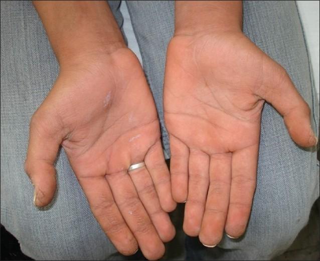
Clinical photograph of the palmar surfaces of both hands demonstrating features consistent with palmoplantar keratoderma (PPK). The image shows bilateral, symmetrical thickening of the skin (hyperkeratosis) with an exaggerated appearance of the dermatoglyphics (skin lines). The palms exhibit a yellowish-orange hue, particularly within the creases and over pressure-bearing areas. Small, scattered white patches or focal areas of desquamation are visible, suggesting scaling or dryness. The texture appears rough and leathery compared to normal palmar skin. No significant fissures or erythematous borders are visible in this specific presentation. This visual finding is characteristic of a group of hereditary or acquired disorders of cornification, often associated with systemic conditions such as Bart-Pumphrey syndrome when occurring alongside leukonychia totalis and sensorineural hearing loss (SNHL).
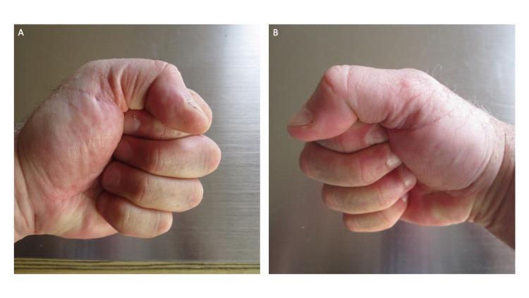
Clinical photograph showing two side-by-side images (A and B) of the left and right hands of a patient, demonstrating a negative 'fist sign.' Both hands are clenched into a tight, complete fist, where the fingertips are in direct contact with the ipsilateral palms and are nearly hidden from view. The metacarpophalangeal (MCP), proximal interphalangeal (PIP), and distal interphalangeal (DIP) joints exhibit a high degree of flexion. The visible skin on the dorsal surfaces and palmar creases appears normal, with no evidence of erythema, blanching, or edema. This clinical presentation illustrates the complete resolution of post-ambulatory swollen hands (POTASH), a condition associated with vigorous physical activity like running. The images serve as an educational comparison to the 'positive fist sign,' where significant hand edema prevents full flexion and contact between digits and the palm.
varicose veins legs abnormal bulging
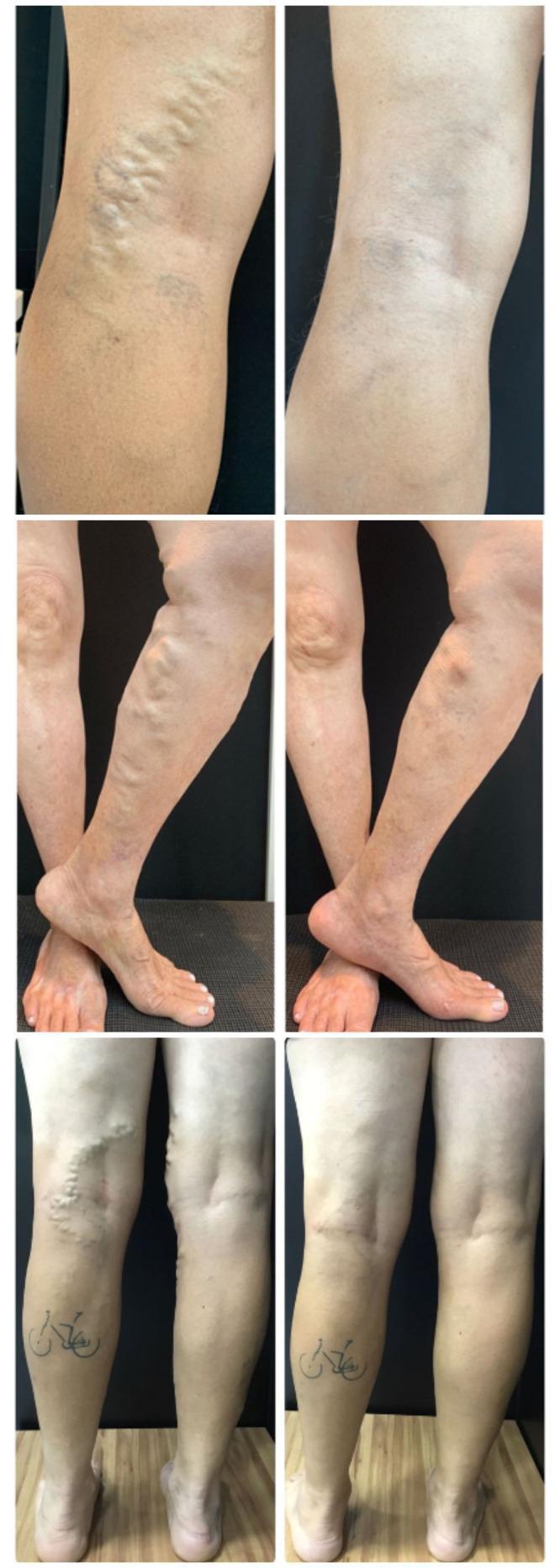
This composite clinical photograph displays three sets of side-by-side 'before' and 'after' images of the lower extremities in patients undergoing assisted total thermal ablation (ATTA) for chronic venous insufficiency. The 'before' images (left) demonstrate classic clinical signs of varicose veins, including prominent, bulging, and tortuous superficial venous segments along the medial thigh, popliteal fossa, and calf. These vessels appear rope-like with visible skin elevation and bluish discoloration. In the corresponding 'after' photographs (right), there is a significant reduction in venous prominence, resulting in a smoother skin contour and improved aesthetic appearance. The secondary and tertiary sets show the resolution of large tributary varicosities on the shins and posterior legs, with one image showing a subtle postoperative scar. These images illustrate the efficacy of endolaser thermal ablation in treating both axial saphenous veins and their associated tributaries, facilitating objective assessment of clinical outcomes in vascular surgery and phlebology.
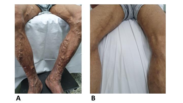
This clinical comparison photograph illustrates the effectiveness of ultrasound-guided foam sclerotherapy for advanced chronic venous insufficiency. Image A (pre-procedure) shows bilateral lower extremities with severe, tortuous varicosities of the great saphenous vein tributaries. Prominent, bulging venous networks are visible from the distal thighs to the ankles, accompanied by cutaneous signs of chronic venous stasis, including hyperpigmentation and skin textural changes. Image B (two weeks post-procedure) demonstrates the same patient's legs after a single session of 3% and 1% polidocanol foam sclerotherapy. There is a marked reduction in the size and prominence of the varicose veins, with a smoother skin surface and decreased venous protrusion. The comparison highlights the rapid clinical involution of symptomatic varicosities following foam injection and subsequent clot drainage. This visual is intended for vascular surgery and dermatology education, focusing on the management of venous disease and minimally invasive treatment outcomes.
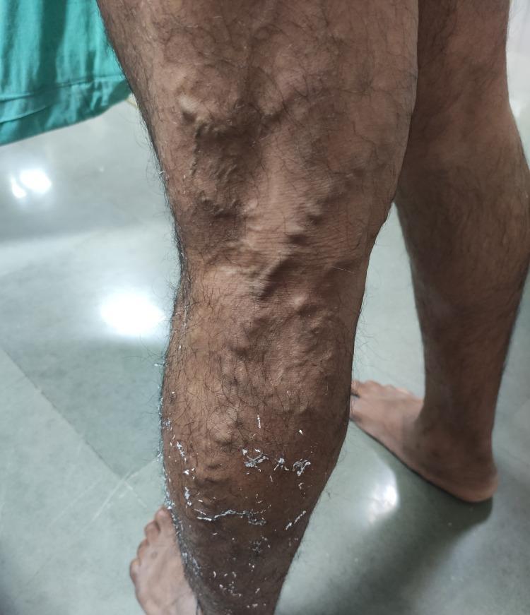
This clinical photograph displays a 50-year-old male's left lower limb with extensive varicose veins. The affected veins are highly tortuous, dilated, and exhibit a prominent, rope-like bulging appearance characteristic of Chronic Venous Insufficiency (CVI). The distribution is primarily located along the anterior and medial aspects of the distal thigh and proximal lower leg. Multiple interconnected, distended venous segments are visible beneath the skin's surface. Additionally, the lower portion of the leg shows secondary skin changes including visible dryness, scaling, and white flakiness, which can be associated with venous stasis dermatitis or the residue of topical treatments. This image serves as a textbook example of CEAP classification grade C2 (varicose veins) and illustrates clinical manifestations often assessed using the Venous Clinical Severity Score (VCSS).
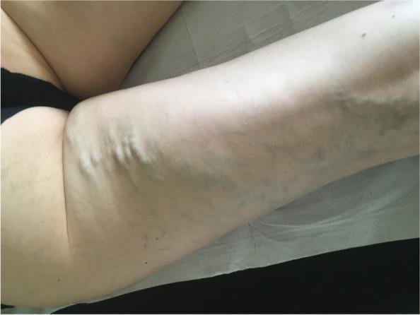
This clinical photograph shows a prone view of a patient’s posterior thigh, highlighting an atypical distribution of varicose veins. The image demonstrates prominent, dilated, and tortuous subcutaneous veins located primarily on the posterior aspect of the upper thigh. These varices exhibit characteristic bulging morphology and a bluish-purple discoloration beneath the skin surface. Unlike common lower extremity varicosities associated with saphenous vein reflux, this specific posterior thigh pattern is a key clinical sign that should raise diagnostic suspicion for a pelvic venous source, such as pelvic congestion syndrome or nutcracker syndrome. The visual evidence emphasizes the importance of a thorough physical examination and the use of cross-sectional imaging (CT or MR venography) to evaluate for proximal venous obstruction or reflux in patients presenting with such atypical venous patterns.
spider veins telangiectasia legs skin
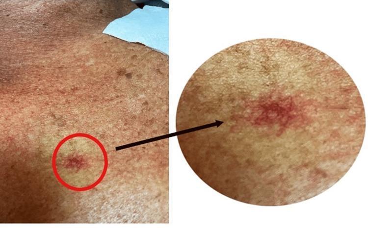
This clinical photograph displays a spider angioma located on the anterior chest wall. The visual consists of a wide-angle view of the skin and a circular magnified inset highlighting the vascular lesion. The spider angioma (also known as spider telangiectasia or nevus araneus) is characterized by a central, prominent red punctum from which small, tortuous capillary vessels radiate outward, mimicking the legs of a spider. The surrounding skin appears tanned and mottled with secondary sun damage and various small hyperpigmented macules. This finding is clinically significant as a cutaneous marker often associated with hyperestrogenic states, such as chronic liver disease (cirrhosis) or pregnancy. The lesion demonstrates blanching upon central pressure, a key diagnostic feature of this vascular malformation. This image is suitable for medical education regarding the physical examination of dermatologic signs of systemic disease and hepatology-related cutaneous manifestations.
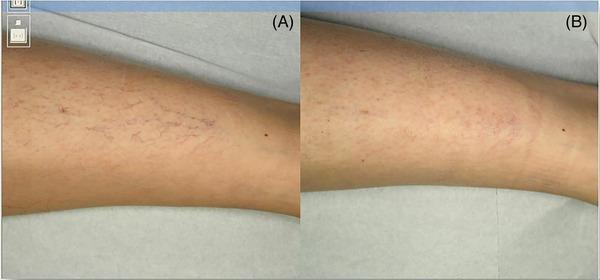
This clinical photograph series is a comparison chart showing the dermatological response to treatment for leg telangiectasia. Image A (baseline) displays a prominent network of fine, branching, dark red and purple vascular lesions on the skin of the lower limb, characteristic of spider veins or telangiectasia. These vessels are sharply defined and show a dense distribution across the cutaneous surface. Image B (6 months post-treatment) illustrates significant clinical improvement, showing the near-complete disappearance of the distinct vascular network. The skin appears smoother with only residual faint erythema or subtle hyperpigmentation where the vessels were previously located. The comparison demonstrates the efficacy of therapeutic intervention (such as sclerotherapy or laser treatment) in reducing the visible manifestation of superficial vascular ectasias. This visual is relevant for dermatology and vascular medicine, specifically for educating on the management of chronic venous insufficiency signs and aesthetic vascular procedures.
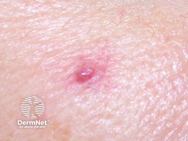
Clinical dermatology photograph of a solitary spider telangiectasia (spider angioma) on the superficial skin. Imaging modality: external macro photography of the skin performed under standard clinical lighting; no dermoscopy or histology. The lesion is a small central erythematous papule (approximately 1–3 mm) from which multiple fine, radiating capillaries extend in a spider-like fashion. The surrounding skin is normal in tone with mild peripheral erythema. On gentle compression the lesion blushes (vascular blanching), confirming superficial dermal vascular origin. Commonly located on the face, neck, upper chest, or proximal limbs; this lesion appears on exposed cutaneous surface consistent with typical sites. Pathophysiology involves dilation of preexisting superficial dermal arterioles and capillaries with a central nidus of vascular supply; estrogen exposure, pregnancy, and liver disease can increase frequency. Clinically, spider angiomas are usually benign; they may be incidental findings or a marker for hormonal influences or hepatic dysfunction in adults. Differential diagnoses include cherry angioma, capillary malformation, rosacea-related telangiectasia, or other vascular malformations. Management is typically conservative; treatment options (laser, electrosurgery) may be considered for cosmetic concerns or symptoms. Clinical relevance includes cutaneous vascular screening for systemic conditions when multiple lesions are present, particularly in middle-aged or older adults.
Normal / Benign Visible Veins
Visible Veins on Palms (Benign Finding)
Thin palms with visible superficial veins - a common normal variant, more noticeable in lean individuals.
Abnormal Veins - When to Be Concerned
1. Varicose Veins (CEAP Grade C2) - Before & After Treatment
2. Severe Varicose Veins with Skin Changes
3. Bilateral Severe Varicose Veins (Before & After Sclerotherapy)

4. Spider Veins / Telangiectasia (CEAP Grade C1)
5. Spider Angioma (Sign of Liver Disease / High Estrogen)
Quick Visual Guide Summary
| What You See | Normal or Abnormal? | Action Needed? |
|---|---|---|
| Flat blue/green veins visible through skin | Normal | No |
| Prominent veins after exercise/heat | Normal | No |
| Fine red/purple web-like lines (spider veins) | Borderline (C1) | Cosmetic only |
| Bulging, rope-like, twisted veins on legs | Abnormal (Varicose C2) | See a doctor |
| Veins + skin darkening/scaling | Abnormal (C4) | See a doctor urgently |
| Multiple spider angiomas on upper body | Abnormal | Rule out liver disease |