
Acid base, nephrotic and nephritic syndrome , casts in urine, acute kidney injury, acute cystitis. I’ve got a pathology exam. Idk anything. Explain me in detail like you’re explaining it to a very dumb person
Reading File
Now let me search the library for all these topics in parallel.
Finding Sources
Finding Sources
Finding Sources
Finding Sources
Finding Sources
Finding Sources
Finding Sources
Finding Sources
Good - now let me read the key sections from the textbooks in parallel.
Reading File
Reading File
Reading File
Reading File
Reading File
Now let me read the acid-base section from the Quick Compendium (most exam-relevant) and the nephrotic/nephritic sections:
Reading File
Reading File
Reading File
Reading File
Now let me get the acid-base disorders section and the nephritic syndrome content:
Reading File
Finding Sources
Reading File
Let me get the nephritic syndrome section and AKI definition:
Reading File
Reading File
Reading File
I now have excellent material from all topics. Let me also fetch the cast image that was referenced:
Loading Image
I have all the material I need. Now I'll write the comprehensive study guide.
Your Complete Pathology Exam Study Guide
I'll explain everything like you've never heard any of this before. Let's go topic by topic.
PART 1: ACID-BASE DISORDERS
The Big Picture First
Your blood pH must stay between 7.35 and 7.45. That's it. Too acidic (pH < 7.35) = acidosis. Too basic/alkaline (pH > 7.45) = alkalosis.
Your body controls pH using two main systems:
- Lungs - control CO2 (a gas that makes the blood acidic when it builds up)
- Kidneys - control bicarbonate (HCO3-, a base that neutralizes acid)
Think of it this way:
CO2 is the acid. HCO3- is the antacid. Lungs blow out CO2. Kidneys hold onto or dump HCO3-.
The 4 Types of Acid-Base Disorders
1. Metabolic Acidosis
What it is: Too much acid (or too little HCO3-) from a metabolic cause (not breathing).
How to spot it: pH is LOW + HCO3- is LOW (they move in the same direction).
The body's response (compensation): Lungs say "let me help" and breathe FASTER to blow off CO2, raising pH back up. For every 1 mEq/L drop in HCO3-, PCO2 drops by 1 mmHg.
The key skill: Calculate the Anion Gap
Anion gap = Na+ - Cl- - HCO3-
Normal anion gap = less than 12
This tells you WHY there's too much acid:
| HIGH Anion Gap (≥12) | NORMAL Anion Gap (<12) |
|---|---|
| MUDPILES mnemonic: | Think: you're losing base, not gaining acid |
| Methanol | Diarrhea (losing HCO3-) |
| Uremia (kidney failure) | Renal tubular acidosis |
| DKA (diabetic ketoacidosis) | Recovery from DKA |
| Paraldehyde | NH4Cl ingestion |
| Isoniazid / Iron | Carbonic anhydrase inhibitors |
| Lactic acidosis | Total parenteral nutrition |
| Ethylene glycol | |
| Salicylates |
Memory trick: High anion gap = some FOREIGN substance (or uremia/lactic acid) is floating in the blood, taking up space where bicarbonate should be.
2. Metabolic Alkalosis
What it is: Too much base (HCO3- is high).
How to spot it: pH HIGH + HCO3- HIGH (same direction again).
Compensation: Lungs breathe SLOWER to retain CO2. For every 0.6 mEq/L rise in HCO3-, PCO2 rises by 1 mmHg.
Causes split by chloride:
| Chloride-Responsive (urine Cl <10) | Chloride-Resistant (urine Cl >10) |
|---|---|
| Vomiting / NG tube suction | Hyperaldosteronism |
| Diuretic therapy | Cushing syndrome |
| Villous adenoma | Exogenous steroids |
| Licorice (glycyrrhizin) |
Easy logic: Vomiting = you lose HCl = blood becomes alkalotic. Give them IV saline (chloride!) and it fixes it. Hyperaldosteronism = aldosterone makes you retain Na and dump K/H = alkalosis that won't fix with just saline.
3. Respiratory Acidosis
What it is: You're not breathing well enough, so CO2 builds up and makes the blood acidic.
How to spot it: pH LOW + PCO2 HIGH (they move in opposite directions).
Causes: Anything that makes you breathe poorly - airway obstruction, pneumonia, neuromuscular disease (ALS, myasthenia gravis), sedation/opioids.
Compensation: Kidneys (slowly) retain more HCO3- to buffer the acid.
- Acute: HCO3- rises by 0.1 mEq/L per 1 mmHg rise in PCO2
- Chronic: HCO3- rises by 0.4 mEq/L per 1 mmHg rise in PCO2
4. Respiratory Alkalosis
What it is: You're breathing too fast (hyperventilating), blowing off too much CO2.
How to spot it: pH HIGH + PCO2 LOW (opposite directions).
Causes: Anxiety, pain, hypoxia (your body tries to compensate), pregnancy, CNS problems, sepsis (early).
Compensation: Kidneys dump HCO3- to lower the pH back down.
How to Read an ABG (Step-by-step)
- Look at the pH - is it acidosis or alkalosis?
- Look at PCO2 (normal: 35-45 mmHg) - if it explains the pH, it's respiratory
- Look at HCO3- (normal: 22-26 mEq/L) - if it explains the pH, it's metabolic
- Ask: is the compensation appropriate? If not, there's a mixed disorder
PART 2: NEPHROTIC vs. NEPHRITIC SYNDROME
This is one of the most commonly tested comparisons in nephrology. Here's the dead-simple way to think about it:
The Kidney Filter Analogy
Your kidney glomerulus is like a coffee filter. It's supposed to let water and small waste products through, but block big proteins (albumin) and blood cells.
- Nephrotic = the filter has holes - proteins leak through (but no blood cells)
- Nephritic = the filter is inflamed/on fire - blood and protein both leak, and pressure builds up
NEPHROTIC SYNDROME
The definition: Protein in urine > 3.5 g/day (or protein:creatinine ratio >2.0 mg/mg)
The 5 classic features (remember "PHELA"):
- Proteinuria (massive - >3.5 g/day)
- Hypoalbuminemia (protein leaking out = low blood protein)
- Edema (severe - low albumin means low oncotic pressure = water leaks into tissues)
- Lipiduria (fat in urine - "oval fat bodies" with Maltese cross under polarized light)
- Hyperlipidemia (liver compensates for low oncotic pressure by making more lipoproteins)
Blood pressure: Usually NORMAL (key exam point!)
Hematuria: May or may not be present
RBC casts: ABSENT (this is the key differentiator from nephritic)
Causes of Nephrotic Syndrome (Primary):
| Disease | Who Gets It | Key Feature |
|---|---|---|
| Minimal Change Disease | Children (#1 cause in kids) | Normal on light microscopy, responds to steroids |
| Focal Segmental Glomerulosclerosis (FSGS) | Adults, African Americans, HIV | Sclerosis (scarring) in some glomeruli |
| Membranous Nephropathy | Adults (#1 cause in adults) | "Spike and dome" on EM, associated with cancer/lupus |
| Membranoproliferative GN | Mixed presentation | Can be nephrotic OR nephritic |
Secondary Causes:
- Diabetes (diabetic nephropathy - most common worldwide)
- Lupus (SLE)
- Amyloidosis
- Drugs (NSAIDs, gold, penicillamine, heroin)
NEPHRITIC SYNDROME
The definition: Glomerular INFLAMMATION - the filter is being attacked.
The classic features (remember "PHARH"):
- Hematuria (blood in urine - brown/cola-colored urine)
- RBC casts (the HALLMARK - red blood cell clumps stuck in tubule molds)
- Proteinuria (but usually <3.5 g/day - non-nephrotic range)
- Hypertension (kidney retains sodium/water, volume overload)
- Oliguria / reduced GFR (kidney function drops)
Blood pressure: RAISED (key exam point!)
Serum albumin: Usually NORMAL (protein loss isn't massive)
Jugular venous pressure: Raised (fluid overloaded)
Key Comparison Table
| Feature | Nephrotic | Nephritic |
|---|---|---|
| Onset | Insidious (slow) | Abrupt |
| Proteinuria | >3.5 g/day (massive) | <3.5 g/day |
| Hematuria | Rare/absent | +++ (hallmark) |
| RBC casts | ABSENT | PRESENT |
| Blood pressure | Normal | HIGH |
| Serum albumin | LOW | Normal/slightly low |
| Edema | Severe (periorbital, ascites) | Mild-moderate |
| JVP | Normal/low | Raised |
Causes of Nephritic Syndrome:
| Disease | Key Association |
|---|---|
| Post-streptococcal GN | 1-3 weeks after strep throat/skin infection in kids |
| IgA nephropathy | Most common primary GN worldwide; hematuria during/after URI |
| Lupus nephritis | SLE patient with renal involvement |
| ANCA-associated vasculitis | Wegener's (GPA), microscopic polyangiitis |
| Anti-GBM disease | Goodpasture syndrome (lung + kidney) |
Rapidly Progressive GN (RPGN)
When nephritic syndrome progresses so fast that kidney function collapses over days to weeks. The biopsy shows crescents in the glomeruli (cells filling up Bowman's space). This is a renal emergency.
PART 3: CASTS IN URINE
Think of casts as molds - they form in the kidney tubules when proteins, cells, or debris solidify in the shape of the tube. They tell you exactly what's happening in the kidney.
All casts are made of Tamm-Horsfall protein (uromodulin) as the scaffold.
Here's a cheat sheet:
Cast Identification Guide
| Cast Type | What's In It | What It Means | Classic Disease |
|---|---|---|---|
| Hyaline | Protein only | Normal OR concentrated urine | Non-specific; seen in healthy people |
| Granular (fine) | Degraded proteins | Usually pathologic | ATN, GN, tubulointerstitial disease |
| Muddy Brown Granular | Pigmented tubular debris | ATN (Acute Tubular Necrosis) | Ischemic or nephrotoxic kidney injury |
| RBC Cast | Red blood cells | Glomerulonephritis | Nephritic syndrome, vasculitis |
| WBC Cast | White blood cells | Infection or inflammation of kidney parenchyma | Pyelonephritis, interstitial nephritis |
| Tubular Cell Cast | Sloughed tubular epithelial cells | Tubular injury | ATN (early) |
| Waxy/Broad Cast | Hyaline + high refractive index | Chronic kidney disease | Dilated, atrophic tubules; CKD |
| Fatty Cast | Lipid droplets (Maltese cross!) | Nephrotic syndrome | Lipiduria |
Here's what actual casts look like under the microscope:
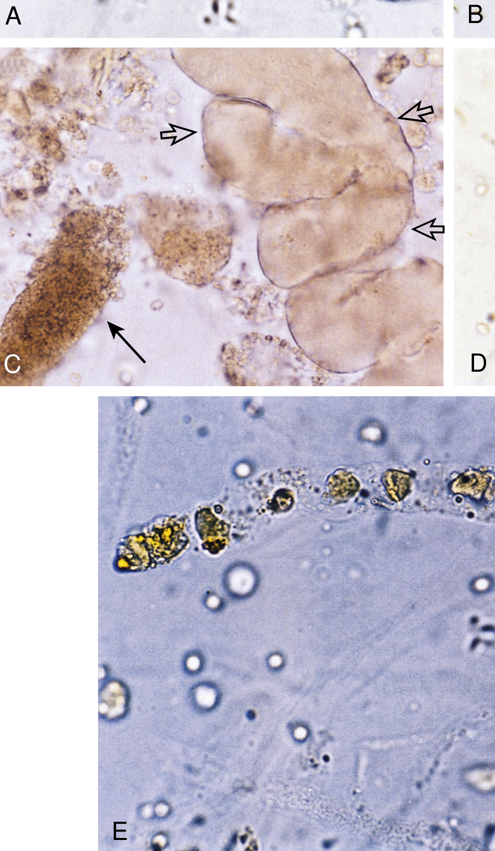
The Memory Trick:
"RBC cast = glomerulonephritis" - This is the single most tested cast. If you see RBC casts, the problem is INSIDE the glomerulus (nephritic syndrome). "Muddy brown cast = ATN" - Patient just had hypotension or a nephrotoxic drug. "WBC cast = pyelonephritis or interstitial nephritis" - Infection or allergy INSIDE the kidney, NOT just a bladder infection. "Waxy/broad cast = chronic kidney disease" - The tubules have given up and gotten fat.
PART 4: ACUTE KIDNEY INJURY (AKI)
What Is AKI?
A sudden drop in kidney function. The kidneys fail to filter waste, and creatinine/BUN builds up in the blood. Defined as:
- Rise in serum creatinine ≥0.3 mg/dL within 48 hours, OR
- Rise to ≥1.5x baseline within 7 days, OR
- Urine output <0.5 mL/kg/hr for >6 hours
The 3 Categories: Where Is the Problem?
Think about it like a highway to a factory and back:
1. PRERENAL AKI (most common - ~60-70%)
Problem: Not enough blood getting TO the kidneys.
The kidney itself is fine - it's just starving for blood flow.
Causes:
- Volume depletion (vomiting, diarrhea, bleeding, burns)
- Low cardiac output (heart failure, cardiogenic shock)
- Vasodilation (sepsis, medications like ACE inhibitors in some contexts)
- NSAIDs (reduce blood flow to glomerulus)
Urine findings:
- Bland sediment - no cells, no casts (or just hyaline casts)
- Concentrated urine (kidney is trying to hold onto every drop)
- Low urine sodium (<20 mEq/L) - kidney is desperately reabsorbing Na
- FENa <1% (fractional excretion of sodium - the kidney is holding onto sodium)
Key: Reversible quickly if you fix the underlying cause (give fluids, etc.)
2. INTRINSIC (RENAL) AKI
Problem: Damage to the kidney itself. This is where it gets interesting.
Sub-categories by what part of the kidney is damaged:
a) Acute Tubular Necrosis (ATN) - most common intrinsic AKI
- Tubules are damaged and die
- Causes: ischemia (prolonged hypotension = "shock kidneys"), nephrotoxins (aminoglycosides, contrast dye, myoglobin from rhabdomyolysis, cisplatin)
- Urine: Muddy brown granular casts + tubular epithelial cell casts
- FENa >2% (tubules can't reabsorb sodium anymore - they're broken)
- Three phases: Oliguric phase → Diuretic phase → Recovery phase
b) Acute Interstitial Nephritis (AIN)
- Inflammation of the space around the tubules (interstitium)
- Causes: Drugs (antibiotics - penicillin, NSAIDs), infections, autoimmune
- Classic triad: Fever + rash + eosinophilia (this is the allergic version)
- Urine: WBC casts + eosinophils in urine (eosinophiluria >5%)
c) Glomerulonephritis
- As discussed above in nephritic syndrome
- Urine: RBC casts + dysmorphic RBCs + proteinuria
3. POSTRENAL AKI (obstruction)
Problem: Urine can't get OUT. Pressure backs up and damages kidneys.
Causes:
- BPH (enlarged prostate)
- Kidney stones
- Tumor compressing ureters
- Strictures
Urine findings:
- Usually bland, or blood if stone is cutting tissue
- May have hematuria
Key: Fix the obstruction, often get recovery.
Urine Sediment Summary for AKI (Exam Gold)
| AKI Type | Urine Sediment |
|---|---|
| Prerenal | Bland (hyaline casts only) |
| ATN | Muddy brown granular casts + tubular cell casts |
| AIN | WBC casts + eosinophils |
| GN (nephritic) | RBC casts + proteinuria + dysmorphic RBCs |
| Postrenal | Bland (or hematuria if stones) |
PART 5: ACUTE CYSTITIS
What Is It?
A bladder infection. Simple as that. Bacteria get into the bladder (usually up the urethra) and infect it.
Most common organism: Escherichia coli (E. coli) - causes ~80% of cases
Who gets it: Mostly women (because their urethra is short and close to the anus). Much less common in men.
Symptoms (the "burning pee" syndrome):
- Dysuria - burning/pain when urinating
- Frequency - peeing constantly, small amounts
- Urgency - sudden strong urge to pee RIGHT NOW
- Suprapubic pain/discomfort
- Turbid (cloudy) or smelly urine
- Occasionally hematuria (blood in urine)
- No fever (if there's fever, think pyelonephritis = kidney infection)
Diagnosis:
- Urinalysis: Pyuria (WBCs in urine), bacteriuria, positive nitrite test, positive leukocyte esterase
- Urine culture: Gold standard - >100,000 colony forming units/mL
Treatment (First-Line Options):
- Nitrofurantoin (monohydrate/macrocrystals) 100 mg twice daily x 5 days - works great for uncomplicated cystitis; low resistance
- TMP-SMX (Trimethoprim-sulfamethoxazole) 1 tab twice daily x 3 days - classic choice; check local resistance (don't use if >20% resistance in your area)
- Fosfomycin 3g single dose - single dose convenience; very low resistance (~2%)
Fluoroquinolones (ciprofloxacin) are no longer recommended as first-line for simple UTIs (save them for complicated UTIs/pyelonephritis; preserve them for resistance reasons).
Uncomplicated vs. Complicated:
- Uncomplicated: Young healthy woman, lower urinary tract only - treat for 3-5 days
- Complicated: Men, pregnancy, diabetes, immunosuppression, structural abnormality, catheter, or pyelonephritis suspected - needs longer treatment + culture guidance
Cystitis vs. Pyelonephritis:
| Cystitis | Pyelonephritis | |
|---|---|---|
| Infection location | Bladder | Kidney |
| Fever | No | YES (systemic symptoms) |
| Flank/CVA tenderness | No | YES |
| Nausea/vomiting | No | Common |
| Treatment duration | 3-5 days oral | 7-14 days (IV if severe) |
THE MASTER CHEAT SHEET
Exam Pattern Recognition
| If you see this... | Think this... |
|---|---|
| Massive proteinuria + edema + low albumin + NO hematuria | Nephrotic syndrome |
| Hematuria + RBC casts + hypertension + oliguria | Nephritic syndrome |
| Muddy brown granular casts + recent hypotension or nephrotoxin | ATN |
| WBC casts + fever + recent antibiotic | Acute interstitial nephritis |
| pH↓ + HCO3-↓ + high anion gap | High-gap metabolic acidosis (MUDPILES) |
| pH↓ + HCO3-↓ + normal anion gap + high Cl- | Non-gap (hyperchloremic) metabolic acidosis |
| pH↑ + PCO2↑ | Metabolic alkalosis |
| pH↓ + PCO2↑ | Respiratory acidosis |
| pH↑ + PCO2↓ | Respiratory alkalosis |
| Dysuria + frequency + no fever | Acute cystitis |
| Dysuria + fever + flank pain | Pyelonephritis |
| Child with edema + proteinuria, responds to steroids | Minimal change disease |
| Adult with proteinuria + "spike and dome" on EM | Membranous nephropathy |
| Post-strep throat + hematuria + hypertension in a child | Post-streptococcal GN |
You've now got everything you need. The biggest exam traps are:
- Nephrotic (NO RBC casts) vs. Nephritic (HAS RBC casts) - this one distinction covers half the nephrology exam
- Prerenal (bland urine, FENa <1%) vs. ATN (muddy brown casts, FENa >2%)
- High anion gap acidosis (MUDPILES) vs. normal anion gap (diarrhea, RTA)
Good luck on your exam! 🎯
*Sources: Quick Compendium of Clinical Pathology 5th ed.; Brenner and Rector's The Kidney; Comprehensive Clinical Nephrology 7th ed.; NKF Primer on Kidney Diseases 8th ed.; Harrison's Principles of Internal Medicine 22nd ed.; Tintinalli's Emergency Medicine.*That's your complete study guide, built straight from authoritative nephrology and pathology textbooks. Here's a quick summary of what's in there:
-
Acid-Base - The 4 disorders (metabolic/respiratory acidosis and alkalosis), how to classify them using pH + HCO3- + PCO2, the anion gap formula with the MUDPILES mnemonic, and compensation formulas.
-
Nephrotic vs. Nephritic - The coffee filter analogy to lock in the concept, complete feature tables, and causes of each. The single biggest exam trap: RBC casts = nephritic, absent = nephrotic.
-
Urine Casts - A complete cast guide with what's inside each cast and what disease it points to, plus an actual microscopy image.
-
Acute Kidney Injury - The prerenal/intrinsic/postrenal classification with urine findings for each, including the ATN muddy brown cast vs. AIN WBC cast distinction.
-
Acute Cystitis - Symptoms, organisms, first-line treatment (nitrofurantoin, TMP-SMX, fosfomycin), and how to distinguish cystitis from pyelonephritis.
The master table at the end is your quick-reference for pattern recognition under exam pressure. You've got this.
This is a shared conversation. Sign in to Orris to start your own chat.