
I have duck feet walk what should I do
duck feet out-toeing gait correction exercises treatment adults
duck feet out-toeing gait posture correction exercises
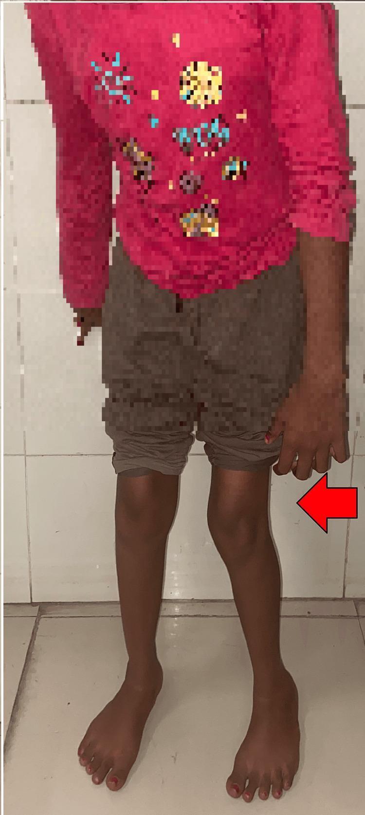
This clinical photograph displays a pediatric patient exhibiting classic postural signs associated with spastic diplegic cerebral palsy. The image focuses on the lower extremities, showing significant internal rotation and adduction at the hips. The knees are positioned close together, and there is evident in-toeing (metatarsus adductus) of the feet, which are oriented toward the body's midline. This characteristic alignment, often described as a 'scissoring' posture, is indicative of underlying spasticity and muscle contractures, specifically in the hip adductors and internal rotators. The red arrow highlights the medial deviation of the left leg. This visual is used in medical education to illustrate the pre-operative clinical presentation of spastic gait abnormalities and pelvic obliquity in neuromusculoskeletal disorders, serving as a baseline for surgical interventions such as adductor tenotomy and gracilis release.
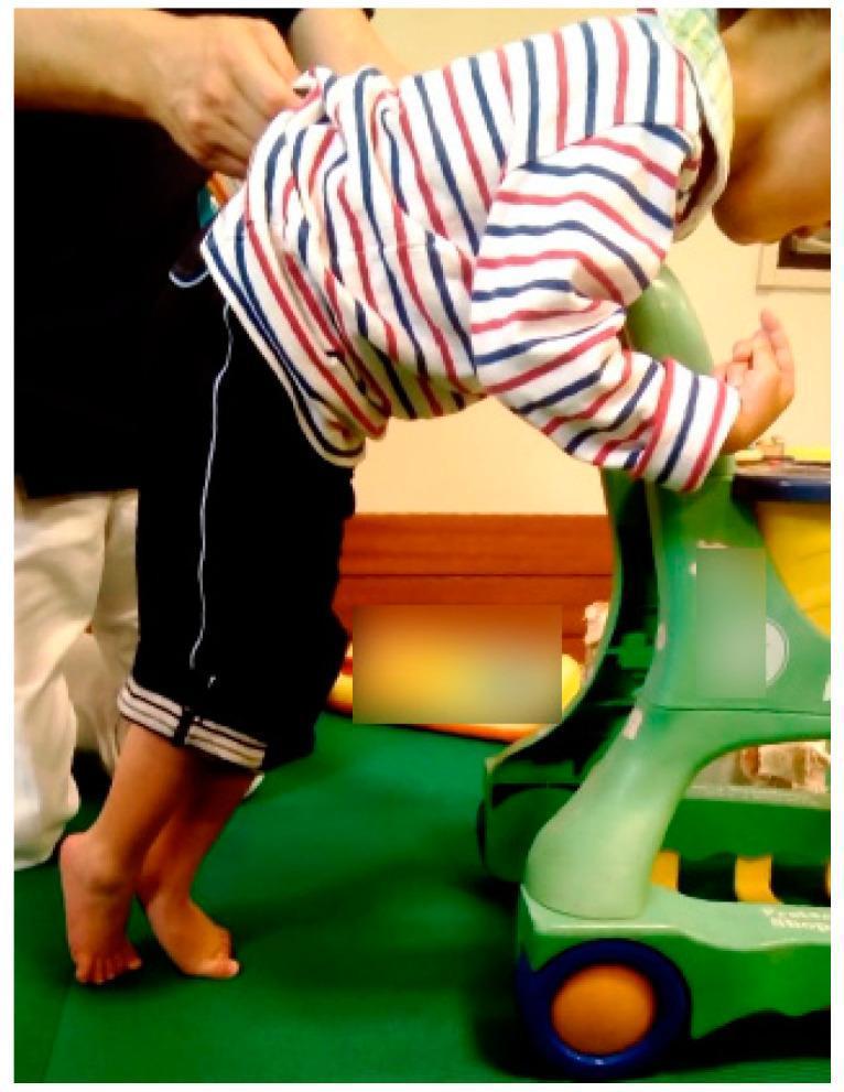
This clinical photograph displays a four-year-old child with spastic cerebral palsy demonstrating a characteristic standing posture while supported by a standing frame and manual assistance. The lower extremities exhibit a classic 'scissors gait' or scissors posture, characterized by hip adduction and internal rotation, causing the legs to cross. The feet are held in a marked equinus position (toe-walking), with the heels significantly elevated off the ground and weight borne on the forefoot. The child shows a forward-leaning trunk posture, utilizing the upper extremities for weight-bearing and balance on a mobility device. This visual illustrates spasticity and motor impairment consistent with Gross Motor Function Classification System (GMFCS) Level IV. The image is an educational example of pediatric neuromotor dysfunction, specifically demonstrating the compensatory postural adaptations and lower limb deformities associated with spastic diplegia.
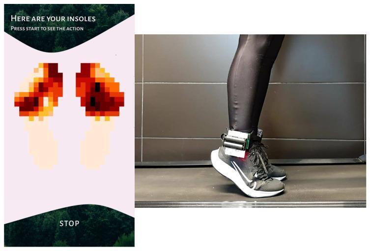
This composite image illustrates a wearable gait analysis system for monitoring plantar pressure distribution during physical activity. On the right, a clinical photograph shows a subject on a treadmill wearing athletic shoes equipped with a prototype hardware module attached at the ankle. The hardware consists of a microcontroller development board and a battery pack connected to a 0.4 mm electronic insole embedded with 16 pressure sensors. On the left, a screenshot of the integrated mobile application displays a real-time heat map of the pressure distribution for both feet. The visualization uses a pixelated 10x22 grid where color gradients (from pale yellow to deep red) represent increasing pressure levels, specifically highlighting high-load areas in the forefoot and midfoot regions during a gait cycle. This system is designed for gait correction, posture control in runners, and physiotherapeutic monitoring of lower limb biomechanics.
What Is "Duck Feet"?
Why Does It Happen?
| Cause | Fixable with Exercise? |
|---|---|
| Tight hip external rotators (piriformis, glutes) | Yes - stretching helps a lot |
| Weak hip internal rotators / glute medius | Yes - strengthening works |
| Habitual posture (lifelong sitting pattern) | Yes - gait retraining helps |
| Femoral retroversion (bony - thigh bone angles backward) | Partially - manage symptoms, not the bone |
| External tibial torsion (bony - shin rotated outward) | Partially - same as above |
| Flat feet / overpronation | Yes - orthotics + arch exercises |
What You Should Do
1. Stretch Tight Hip External Rotators
- Lie on your back, cross one ankle over the opposite knee (figure-4 position)
- Pull the uncrossed leg toward your chest until you feel a stretch in the crossed-leg glute
- Hold 30-45 seconds, 3 sets per side daily
- Kneeling lunge, push hips forward
- Tilt your body slightly away from the kneeling-side knee
- Hold 30-45 seconds per side
2. Strengthen Hip Internal Rotators & Glute Medius
- Lie on your side, knees bent at 90°, feet together
- Lift the top knee like a clamshell opening - keep feet together
- 3 sets x 15-20 reps per side
- Same position, but lift the whole top leg toward the ceiling with foot flexed
- 3 sets x 15 reps per side
- Lie on back, one knee bent, one leg straight
- Drive through the bent leg's heel to lift hips - squeeze glute at top
- 3 sets x 10-12 reps per side
- Band around ankles, slight squat position
- Take 10 steps sideways - keep feet pointing forward
- 3 sets x 10 steps each direction
3. Gait Retraining (Conscious Habit Change)
- When standing, consciously point your feet straight forward or just slightly out (~5°)
- Walking: Focus on pushing off through your big toe, not the outer edge of your foot
- Cue yourself: Think "toes forward" with each step; set reminders if needed
- Practice walking in front of a mirror or filming yourself to see progress
4. Ankle and Foot Work
- Big toe extension exercises: Spread toes, lift big toe while keeping others down - improves push-off mechanics
- Arch strengthening (short foot exercise): While seated, try to "shorten" your foot by doming the arch without curling toes
- Tennis ball roll: Roll under each foot for 2 minutes to release the plantar fascia
5. Orthotics and Footwear
- Arch-supporting orthotics can help if flat feet are contributing
- Avoid high heels and stiff-soled shoes - they worsen alignment
- Look for shoes with neutral or slight motion-control properties
6. When to See a Professional
- You have knee, hip, or lower back pain alongside your gait issue
- Home exercises haven't helped after 6-8 weeks
- You want a proper gait analysis to identify the exact cause
- The issue is severe or causing you to trip/fall
Realistic Expectations
- Habitual/muscular causes: With consistent daily work, most people see noticeable improvement in 4-8 weeks
- Bony/structural causes (retroversion, tibial torsion): Exercise won't change the bone angle, but strengthening the glutes and hips still reduces knee strain and back pain that out-toeing causes - so it's still worth doing
- Surgery is only considered in severe cases causing significant functional problems, usually in children