
Explain muscle of upper limb
Reading File
Now let me search the medical library for upper limb muscles.
Finding Sources
Finding Sources
Finding Sources
Reading File
Reading File
Finding Sources
Reading File
Reading File
Reading File
Reading File
Reading File
Now I have comprehensive data from the THIEME Atlas. Let me also fetch a couple of key anatomical diagrams to include.
Loading Image
Loading Image
Reading File
I now have extensive content from the THIEME Atlas of Anatomy. Let me compile the comprehensive answer.
Muscles of the Upper Limb
The upper limb muscles are classified by their topographical location and function. They span four regions: the shoulder girdle, the shoulder joint, the arm, the forearm, and the hand.
1. Classification Principles
Muscles of the upper limb can be grouped by:
- Origin (trunk-derived vs. limb bud)
- Topography (where they sit)
- Function (flexors vs. extensors)
- Innervation - the most clinically useful grouping
The nerves for extensors (radial and axillary nerves) arise from the posterior divisions of the brachial plexus, while nerves for flexors (musculocutaneous, ulnar, median) arise from the anterior divisions.
2. Muscles of the Shoulder Girdle
These muscles move and stabilize the scapula.
Muscles that migrated from the head (CN XI innervated)
| Muscle | Action | Innervation |
|---|---|---|
| Trapezius | Elevates/retracts/depresses scapula; tilts head | Accessory nerve (CN XI), C2-C4 |
| Sternocleidomastoid | Turns head; elevates sternum in breathing | Accessory nerve (CN XI) |
Trapezius has three parts:
- Descending part - draws scapula obliquely upward, tilts head to same side
- Transverse part - draws scapula medially
- Ascending part - draws scapula medially downward
Posterior trunk/shoulder girdle muscles
| Muscle | Origin | Insertion | Action | Nerve |
|---|---|---|---|---|
| Levator scapulae | Transverse processes C1-C4 | Superior angle of scapula | Elevates scapula medially; inclines neck | Dorsal scapular nerve, C3-C4 |
| Rhomboid minor | Spinous processes C6-C7 | Medial border of scapula (above spine) | Steadies scapula; draws it medially upward | Dorsal scapular nerve (C4, C5) |
| Rhomboid major | Spinous processes T1-T4 | Medial border of scapula (below spine) | Steadies scapula; draws it medially upward | Dorsal scapular nerve (C4, C5) |
Anterior trunk/shoulder girdle muscles
| Muscle | Action | Nerve |
|---|---|---|
| Subclavius | Stabilizes clavicle; depresses shoulder | Subclavian nerve (C5, C6) |
| Pectoralis minor | Draws scapula downward/forward; assists in inspiration | Medial pectoral nerve (C8, T1) |
| Serratus anterior | Protracts and rotates scapula (critical for arm elevation); holds scapula against thorax | Long thoracic nerve (C5-C7) |
3. Muscles of the Shoulder Joint
Rotator Cuff (SITS) - stabilize the glenohumeral joint
| Muscle | Origin | Insertion | Action | Nerve |
|---|---|---|---|---|
| Supraspinatus | Supraspinous fossa | Greater tubercle of humerus | Initiates abduction (0-15°); stabilizes head | Suprascapular nerve (C5, C6) |
| Infraspinatus | Infraspinous fossa | Greater tubercle | External rotation; stabilizes head | Suprascapular nerve (C5, C6) |
| Teres minor | Lateral border of scapula | Greater tubercle | External rotation; adduction | Axillary nerve (C5, C6) |
| Subscapularis | Subscapular fossa | Lesser tubercle | Internal rotation; adduction | Subscapular nerve (C5-C7) |
Other shoulder joint muscles
| Muscle | Origin | Insertion | Action | Nerve |
|---|---|---|---|---|
| Deltoid | Lateral clavicle, acromion, scapular spine | Deltoid tuberosity of humerus | Abduction (after supraspinatus initiates); flexion (anterior part); extension (posterior part) | Axillary nerve (C5, C6) |
| Latissimus dorsi | Spinous processes T7-T12, thoracolumbar fascia, iliac crest, lower ribs | Floor of intertubercular groove | Internal rotation, adduction, extension; expiration | Thoracodorsal nerve (C6-C8) |
| Teres major | Inferior angle of scapula | Crest of lesser tubercle | Internal rotation, adduction, extension | Lower subscapular nerve (C5-C7) |
| Pectoralis major | Clavicle, sternum, costal cartilages | Lateral lip of intertubercular groove | Adduction, internal rotation, flexion (clavicular head) | Medial and lateral pectoral nerves |
| Coracobrachialis | Coracoid process | Middle third of humerus | Flexion and adduction of arm | Musculocutaneous nerve (C5-C7) |
4. Muscles of the Arm
Anterior (Flexor) Compartment
| Muscle | Origin | Insertion | Action | Nerve |
|---|---|---|---|---|
| Biceps brachii (long head) | Supraglenoid tubercle | Radial tuberosity + bicipital aponeurosis | Elbow flexion; supination (most powerful when elbow is flexed); shoulder stabilization | Musculocutaneous nerve (C5-C7) |
| Biceps brachii (short head) | Coracoid process | Same as long head | Same as long head | Musculocutaneous nerve |
| Brachialis | Distal half of anterior humerus | Ulnar tuberosity | Pure elbow flexor (most powerful flexor) | Musculocutaneous nerve + radial nerve (C5-C7) |
Posterior (Extensor) Compartment
| Muscle | Origin | Insertion | Action | Nerve |
|---|---|---|---|---|
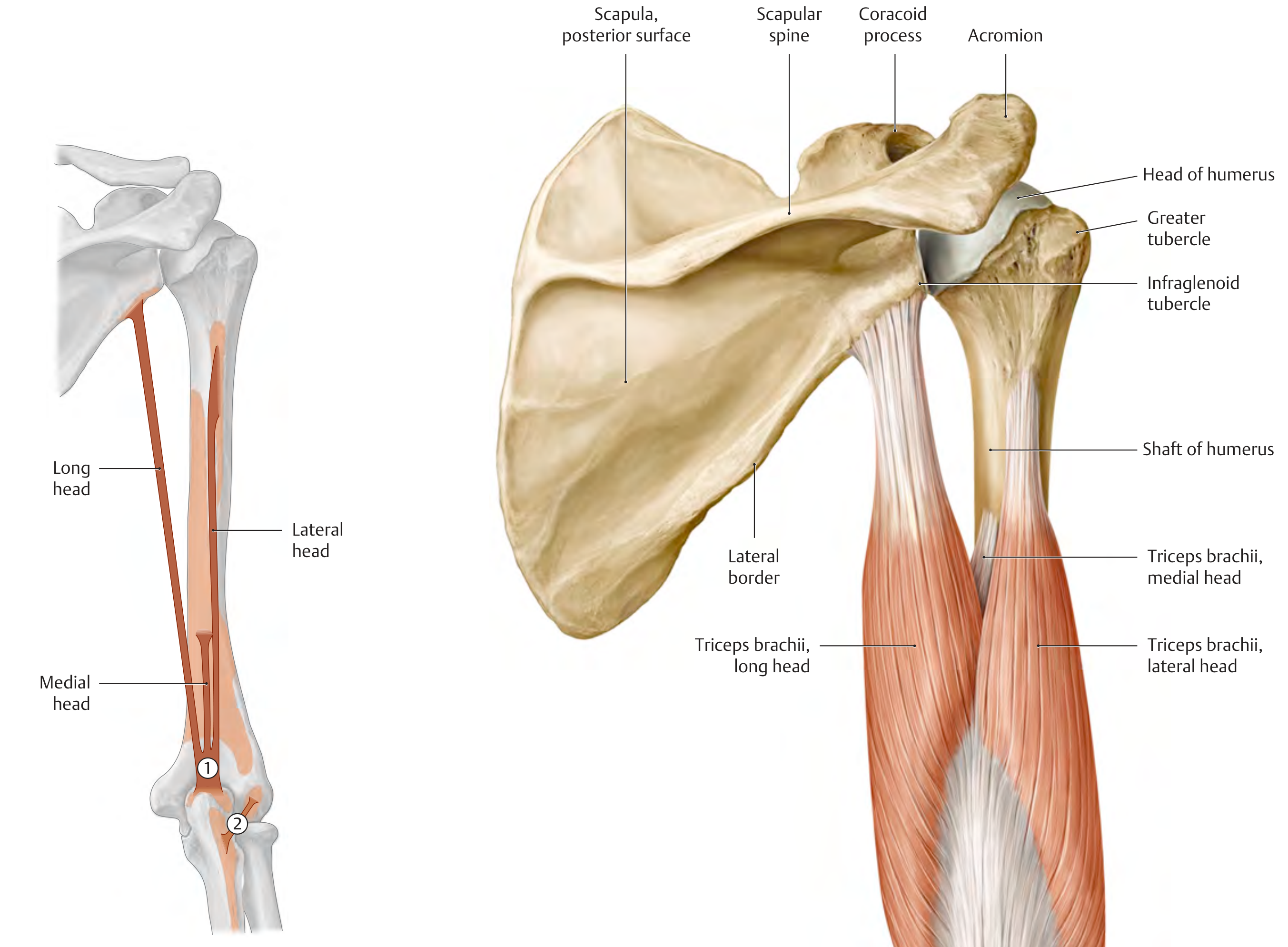
| Triceps brachii (long head) | Infraglenoid tubercle of scapula | Olecranon of ulna | Elbow extension; shoulder adduction/extension | Radial nerve (C6-C8) |
| Triceps brachii (medial head) | Posterior humerus distal to radial groove | Olecranon | Elbow extension | Radial nerve |
| Triceps brachii (lateral head) | Posterior humerus proximal to radial groove | Olecranon | Elbow extension | Radial nerve |
| Anconeus | Lateral epicondyle of humerus | Radial surface of olecranon | Extends elbow; tightens joint capsule | Radial nerve (C6-C8) |
Clinical note: The radial nerve runs in the radial groove (spiral groove) of the humerus. A mid-shaft humeral fracture can damage it, causing wrist drop (loss of wrist and finger extension).
5. Muscles of the Forearm
Radialis (Lateral) Group
| Muscle | Action | Nerve |
|---|---|---|
| Brachioradialis | Elbow flexion; semipronation | Radial nerve (C5, C6) |
| Extensor carpi radialis longus | Wrist extension + radial deviation | Radial nerve (C6, C7) |
| Extensor carpi radialis brevis | Wrist extension + radial deviation | Radial nerve (C7, C8) |
Anterior (Flexor) Compartment of Forearm
Superficial layer (all from medial epicondyle via common flexor origin):
| Muscle | Main Action | Nerve |
|---|---|---|
| Pronator teres | Pronation; elbow flexion | Median nerve (C6, C7) |
| Flexor carpi radialis | Wrist flexion + radial deviation | Median nerve (C6, C7) |
| Palmaris longus | Wrist flexion; tightens palmar aponeurosis | Median nerve (C7, C8) |
| Flexor carpi ulnaris | Wrist flexion + ulnar deviation | Ulnar nerve (C7, C8) |
| Flexor digitorum superficialis | Flexion at PIP joints of digits 2-5 | Median nerve (C7, C8, T1) |
Deep layer:
| Muscle | Main Action | Nerve |
|---|---|---|
| Flexor digitorum profundus | Flexion at DIP joints digits 2-5 | Median nerve (lateral half); Ulnar nerve (medial half) |
| Flexor pollicis longus | Flexion of thumb (IP joint) | Median nerve (C8, T1) |
| Pronator quadratus | Pronation | Anterior interosseous branch of median nerve (C8, T1) |
Posterior (Extensor) Compartment of Forearm
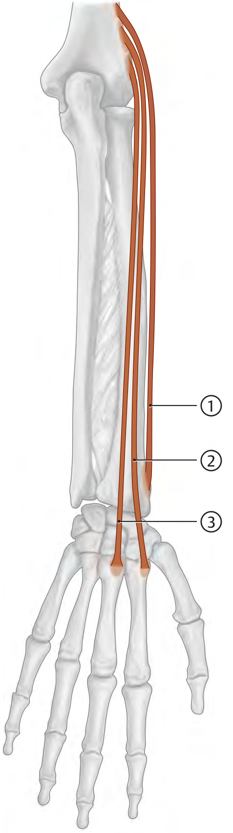
Superficial extensors (from lateral epicondyle - common extensor origin):
| Muscle | Action | Nerve |
|---|---|---|
| Extensor digitorum | Extends digits 2-5 at MCP joints | Radial nerve - posterior interosseous (C7, C8) |
| Extensor digiti minimi | Extends little finger | Posterior interosseous nerve |
| Extensor carpi ulnaris | Wrist extension + ulnar deviation | Posterior interosseous nerve |
Deep extensors:
| Muscle | Action | Nerve |
|---|---|---|
| Supinator | Supination of forearm | Posterior interosseous nerve (C5-C7) |
| Abductor pollicis longus | Abducts and extends thumb | Posterior interosseous nerve |
| Extensor pollicis brevis | Extends thumb MCP joint | Posterior interosseous nerve |
| Extensor pollicis longus | Extends thumb IP joint (forms "anatomical snuffbox" border) | Posterior interosseous nerve |
| Extensor indicis | Extends index finger | Posterior interosseous nerve |
6. Muscles of the Hand
Thenar Muscles (thumb eminence - all median nerve, C8-T1)
| Muscle | Action |
|---|---|
| Abductor pollicis brevis | Abducts thumb at MCP joint |
| Flexor pollicis brevis | Flexes thumb at MCP joint (superficial head: median; deep head: ulnar) |
| Opponens pollicis | Opposition of thumb (most functionally important) |
| Adductor pollicis | Adducts thumb - innervated by ulnar nerve (exception!) |
Hypothenar Muscles (little finger eminence - all ulnar nerve, C8-T1)
| Muscle | Action |
|---|---|
| Abductor digiti minimi | Abducts little finger; flexes at MCP |
| Flexor digiti minimi brevis | Flexes little finger at MCP |
| Opponens digiti minimi | Opposition of little finger |
| Palmaris brevis | Tightens palmar aponeurosis (protective) |
Intrinsic Hand Muscles
| Muscle | Number | Action | Nerve |
|---|---|---|---|
| Lumbricals | 4 | Flex MCP joints; extend PIP/DIP joints | Lateral two: Median; Medial two: Ulnar |
| Dorsal interossei | 4 | Abduct fingers (DAB); flex MCP; extend IP | Ulnar nerve |
| Palmar interossei | 3 | Adduct fingers (PAD); flex MCP; extend IP | Ulnar nerve |
7. Innervation Summary
| Nerve | Compartments Supplied |
|---|---|
| Axillary nerve (C5, C6) | Deltoid, teres minor |
| Musculocutaneous nerve (C5-C7) | Anterior arm (biceps, brachialis, coracobrachialis) |
| Radial nerve (C5-C8) | Posterior arm + forearm (all extensors, brachioradialis, anconeus) |
| Median nerve (C6-T1) | Most anterior forearm flexors; lateral thenar; lateral two lumbricals |
| Ulnar nerve (C7-T1) | Flexor carpi ulnaris, medial FDP; all intrinsics except lateral thenar |
Memory aid for hand intrinsics: "All intrinsics of the hand are ulnar, EXCEPT the LOAF muscles" - Lumbricals (lateral 2), Opponens pollicis, Abductor pollicis brevis, Flexor pollicis brevis = Median nerve.
Source: General Anatomy and Musculoskeletal System, THIEME Atlas of Anatomy
This is a shared conversation. Sign in to Orris to start your own chat.