
In which GI issue the pain goes after meal ?
Duodenal ulcer is the classic GI condition where pain is relieved by eating - food acts as a buffer against acid, so the pain goes away after meals (only to return 2-3 hours later when the stomach empties again).
Searching sources...
Reading File
Finding Sources
Finding Sources
Finding Sources
The textbooks confirm the answer clearly. Here is a full explanation:
GI Pain Relieved by Eating: Duodenal Ulcer
Duodenal ulcer is the classic GI condition where pain is relieved after eating a meal.
Why does food relieve the pain?
The duodenum is the first part of the small intestine, just past the stomach. When the stomach is empty, acid pours directly onto the ulcerated mucosa of the duodenum, causing pain. When food enters the stomach:
- Food acts as a chemical buffer, neutralizing gastric acid
- This reduces the acid load reaching the duodenal ulcer
- The pain therefore eases after eating
This is sometimes called "hunger pain" - the pain typically comes on 2-4 hours after a meal (when the stomach empties again), is relieved by eating, and is often worse at night.
Classic Pain Pattern of Duodenal Ulcer:
| Feature | Duodenal Ulcer |
|---|---|
| Location | Mid-epigastric, may radiate to back |
| Timing | 2-4 hours after meals, nocturnal |
| Effect of food | Relieves the pain |
| Effect of antacids | Relieves pain |
| Character | Burning, "empty/hungry" feeling |
Contrast with Gastric Ulcer:
In gastric ulcer, the pain is typically worsened by food, because eating stimulates more acid secretion directly onto the ulcer in the stomach. Anorexia and weight loss are also more common in gastric ulcers.
As stated in S Das: A Manual on Clinical Surgery, "in duodenal ulcer, food relieves pain (hunger pain)." - and Tintinalli's Emergency Medicine confirms: "Duodenal ulcer pain may be relieved after eating food, whereas gastric ulcer pain is often exacerbated by eating."
One Special Case - Zollinger-Ellison Syndrome:
In this rare condition (gastrin-secreting tumor), duodenal ulcers form but are multiple and refractory. The pain pattern may still include partial relief with food/antacids, but ulcers are resistant to standard treatment.
In summary: Duodenal ulcer is the GI condition where pain characteristically goes away after eating a meal, due to the acid-buffering effect of food.
How to diagnose duodenal ulcer ?
Duodenal ulcer is diagnosed through a combination of clinical history, endoscopy (gold standard), H. pylori testing, and sometimes barium studies. Endoscopy directly visualizes the ulcer and allows biopsy.
Searching sources...
Finding Sources
Finding Sources
Finding Sources
Finding Sources
Reading File
Reading File
Finding Sources
Finding Sources
Reading File
Finding Sources
Excellent - I have comprehensive information from the textbooks. Here is a detailed answer:
How to Diagnose Duodenal Ulcer
Diagnosis is based on a combination of clinical assessment, endoscopy (gold standard), and H. pylori testing.
1. Clinical History (Symptoms)
The diagnosis cannot be confirmed on clinical grounds alone, but a "classic" history raises strong suspicion:
- Epigastric burning/gnawing pain - midepigastric, sometimes radiating to the back
- Pain relieved by food, milk, or antacids (hunger pain)
- Nocturnal pain - waking the patient at night (2-4 AM)
- Periodic pattern - weeks of pain followed by pain-free intervals
- Absent weight loss (unlike gastric ulcer or malignancy)
Alarm features requiring urgent investigation:
- Unintentional weight loss
- Dysphagia or persistent vomiting
- GI bleeding (hematemesis, melena, iron-deficiency anaemia)
- Epigastric mass
- Failure to respond to PPI therapy
- Family history of GI cancer
2. Physical Examination
Usually unremarkable in uncomplicated duodenal ulcer. Findings may include:
- Mild epigastric tenderness on deep palpation
- Signs of complications: board-like rigidity (perforation), pallor (bleeding)
3. Investigations
A. Endoscopy (Gold Standard - EGD/OGD)
- Esophagogastroduodenoscopy (EGD) is the definitive investigation
- Provides direct visualization of the ulcer in the duodenum (usually first part/D1)
- Allows tissue biopsy for H. pylori testing and to exclude malignancy
- Identifies complications (bleeding, deformity)
- Sensitivity and specificity are very high
B. H. pylori Testing
Since ~90% of duodenal ulcers are caused by H. pylori, testing is mandatory:
| Test | Type | Notes |
|---|---|---|
| Urea Breath Test (UBT) | Non-invasive | Most accurate non-invasive test; sensitivity & specificity ~95%. Best for confirming eradication |
| Stool H. pylori Antigen | Non-invasive | Good sensitivity & specificity; also used to confirm eradication |
| Rapid Urease Test (CLO test) | Invasive (biopsy at endoscopy) | Quick; may be false-negative if on PPI |
| Histopathology (biopsy) | Invasive | Simultaneous diagnosis of ulcer + H. pylori; gold standard for H. pylori |
| Serum H. pylori Antibody | Non-invasive | Indicates past or present infection only; cannot confirm eradication (stays positive up to 18 months post-treatment) |
Note: PPIs can suppress H. pylori tests - stop PPIs 2 weeks before testing where possible.
C. Barium Meal (Upper GI Series)
- Older technique, still used if endoscopy is not tolerated or fails
- Barium within an ulcer crater is diagnostic
- Secondary changes (edema, scarring, deformity) may also be seen
- Less sensitive than endoscopy; cannot biopsy
D. CT Scan
- Not the test of choice for duodenal ulcer
- Occasionally demonstrates ulceration but is much less sensitive
- Useful for detecting complications like perforation (free air under diaphragm)
4. Summary Diagnostic Approach
Suspect Duodenal Ulcer (classic history)
         |
         ↓
Any ALARM features?
   YES → Urgent EGD
   NO  → Age < 60 with no alarm features?
              ↓
      Test & Treat strategy:
      Non-invasive H. pylori test (UBT or stool antigen)
      If positive → treat
      If no response → EGD
Sources: Washington Manual of Medical Therapeutics; Yamada's Textbook of Gastroenterology, 7th ed.; Tintinalli's Emergency Medicine
What's the treatment for duodenal ulcers ?
Treatment of duodenal ulcer centers on acid suppression (PPIs), H. pylori eradication, and removing causative factors like NSAIDs. Surgery is reserved for complications.
Searching sources...
Finding Sources
Finding Sources
Reading File
Reading File
Reading File
Finding Sources
Reading File
Finding Sources
I now have comprehensive information. Here is the full treatment answer:
Treatment of Duodenal Ulcer
Treatment has four main pillars: lifestyle changes, acid suppression, H. pylori eradication, and management of complications.
1. Lifestyle Modifications
- Stop NSAIDs/aspirin - a major causative factor; if aspirin is essential for cardiovascular protection, co-prescribe a daily PPI
- Smoking cessation - smoking doubles the risk and delays ulcer healing
- Avoid alcohol - irritates the mucosa
- Dietary changes - avoid foods that worsen symptoms (spicy food, caffeine)
2. Acid Suppression (Antisecretory Therapy)
A. Proton Pump Inhibitors (PPIs) - First-Line
PPIs are the most potent and preferred antisecretory agents:
- Irreversibly block H⁺/K⁺-ATPase (proton pump) on parietal cells
- Healing rates: 85% at 4 weeks, 96% at 8 weeks
- Duodenal ulcers are treated for 8 weeks (gastric ulcers need 12 weeks)
- Examples: Omeprazole, Esomeprazole, Pantoprazole, Lansoprazole, Rabeprazole
Maintenance PPI is considered in:
- Large ulcers (>2 cm)
- Refractory or frequently recurring PUD
- Failed H. pylori eradication
- Patients who cannot stop NSAID/aspirin use
B. H2 Receptor Antagonists (H2RAs) - Second-line
- Block histamine H2 receptors on parietal cells
- Less potent than PPIs - largely replaced by PPIs
- Still useful when PPIs are not tolerated
- Examples: Famotidine (most potent), Ranitidine, Cimetidine
- Dose adjustment needed in renal insufficiency
C. Antacids
- Oldest form of therapy; neutralize gastric acid chemically
- Used for symptomatic relief, not definitive treatment
- Types: Aluminum hydroxide, Magnesium trisilicate, Calcium carbonate
- Do not combine with PPIs - an alkaline environment prevents PPI activation
D. Sucralfate
- Mucosal protective agent; forms a protective coating over the ulcer
- Adjunct therapy to promote healing
3. H. pylori Eradication (Core of Treatment)
Since ~90% of duodenal ulcers are caused by H. pylori, eradication is mandatory when infection is confirmed. Eradication reduces recurrence rates to just 2% (vs. 60-80% without eradication).
First-Line Regimens (14-day course recommended)
Selection is guided by penicillin allergy, prior macrolide exposure, and local clarithromycin resistance:
| Scenario | Regimen |
|---|---|
| No penicillin allergy, no prior macrolide exposure, low clarithromycin resistance | Clarithromycin Triple Therapy: PPI + Amoxicillin 1g BD + Clarithromycin 500mg BD |
| No penicillin allergy, prior macrolide exposure OR clarithromycin resistance >15% | Bismuth Quadruple Therapy: PPI + Bismuth + Tetracycline + Metronidazole |
| Penicillin allergy, no prior macrolide exposure | Bismuth Quadruple Therapy |
| Penicillin allergy + prior macrolide exposure | Bismuth Quadruple Therapy |
| Alternative option | Levofloxacin Triple Therapy: PPI + Levofloxacin + Amoxicillin |
Note: Clarithromycin resistance is rising globally (20-35% worldwide). Bismuth quadruple therapy is now preferred in many regions as first-line.
Second-Line (if first-line fails)
- Bismuth quadruple therapy (if not used first-line)
- Levofloxacin triple therapy
- Always avoid repeating antibiotics that were used in the failed first-line regimen
Third-Line / Salvage
- Culture and sensitivity-guided therapy
- Rifabutin-based regimens
- Multidrug regimens guided by a gastroenterologist
Confirm Eradication
Test at 4-6 weeks after completing therapy using:
- Urea Breath Test (UBT) - preferred
- Stool H. pylori antigen test
- Do NOT use serology - antibodies persist for up to 18 months
4. Surgical Treatment
Surgery is rarely needed in the modern era due to excellent medical therapy. It is reserved for complications:
| Complication | Surgical Procedure |
|---|---|
| Bleeding duodenal ulcer | Oversewing of the bleeding vessel + H. pylori treatment |
| Perforation | Omental patch closure (Graham patch) + H. pylori treatment |
| Gastric outlet obstruction | Rule out malignancy, then gastrojejunostomy |
| Intractable (failed all medical therapy) | Truncal vagotomy ± antrectomy |
Vagotomy reduces acid secretion by denervating parietal cells - historically the mainstay of surgical PUD management, but now rarely performed.
Treatment Summary Flow
Confirmed Duodenal Ulcer
        |
        ↓
Test for H. pylori
   Positive → PPI + Antibiotics (eradication regimen x14 days)
              + Confirm eradication at 4-6 weeks
   Negative → PPI alone x 8 weeks
              + Stop NSAIDs + lifestyle changes
        |
        ↓
Complications? → Endoscopic or surgical intervention
Sources: Sabiston Textbook of Surgery, 21st ed. - Yamada's Textbook of Gastroenterology, 7th ed. - Washington Manual of Medical Therapeutics - ACG Clinical Guideline: Treatment of H. pylori Infection 2017
How to identify peptic ulcers
Peptic ulcers are identified through clinical history, physical examination, endoscopy (gold standard), H. pylori testing, and sometimes imaging. Endoscopy directly visualizes the ulcer and allows biopsy.
Searching sources...
Finding Sources
Finding Sources
Finding Sources
Finding Sources
Reading File
Finding Sources
Finding Sources
Finding Sources
Reading File
Reading File
Now let me fetch the radiology image of a benign gastric ulcer that was found in the textbook:
Loading Image
Finding Sources
I now have a comprehensive picture from the textbooks. Here is the complete answer:
How to Identify Peptic Ulcers
Peptic ulcer identification involves three layers: clinical recognition, investigations, and endoscopic/radiological characterization.
1. Clinical Identification (History & Symptoms)
A "classic" history strongly suggests peptic ulcer disease, though symptoms alone cannot confirm the diagnosis:
| Feature | Duodenal Ulcer | Gastric Ulcer |
|---|---|---|
| Pain location | Mid-epigastric | Mid-epigastric |
| Effect of food | Relieved by eating | Worsened by eating |
| Timing | 2-4 hrs after meals, nocturnal | Soon after meals |
| Weight | Maintained | May lose weight |
| Character | Burning, "hungry/empty" feeling | Burning, dull ache |
| Antacid relief | Yes | Yes |
Alarm features - these always require urgent endoscopy:
- Unexplained weight loss
- Dysphagia (difficulty swallowing)
- Persistent vomiting
- GI bleeding (hematemesis, melena, iron-deficiency anaemia)
- Palpable epigastric mass
- Failure to respond to PPI after 4-6 weeks
- Age >55 with new dyspepsia
2. Physical Examination
Usually normal in uncomplicated peptic ulcer. May find:
- Mild epigastric tenderness on deep palpation
- Succussion splash - suggests gastric outlet obstruction
- Signs of complications: board-like rigidity (perforation), pallor/tachycardia (bleeding)
3. Definitive Investigation - Endoscopy (Gold Standard)
Upper GI Endoscopy (EGD/OGD) is the test of choice:
- Provides direct visualization of the ulcer
- Identifies the location (stomach vs. duodenum), size, depth, and edges
- Allows biopsy - mandatory for all gastric ulcers to exclude malignancy
- Simultaneous H. pylori testing via rapid urease (CLO) test or histology
- Identifies active bleeding and stigmata of hemorrhage (Forrest classification)
Endoscopic Appearance - Benign vs. Malignant Gastric Ulcer
| Feature | Benign Ulcer | Malignant Ulcer |
|---|---|---|
| Ulcer crater | Round or ovoid, smooth | Irregular crater |
| Edges | Smooth, raised, erythematous; covered by gray fibrous exudate | Nodular, heaped-up, irregular |
| Gastric folds | Radiate smoothly to the crater edge | Clubbed or fused, stop before the ulcer |
| Wall | Projects beyond the gastric contour | Projects inside the gastric lumen |
| Location | Lesser curvature, posterior wall; within 2 cm of antral-fundal junction | Variable |
The visual appearance of benign and malignant gastric ulcers may be identical - differentiation can only be confirmed by biopsy. Multiple biopsies from the perimeter of the lesion + brushings increase diagnostic accuracy to ~95%.
4. H. pylori Testing
Since H. pylori causes ~90% of peptic ulcers, testing is a key part of identification:
| Test | Method | Notes |
|---|---|---|
| Urea Breath Test | Non-invasive | Sensitivity/specificity ~95%; best for confirmation and test-of-cure |
| Stool Antigen Test | Non-invasive | Good sensitivity & specificity |
| Rapid Urease (CLO) Test | At endoscopy (biopsy) | Quick; may be false-negative on PPIs |
| Histopathology | At endoscopy (biopsy) | Gold standard for H. pylori; also diagnoses ulcer |
| Serology (IgG antibody) | Blood test | Only shows past or current exposure; cannot confirm eradication |
5. Barium Meal (Upper GI Series)
Used when endoscopy is not tolerated or has failed:
- Barium within an ulcer crater is diagnostic
- Benign gastric ulcer appears as a round/ovoid barium collection with smooth radiating folds projecting beyond the gastric wall contour
- Malignant ulcer projects inside the lumen within a tumor mass, with nodular folds and acute angles at the wall
- Secondary changes from edema, scarring, or deformity may also be visible
Barium meal image - Benign Gastric Ulcer:
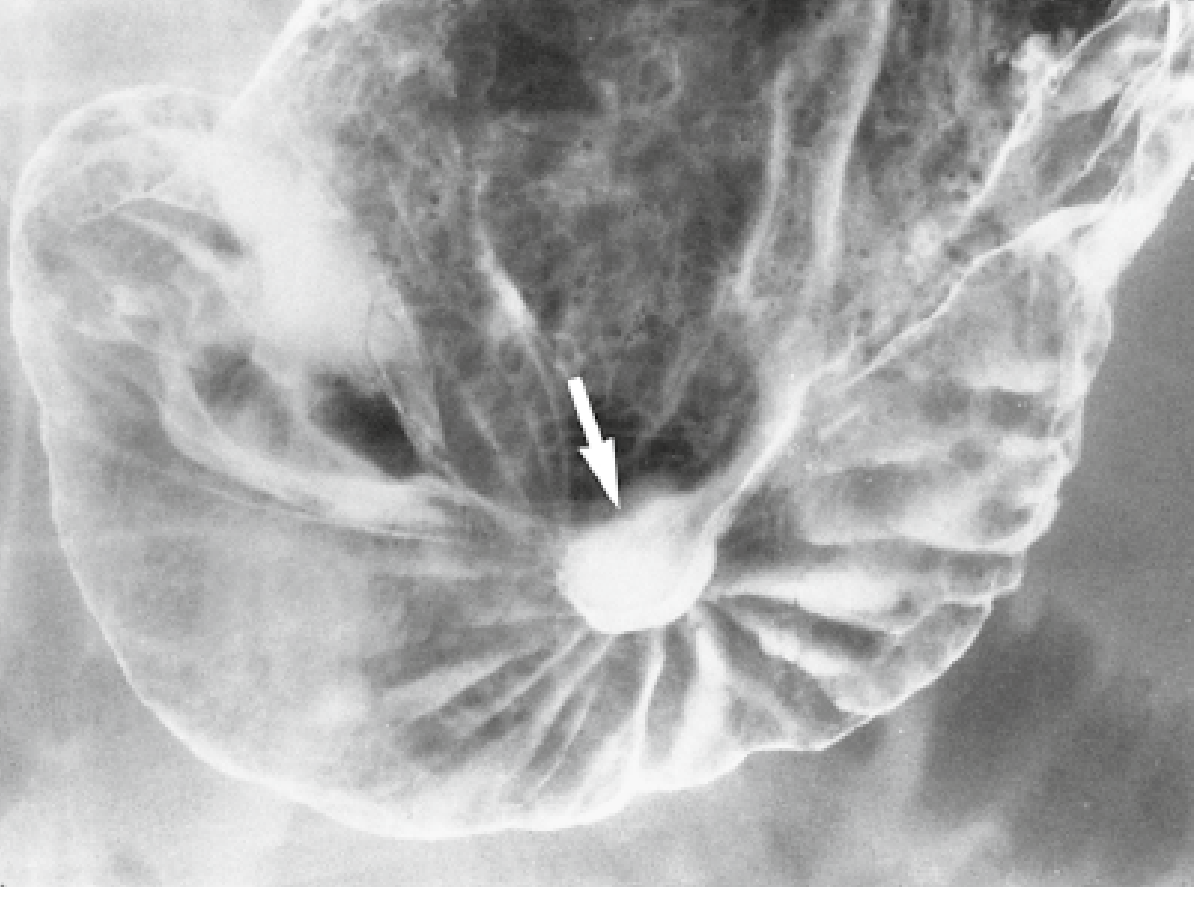
Arrow shows a round, symmetrical benign gastric ulcer on the posterior wall with smooth folds radiating to the ulcer crater edge - classic benign radiological appearance. (Yamada's Textbook of Gastroenterology, 7th ed.)
6. CT Scan
- Not the primary test for peptic ulcer identification
- Much less sensitive than endoscopy or barium studies
- Useful for detecting complications: free air (perforation), abscesses, fistulas
Summary: Identification Approach
Dyspepsia / epigastric symptoms
            |
    Alarm features present?
    YES → Urgent EGD (endoscopy)
    NO  → Age < 55?
              YES → Non-invasive H. pylori test
                    Positive → Test & Treat
                    Negative → Trial of PPI
                    No response → EGD
              NO  → EGD (higher cancer risk)
Sources: Yamada's Textbook of Gastroenterology, 7th ed. - Maingot's Abdominal Operations - Washington Manual of Medical Therapeutics - Tintinalli's Emergency Medicine
Compare symptoms of Peptic ulcers to duodenal ulcers
A duodenal ulcer IS a type of peptic ulcer - so their symptoms overlap significantly, but there are key distinguishing features worth comparing.
Searching sources...
Finding Sources
Finding Sources
Finding Sources
Finding Sources
Finding Sources
Finding Sources
Reading File
Reading File
Excellent - the textbooks provided a detailed symptom frequency table. Here is the complete comparison:
Peptic Ulcer vs. Duodenal Ulcer: Symptom Comparison
First, an Important Clarification
A duodenal ulcer is a subtype of peptic ulcer disease (PUD). "Peptic ulcer" is the umbrella term covering ulcers in the stomach (gastric ulcer) and the first part of the small intestine (duodenal ulcer). So when comparing "peptic ulcer symptoms" vs. "duodenal ulcer symptoms," the most clinically useful comparison is gastric ulcer vs. duodenal ulcer.
Shared Symptoms (Present in All Peptic Ulcers)
Both gastric and duodenal ulcers share these core features:
- Epigastric (upper abdominal) burning/gnawing pain - the hallmark symptom
- Nausea
- Vomiting
- Bloating and belching
- Relief with antacids
- Episodic pattern - weeks of pain, then pain-free intervals
- Nocturnal pain - waking the patient at night
- Silent/asymptomatic presentation - common in both
Key Differences: Gastric Ulcer vs. Duodenal Ulcer
| Symptom / Feature | Gastric Ulcer | Duodenal Ulcer |
|---|---|---|
| Effect of food on pain | Food worsens pain (postprandial pain) | Food relieves pain (hunger pain) |
| Pain timing | Pain within 30-60 min of eating | Pain 2-4 hrs after meals; nocturnal |
| Nocturnal pain | 32-43% | 50-88% (more common) |
| Pain severity | Frequently severe (68%) | Frequently severe (53%) |
| Anorexia | 46-57% (more common) | 25-36% |
| Weight loss | 24-61% (more common) | 19-45% |
| Vomiting | 38-73% (more common) | 25-57% |
| Nausea | 54-70% | 49-59% |
| Increased appetite | Not typical | 19% (hunger pain drives eating) |
| Heartburn | 19% | 27-59% (more common) |
| Pain clusters (episodic) | 16% | 56% (much more periodic) |
| Food relief of pain | 2-48% | 20-63% (more reliably relieved) |
| Radiation to back | 34% | 20-31% |
| Fatty food intolerance | - | 41-72% |
| Typical patient | Older (50-70s), M > F | Younger (30-50s), M > F |
| Relation to H. pylori | ~70-80% | ~90-95% |
| NSAID association | Strong | Moderate |
| Malignancy risk | Must always exclude cancer | Very rare malignant transformation |
Frequency data from Yamada's Textbook of Gastroenterology, 7th ed., Table 49.3
The Classic Pain Pattern - Side by Side
GASTRIC ULCER                    DUODENAL ULCER
──────────────────────           ──────────────────────
Eat meal                         Eat meal
   ↓                                ↓
Pain STARTS or WORSENS           Pain RELIEVED (food buffers acid)
   ↓                                ↓
Avoids food → loses weight       2-4 hours later: stomach empties
                                    ↓
                                 Acid hits ulcer → Pain RETURNS
                                    ↓
                                 Eats again → Pain relieved again
                                 (cycle repeats, gains/maintains weight)
Important Caveats
- Symptom overlap is high - distinguishing gastric from duodenal ulcer on symptoms alone is unreliable; endoscopy is needed
- Elderly patients (>65 yrs) often have no symptoms at all or only atypical symptoms (nausea, weight loss, bleeding) regardless of ulcer type
- Asymptomatic "silent" ulcers are common in both types, especially in NSAID users
- Alarm symptoms (unexplained weight loss, vomiting, bleeding, dysphagia, epigastric mass) in either type require urgent endoscopy to rule out malignancy
Sources: Yamada's Textbook of Gastroenterology, 7th ed. - Tintinalli's Emergency Medicine - Swanson's Family Medicine Review
This is a shared conversation. Sign in to Orris to start your own chat.