
Venous vs arterial vs neurogenic ulcers
"leg ulcer"[MeSH Terms] AND "diagnosis"
venous ulcer gaiter area lipodermatosclerosis
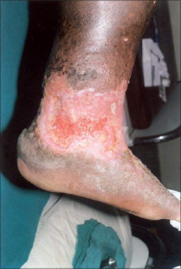
Clinical photograph of the medial aspect of the lower leg, ankle, and foot of a patient, demonstrating a large, chronic venous stasis ulcer. The ulcer features highly irregular borders and a base covered with mixed granulation tissue and yellowish fibrinous slough. The surrounding skin exhibits significant dermatological changes characteristic of long-standing venous insufficiency and chronic venous hypertension, including hyperpigmentation (stasis dermatitis) and induration. The texture of the peri-ulcer skin is suggestive of lipodermatosclerosis, characterized by a 'woody' or thickened appearance and atrophy of the subcutaneous fat. The ulcer's location in the 'gaiter area'—the lower third of the leg above the medial malleolus—is a hallmark clinical finding for venous ulcers. This image serves as an educational example of advanced chronic venous disease and the resulting cutaneous manifestations and ulceration.
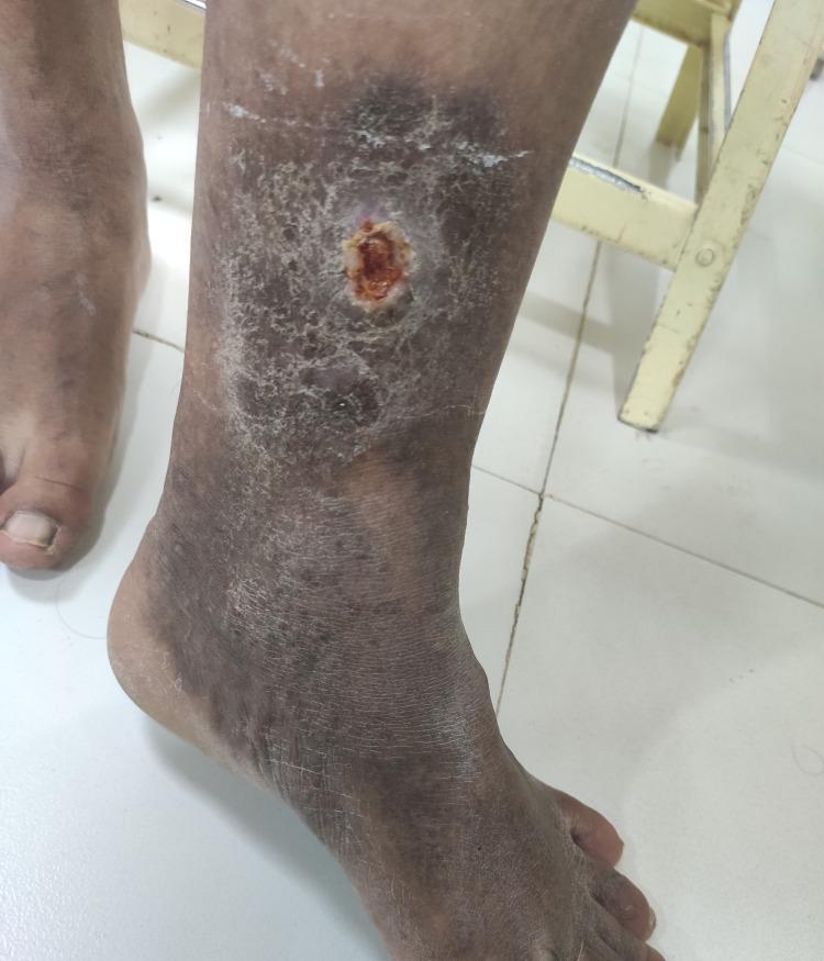
This clinical photograph displays a chronic venous leg ulcer located superior to the left medial malleolus in an elderly male patient. The ulcer is approximately 2 cm in diameter, circular, with a central reddish-brown granulating base and a surrounding pale ring. The adjacent lower leg skin exhibits classic signs of advanced chronic venous insufficiency (CVI), specifically lipodermatosclerosis and stasis dermatitis. Notable features include diffuse hyperpigmentation (hemosiderin staining), induration, and a mottled, scaly texture. Fine white reticulated lines, characteristic of atrophie blanche, are visible within the hyperpigmented region. The distal foot and toes appear relatively spared compared to the gaiter area of the leg. This image serves as a clinical example of CEAP grade C6 venous disease, illustrating the cutaneous manifestations of long-standing venous hypertension and its progression to active ulceration.
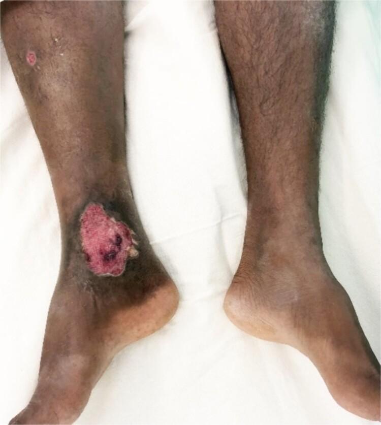
Clinical photograph showing a comparative view of the lower limbs, highlighting chronic cutaneous changes on the right leg. The most prominent feature is a large, irregularly shaped ulcer located in the right medial gaiter area, just above the ankle. The ulcer bed contains reddish-pink granulation tissue with focal dark, necrotic areas and distinct white, thickened borders. Surrounding the ulcer is extensive, circumferential hyperpigmentation and lipodermatosclerosis-like skin changes, characteristic of chronic venous insufficiency or stasis. A smaller, secondary circular ulcer is visible higher on the anterior shin of the same limb. In contrast to the relatively normal hair distribution and skin tone of the left leg, the right lower limb exhibits significant alopecia (loss of hair) and trophic skin changes. This visual evidence supports a diagnosis of a chronic venous ulcer, potentially secondary to underlying vascular pathology such as an arteriovenous fistula (AVF) or post-thrombotic syndrome.
Venous vs arterial vs neurogenic (neuropathic) ulcers
| Feature | Venous | Arterial (ischaemic) | Neuropathic |
|---|---|---|---|
| Frequency | About 85% of chronic leg ulcers in resource-rich countries (Bailey & Love, p. 1060) | Less common | Common in diabetes |
| Cause | Venous hypertension from superficial, deep or perforator incompetence, or obstruction | Atherosclerotic occlusion (smoking, diabetes, hypertension, hyperlipidaemia), Buerger disease, cholesterol emboli | Sensory (plus motor and autonomic) neuropathy causing repetitive unnoticed pressure or trauma |
| Site | Gaiter area, above the medial malleolus | Toes, pressure points, lateral ankle, dorsum of foot, or sites of shoe friction | Plantar surface: metatarsal heads, heel, pressure points over deformities |
| Pain | Mild to moderate, eased by elevation | Severe, worse on elevation and at night, relieved by dependency | Painless |
| Margin | Irregular, sloping, shallow | Punched-out or irregular, well demarcated | Punched-out with regular margin, surrounded by callus |
| Base | Granulation tissue and yellow slough, exudative | Pale, minimal granulation, necrotic or with dry eschar | Deep, may track to tendon or bone (osteomyelitis risk) |
| Surrounding skin | Hyperpigmentation (haemosiderin), lipodermatosclerosis, stasis eczema, atrophie blanche, oedema, varicose veins | Thin, shiny, hairless, pale or cyanotic, dependent rubor | Calluses, dry skin, bony deformity (Charcot, claw toes) |
| Temperature | Warm | Cold | Warm, dry |
| Pulses | Present (but 15-30% have coexisting arterial disease, a "mixed" ulcer) | Reduced or absent, with a low ABI | Present |
| Veins | Varicose or dilated | Collapsed | Dilated (from arteriovenous shunting) |
| Sensation | Normal | Variable | Lost: monofilament, vibration, reflexes |
Venous ulcer
- Diagnosis: Duplex ultrasound maps reflux and obstruction. Check the ABI before compression, because 15-30% of "venous" ulcers have coexisting arterial disease.
- Treatment: Multilayer graduated compression (4-layer, about 40 mmHg at the ankle) is the mainstay once arterial disease is excluded. Treat superficial reflux with endovenous ablation, which reduces recurrence. Add leg elevation and exercise.
Arterial ulcer
- Presentation: A red, painful plaque breaks down into a painful ulcer with surrounding purpuric erythema. Granulation is minimal and there is often little infection. A dry, inactive eschar may form (Andrews').
- Signs of arterial cause:
- skin thinning and hair loss
- reduced or absent pulses
- pallor on elevation
- a cool limb
- dependent rubor
- claudication
- night pain relieved by hanging the foot down
- Cholesterol emboli: Suspect these if there is sudden onset, eosinophilia, renal insufficiency and palpable pulses.
- ABI: Below 0.75 indicates arterial insufficiency and below 0.5 indicates severe insufficiency (Andrews'). Falsely high values occur in calcified vessels, as in diabetes or renal failure, so use toe pressures or TcPO2 there.
- Treatment:
- Revascularisation (endovascular or bypass) is the key step.
- Do not apply compression to a limb with significant arterial disease.
- Control risk factors and stop smoking.
- Protect the area from injury and cold.
- An ischaemic ulcer marks a limb at increased risk of loss.
Neuropathic ulcer
- Mechanism: Loss of protective sensation, with motor neuropathy causing deformity and high focal pressure. Autonomic neuropathy causes dry, fissured skin. The foot is warm with bounding pulses (Schwartz's).
- Treatment: Offloading is the primary principle (Andrews'). Total contact cast or removable walker are options, and shoes or padded boots can offload different areas.
- Debride necrotic tissue back to bleeding viable tissue. This is usually painless because the foot is insensate.
- Treat clinical infection, but colonisation alone usually does not need treatment.
- Osteomyelitis is best treated by removing the infected bone.
- After healing, use appropriately fitting depth and width shoes, inspect the feet regularly for hot spots, and debride dystrophic nails to prevent recurrence.
- Neuroischaemic foot: Diabetic feet often have both neuropathy and PAD. Always check perfusion.
Practical points
- Check pulses and the ABI in every leg ulcer, and add duplex if venous disease is suspected.
- Painless plantar ulcer with callus and good pulses points to neuropathic. Painful toe or ankle ulcer with absent pulses points to arterial. Gaiter-area ulcer with pigmentation and varicosities points to venous.
- Biopsy any non-healing or atypical ulcer to exclude malignancy such as squamous cell carcinoma (Marjolin ulcer). Also consider vasculitis and pyoderma gangrenosum.
Recent evidence check
History taking of digital pitting in raynauds
"Raynaud Disease"[MeSH Terms] AND "digital ulcers"
History taking: digital pitting in Raynaud's
- characterise the Raynaud's
- characterise the pitting and any ulcers
- look for an underlying connective tissue disease (CTD) or other cause
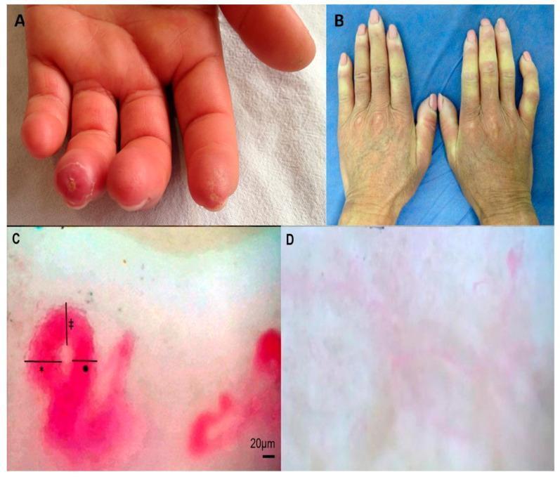
1. Presenting complaint and Raynaud's characterisation
- Did the fingers go white, then blue, then red? Raynaud's is clinically a triphasic attack: painful pallor or ischaemia, then cyanosis in some, then reactive hyperaemia on rewarming (Fitzpatrick's Dermatology, p. 1119).
- Is the change sharply demarcated? Which digits are involved? Does it spare the thumbs?
- Pallor alone, or colour change in the toes, ears, nose or lips, also matters.
- Are attacks provoked by cold or by emotional stress?
- What is the frequency, duration and severity of attacks? Do they happen in summer?
- When did the first attack occur?
- Primary Raynaud's typically starts before age 30.
- Onset after age 30-40, rapid worsening, or attacks that are asymmetric, severe or painful point to a secondary cause.
- Is there pain between attacks or at rest?
- Have you had fingertip ulcers, sores or gangrene?
- Any digital pulp loss or tightening?
2. The pitting and digital ulcers
- When and where: When was the pitting first noticed? Which fingers? Is it on the fingertip pulp only, or also over the extensor surfaces of the joints? Extensor ulcers over contractures are typical of diffuse SSc (Rheumatology, 2022).
- Ulcer history: Have there been ulcers before the pitting, and how many in total? How long did they take to heal? Have any recurred or become chronic? Chronic or recurrent DU carry the greatest risk and burden (Rheumatology, 2022).
- Complications:
- infection, discharge or antibiotic courses
- bone infection or hospital admission
- gangrene, autoamputation or surgery
- sudden severe persistent ischaemia with abrupt onset, which is a medical emergency (Rheumatology, 2022)
- Pain and function: How severe is the pain? Is there trouble with grip, buttons, writing or work?
- Trauma: Was there any injury or pressure at the site?
3. Screening for an underlying CTD (systems review)
- Puffy fingers or tight, thick skin? Where does the tightness extend (fingers only, forearm, face, trunk)? Have rings stopped fitting?
- Calcium lumps (calcinosis) or small red spots (telangiectasia)?
- Mouth tightness or reduced mouth opening?
- Rash, photosensitivity or hair loss (lupus)? Heliotrope or Gottron-type rash (dermatomyositis)?
- Joint pain or swelling, morning stiffness, proximal muscle weakness (overlap or mixed CTD)?
- Heartburn, dysphagia, early satiety, bloating, constipation or diarrhoea?
- Breathlessness, dry cough, exertional symptoms or syncope, which suggest interstitial lung disease or pulmonary hypertension?
- Chest pain, palpitations or oedema?
- New severe headache, visual change or hypertension, which may signal scleroderma renal crisis?
- Dry eyes or dry mouth?
- Recurrent oral or nasal ulcers? Pleuritic pain?
| Antibody | Associated features |
|---|---|
| Centromere | Limited skin sclerosis, severe gut disease, isolated PAH, calcinosis |
| Scl-70 | Diffuse skin sclerosis, pulmonary fibrosis |
| RNA polymerase III | Diffuse skin sclerosis, renal crisis |
| U1RNP | Overlap features, arthritis |
4. Other causes of Raynaud's and digital ischaemia
- Beta-blockers
- Ergotamine and triptans
- Stimulants (ADHD medication, amphetamines, cocaine)
- Chemotherapy (bleomycin, cisplatin)
- Interferon
- Nicotine
- Vibrating tools (vibration white finger)
- Vinyl chloride
- Repetitive hand trauma (hypothenar hammer syndrome)
- Cold exposure
- Cervical rib or thoracic outlet symptoms
- Atrial fibrillation
- Antiphospholipid syndrome (clots, miscarriages)
- Cryoglobulins or cold agglutinins (cold-induced purpura, hepatitis C)
- Malignancy
- Paraproteinaemia
- Heavy smoking with a young male patient points to Buerger disease. Arterial causes of digital ulceration are suggested by arm claudication, rest pain and tissue loss (Current Surgical Therapy 14e).
5. Other history
- Past medical history: Atherosclerotic risk factors, diabetes and previous vascular disease.
- Family history: CTD, Raynaud's, thyroid disease.
- Smoking: This worsens vasospasm and impairs ulcer healing, so ask about it directly.
- Medicines: Include over-the-counter products. Ask about any vasodilators or antiplatelets already used and whether they worked.
- Pregnancy plans: Relevant for drug choice, particularly endothelin antagonists, which are contraindicated in pregnancy.
- Social: Occupation, hobbies and impact on daily life.
Red flags to escalate
- Abrupt, severe, persistent ischaemia with threatened tissue (critical digital ischaemia)
- Signs of infection (spreading erythema, discharge, fever)
- New breathlessness or hypertension with headache
- Rapidly progressive skin tightening
Likely next steps
- Examination of hands, skin, nailfolds, pulses and blood pressure
- Nailfold capillaroscopy
- ANA with ENA panel, including centromere, Scl-70 and RNAP III antibodies
- Baseline FBC, renal function and urine dipstick
- Echocardiogram and lung function tests as directed by symptoms