
Concise this 4 metal posions pdf into a single pdf so for last minute revision it's much efficient for exam include a to z all details u can add images for better and easy understanding certain new pneumonics
mkdir -p /tmp/workspace/metal_poisons/{src,output,assets} && cd /tmp/workspace/metal_poisons && python3 -c " import pdfplumber, os files = { 'arsenic': '/tmp/workspace/attachments/3d6c6674-ef18-40c3-b7c2-735a6b9b62c6/10.1.-Arsenic.pdf', 'lead': '/tmp/workspace/attachments/3d6c6674-ef18-40c3-b7c2-735a6b9b62c6/10.3.- Lead poisoning.pdf', 'copper': '/tmp/workspace/attachments/3d6c6674-ef18-40c3-b7c2-735a6b9b62c6/10.4.-Copper poisoning.pdf', 'mercury': '/tmp/workspace/attachments/3d6c6674-ef18-40c3-b7c2-735a6b9b62c6/Mercury.pdf', } for name, path in files.items(): out = f'/tmp/workspace/metal_poisons/src/{name}.txt' with pdfplumber.open(path) as pdf: text = '' for i, page in enumerate(pdf.pages): t = page.extract_text() or '' text += f'--- page {i+1} ---\n' + t + '\n' with open(out, 'w') as f: f.write(text) print(name, 'pages:', len(pdf.pages) if False else '', 'chars:', len(text)) "
lead poisoning burton line gums basophilic stippling
Kayser-Fleischer ring copper Wilson disease
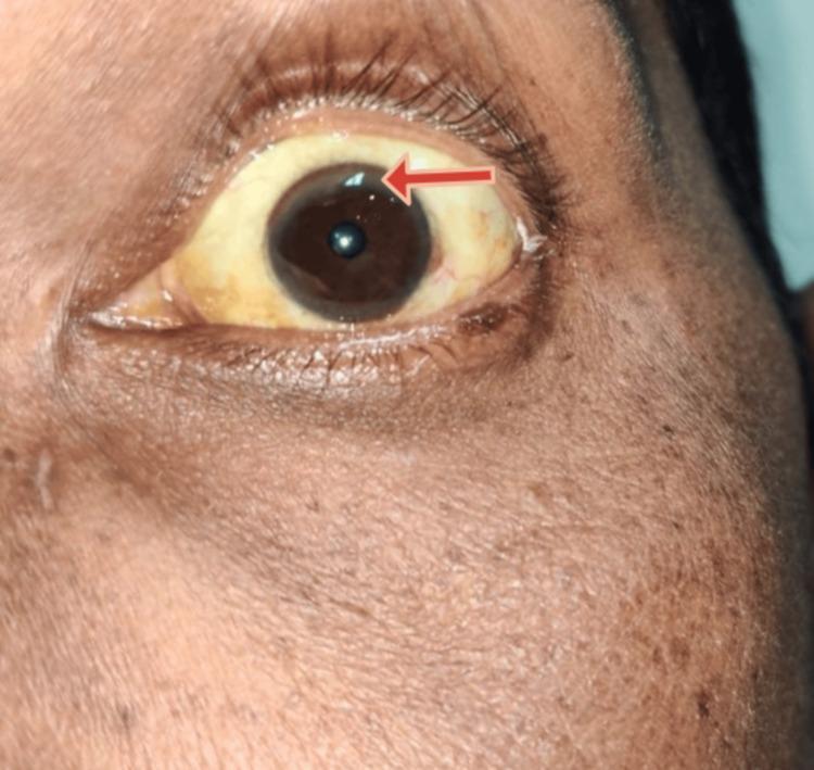
A close-up clinical photograph of a left eye and periorbital region demonstrating characteristic signs of hepatolenticular degeneration (Wilson disease). The sclera exhibits marked icterus (yellowish discoloration), consistent with hyperbilirubinemia or underlying chronic liver disease. At the peripheral margin of the cornea, a brownish-yellow Kayser-Fleischer (KF) ring is visible, particularly highlighted by a red arrow in the superior-temporal quadrant. This ring represents copper deposition in Descemet's membrane. The iris is dark brown, and the surrounding skin shows a slightly uneven pigmentation. The image serves as an educational example of ophthalmologic manifestations associated with systemic metabolic disorders and copper metabolism dysfunction.
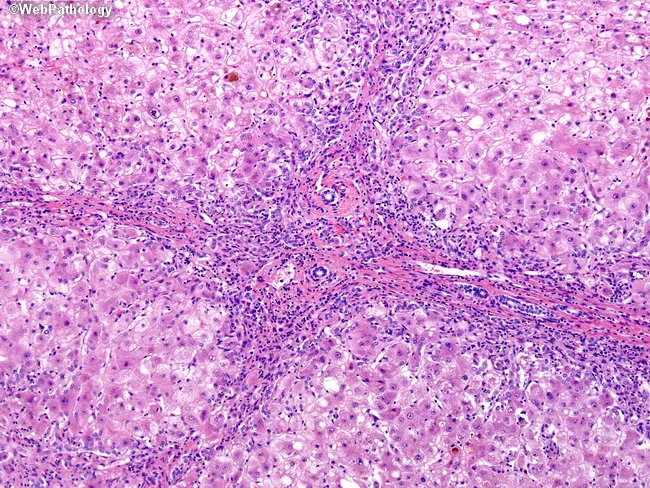
Light microscopy of a formalin-fixed liver biopsy stained with Hematoxylin and Eosin demonstrates advanced Wilson disease–related hepatic injury. The hepatic architecture is distorted by micronodular to early macronodular cirrhosis with regenerative nodules separated by thickened fibrous septa containing lymphoplasmacytic inflammatory cells and ductular reactions. Within the parenchyma, widespread macrovesicular steatosis and hepatocellular ballooning degeneration are evident, accompanied by focal dense eosinophilic cytoplasm and cholestatic changes. Portal tracts show chronic inflammatory infiltrates, interface activity, and occasionally bile duct proliferation. The combination of steatosis, ballooning degeneration, cholestasis, and progressive fibrotic remodeling reflects copper-induced hepatotoxicity in untreated disease. Copper accumulation in hepatocytes drives oxidative injury and disrupted biliary copper export, contributing to hepatocellular damage and fibrosis. These histologic features correlate with clinical laboratory abnormalities and can inform diagnosis, disease staging, and prognosis. In differential diagnosis, similar features can appear in nonalcoholic fatty liver disease or autoimmune hepatitis but the context of Wilson disease (ATP7B mutation) and possible hepatic copper quantification supports etiologic attribution. Clinically, histology complements genetic testing, ceruloplasmin levels, and Kayser-Fleischer ring assessment, guiding chelation therapy and monitoring for progression to cirrhosis, portal hypertension, and hepatocellular carcinoma risk. This histology supports timely intervention and guides multidisciplinary management, including chelation and surveillance strategies.
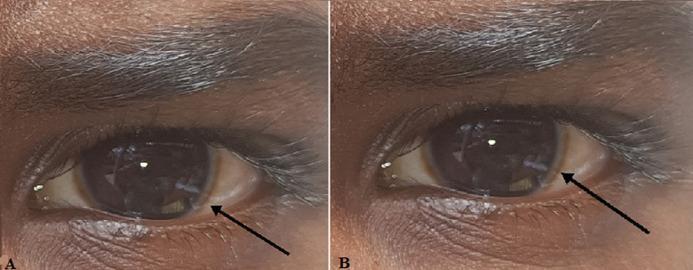
This clinical photograph consists of two panels (A and B) showing the right and left eyes of a patient with dark-pigmented skin. The primary pathological finding is a Kayser-Fleischer ring, visible as a distinct brownish-golden or greenish-brown discoloration at the periphery of the cornea near the limbus. The ring is most prominent in the superior and inferior poles, appearing as a circumferential band that encircles the dark brown iris. The sclera shows mild conjunctival vascularity but is otherwise white. The pupils are round and reactive to light. This ocular sign is a hallmark clinical manifestation of Wilson disease, resulting from copper deposition in Descemet's membrane. The image serves as a classic educational example for hepatology, neurology, and ophthalmology, illustrating the diagnostic physical exam findings associated with impaired copper metabolism.
mercury poisoning acrodynia erethism
Mees lines transverse white lines fingernails arsenic
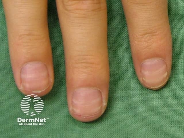
Clinical photograph of fingernails showing transverse leukonychia (leukonychia striata). High-resolution dorsal view demonstrates white horizontal bands across the nail plates, parallel to the lunula, varying in width but generally uniform across affected digits. The bands reflect disruption of nail plate translucency with preserved underlying nail bed color. Etiology ranges from minor trauma and filing to systemic conditions; classic Mees lines are transverse leukonychia associated with arsenic exposure, whereas Beau’s lines are ridges due to growth arrest. In this image, the bands are discrete, smooth-edged, and do not extend proximally beyond the nail fold, suggesting leukonychia of the nail plate or matrix involvement rather than subungual pathology. The finding is often benign but may indicate nutritional deficiencies (zinc, calcium), nutritional status, or toxin exposure; clinical correlation is essential. The differential includes traumatic leukonychia, zinc deficiency, arsenic poisoning, drug-induced nail changes, and systemic illness. The diagnostic significance lies in recognizing potential clues to toxicity or malnutrition, prompting history taking and laboratory evaluation if new or progressive. This image is valuable for dermatology training, medical education, and clinical toxicology, enabling rapid recognition of transverse bands and informing subsequent assessment. Accurate interpretation supports early intervention and improves patient outcomes including toxin screening when indicated.
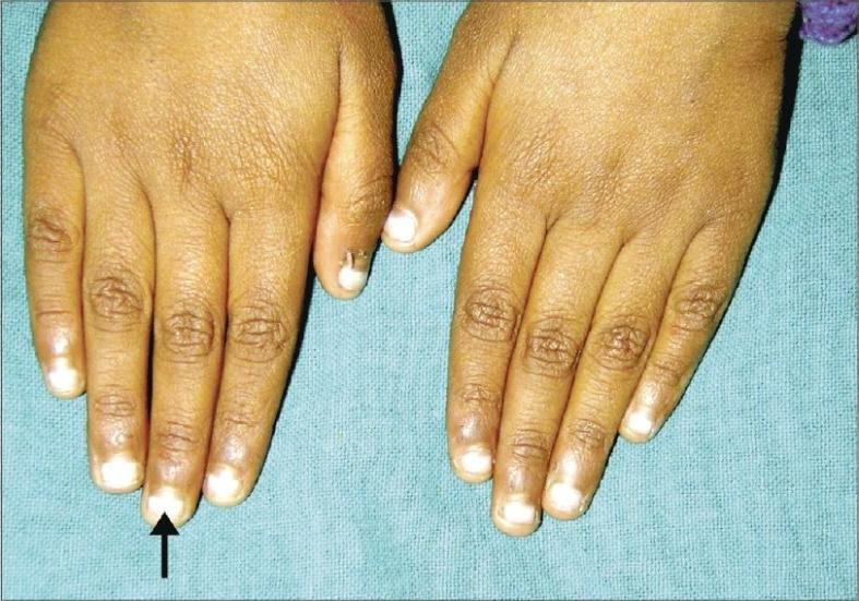
Clinical photograph of the dorsal aspect of both hands in a pediatric patient, demonstrating Mees' lines (true leukonychia). The image shows multiple fingernails with well-defined, single, white transverse bands approximately 2 mm in width. These hypopigmented bands span the entire breadth of the nail plate and run parallel to the lunula. The overlying nail surface is smooth and intact, indicating the pathology is within the nail plate substance rather than a surface deformity. This clinical sign is classically associated with systemic insults such as arsenic poisoning, heavy metal toxicity, or, as in this context, certain chemotherapy regimens for acute lymphoblastic leukemia. The distribution across multiple digits suggests a systemic etiology affecting the nail matrix during a specific period of growth. The presence of these non-blanchable lines is an important diagnostic indicator in dermatology and oncology for tracking toxic exposures or treatment side effects.
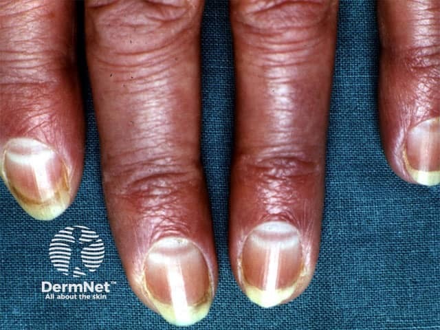
Clinical photography of the nails demonstrating Muehrcke lines. Anterolateral view of several fingernails (index, middle, ring, and little fingers) on both hands. The nail plates show paired, parallel, transverse white bands crossing the lunula region and extending toward the distal edge. These lines are non-palpable and lie within the nail bed/plate interface, giving the appearance of alternating light bands against the surrounding pinkish nail bed. The bands do not accompany surface textural changes and disappear with recovery of serum albumin; they are not true leukonychia. The finding is best explained by hypoalbuminemia leading to edema of the nail bed and reduced capillary perfusion, producing transverse opacities. Associated clinical context includes liver dysfunction, nephrotic syndrome, malnutrition, inflammatory states, or protein loss; systemic edema may be present. Distinguish from Beau lines (transverse ridges within the nail plate from growth arrest) and Mees’ lines (arsenic poisoning) which are embedded in the nail plate rather than the nail bed. This image underscores the dermatologic sign’s diagnostic significance as a noninvasive clue to systemic protein deficiency. Potential clinical use cases include screening for hypoalbuminemia in hospitalized patients, monitoring nutritional status in malnourished individuals, and differential diagnosis of white nail banding in clinical dermatology in practice.
blue line gums lead poisoning Burton line
acrodynia pink disease children hands feet mercury
lead line gingiva blue gray discoloration
basophilic stippling red blood cells lead poisoning peripheral smear
mkdir -p /tmp/workspace/metal_poisons/assets && cd /tmp/workspace/metal_poisons/assets && \ curl -sL -o mees_lines.jpg "https://cdn.orris.care/cdss_images/pmc_clinical_VQA_4548eb1327f66f2f20282b30c9c29600b2dc9323fbad818b8b22b3385914583e.jpg" && \ curl -sL -o kf_ring.jpg "https://cdn.orris.care/cdss_images/pmc_clinical_VQA_68aa3a115fc7d3e4b66c88600a70e628e87afcd87bc6b5a5c0d7517042ee676d.jpg" && \ curl -sL -o wilson_liver.jpg "https://cdn.orris.care/cdss_images/Pathology_1760053706179_df123ed9-7eee-4d5d-a1e3-a138e8ad4ae6.jpg" && \ ls -la
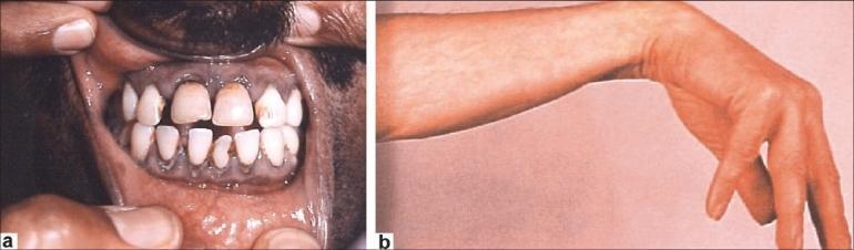
cd /tmp/workspace/metal_poisons/assets && curl -sL -o lead_burton_wristdrop.jpg "https://cdn.orris.care/cdss_images/pmc_clinical_VQA_20c20af1b2527269a0b9c89d2661db20f16596426d9a177e3d7e59ba1d6e66d2.jpg" && ls -la