
Cardio myopathy 4m
cardiomyopathy types dilated hypertrophic restrictive pathology
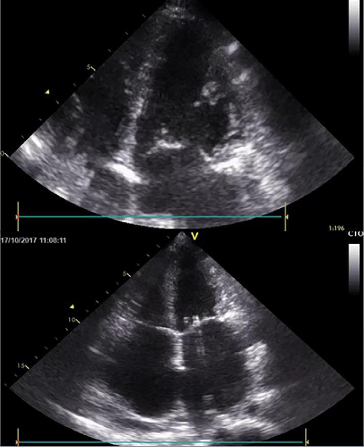
This diagnostic image consists of two grayscale transthoracic echocardiogram stills presented in an apical four-chamber (A4C) view. The visuals demonstrate a classic presentation of restrictive cardiomyopathy. Key anatomical features include severe biatrial enlargement, where both the left and right atria appear significantly dilated and disproportionately larger than the ventricles. In contrast, the left and right ventricular volumes remain within a relatively normal range with preserved cavity dimensions. The atrioventricular valves (mitral and tricuspid) are visible in their expected anatomical positions between the enlarged atria and the ventricles. The myocardium shows characteristic echogenicity associated with restrictive pathology. This visual is intended for medical education regarding heart failure with preserved ejection fraction (HFpEF) and the morphological differentiation between restrictive, dilated, and hypertrophic cardiomyopathies. It highlights the clinical significance of biatrial dilation as a hallmark of impaired ventricular filling and elevated filling pressures.
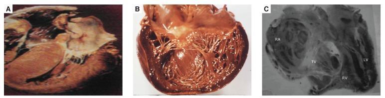
A comparative series of three pathological specimens (gross pathology and diagnostic imaging) illustrating distinct forms of cardiomyopathy. Panel A (Hypertrophic Cardiomyopathy) displays significant concentric hypertrophy of the myocardium, particularly involving the interventricular septum, resulting in a markedly narrowed, slit-like ventricular cavity. Panel B (Dilated Cardiomyopathy) shows a transversely sectioned heart with global ventricular dilation and attenuated, thinned myocardial walls; the trabeculae carneae and papillary muscles are visible within the expanded chambers. Panel C (Endomyocardial Fibrosis) is a radiographic or anatomical section showing structural distortion with characteristic subendocardial thickening and fibrosis. Labeled structures include the right atrium (RA), tricuspid valve (TV), right ventricle (RV), and left ventricle (LV). This panel specifically demonstrates apical obliteration and fibrotic replacement of the endocardium, leading to restrictive physiology. This comparison highlights morphological differences in chamber volume and wall thickness essential for diagnosing heart failure etiologies.
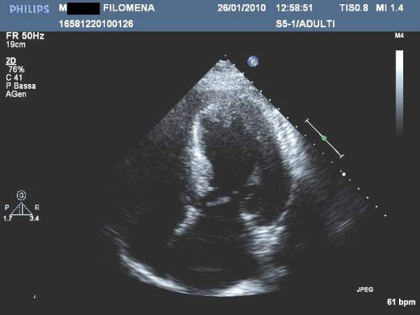
**Imaging Modality:** 2D Transthoracic Echocardiogram (TTE). **Anatomical View:** Apical four-chamber view. **Observed Pathology:** End-stage hypertrophic cardiomyopathy (HCM) characterized by significant, diffuse left ventricular (LV) hypertrophy. Unlike the typical progression to a "burnt-out" phase involving wall thinning and cavity dilation, this image demonstrates persistent, marked concentric wall thickening of the interventricular septum and lateral wall. **Visual Features:** - **Ventricular Morphology:** Prominent myocardial thickening with a non-dilated left ventricular cavity. - **Myocardial Texture:** Increased echogenicity of the myocardium, suggesting possible replacement fibrosis. - **Cardiac Function:** Impaired systolic function is evidenced by reduced end-systolic inward movement and wall thickening (reported LVEF 40%). - **Landmarks:** Visible left atrium, right atrium, and right ventricle appearing proportionately smaller than the hypertrophied left ventricle. **Diagnostic Significance:** This image represents the hypokinetic-restrictive stage of HCM. The key differentiating feature is the presence of moderate systolic dysfunction (EF <50%) without the ventricular wall thinning usually associated with dilated cardiomyopathy or late-stage remodeling, signifying advanced disease progression.
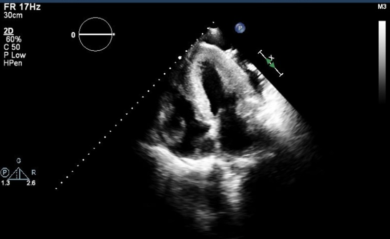
**Imaging Modality:** Transthoracic Echocardiogram (TTE), 2D grayscale. **Anatomical View:** Apical four-chamber (A4C) view. **Observed Pathology:** The image demonstrates severe concentric biventricular hypertrophy. There is marked, symmetrical thickening of the left ventricular (LV) walls, including the interventricular septum and the lateral wall, leading to a significantly reduced LV cavity size. Parallel thickening is also noted in the right ventricular (RV) free wall, indicating global myocardial involvement. **Characteristic Visual Features:** * **Myocardial Texture:** The myocardium exhibits an increased echogenicity with a "speckled" or granular appearance, often associated with infiltrative cardiomyopathies such as cardiac amyloidosis. * **Chamber Morphology:** Both ventricles appear small and restrictive. Both the left and right atria appear relatively dilated compared to the thickened ventricles. * **Valve Morphology:** The atrioventricular valves (mitral and tricuspid) are visible, with the mitral valve leaflets appearing slightly thickened. **Diagnostic Significance:** These findings are characteristic of severe restrictive or infiltrative cardiomyopathy. The combination of concentric biventricular wall thickening and biatrial enlargement are key diagnostic markers for advanced hypertrophic or infiltrative disease processes.
![This set of four Cardiovascular Magnetic Resonance (CMR) images illustrates different cardiomyopathy phenotypes. Panel A (balanced-SSFP, 4-chamber view) displays hypertrophic cardiomyopathy with asymmetric septal hypertrophy causing marked right ventricular (RV) cavity compression. Panel B (balanced-SSFP, 4-chamber view) shows left ventricular non-compaction, with white arrowheads highlighting the characteristic hyper-trabeculated endocardial layer and thin compacted epicardial myocardium. Panel C (Late Gadolinium Enhancement [LGE], 4-chamber view) shows idiopathic dilated cardiomyopathy with enlarged cardiac chambers and an absence of focal LGE, suggesting no replacement fibrosis. Panel D (LGE, short-axis view) demonstrates restrictive cardiomyopathy secondary to chronic pressure overload (aortic stenosis); the hyper-intense (white) signal in the sub-endocardial region indicates global sub-endocardial fibrosis. These images serve as a diagnostic comparison for identifying structural myocardial abnormalities, varying wall thicknesses, and tissue characterization via gadolinium enhancement in a pediatric or adult cardiology context.](https://cdn.orris.care/cdss_images/pmc_clinical_VQA_a930e972c0e89ae4bbcd769b8fcc7883df22a1fa4cc534fcc1fece3955ef0a5c.jpg)
This set of four Cardiovascular Magnetic Resonance (CMR) images illustrates different cardiomyopathy phenotypes. Panel A (balanced-SSFP, 4-chamber view) displays hypertrophic cardiomyopathy with asymmetric septal hypertrophy causing marked right ventricular (RV) cavity compression. Panel B (balanced-SSFP, 4-chamber view) shows left ventricular non-compaction, with white arrowheads highlighting the characteristic hyper-trabeculated endocardial layer and thin compacted epicardial myocardium. Panel C (Late Gadolinium Enhancement [LGE], 4-chamber view) shows idiopathic dilated cardiomyopathy with enlarged cardiac chambers and an absence of focal LGE, suggesting no replacement fibrosis. Panel D (LGE, short-axis view) demonstrates restrictive cardiomyopathy secondary to chronic pressure overload (aortic stenosis); the hyper-intense (white) signal in the sub-endocardial region indicates global sub-endocardial fibrosis. These images serve as a diagnostic comparison for identifying structural myocardial abnormalities, varying wall thicknesses, and tissue characterization via gadolinium enhancement in a pediatric or adult cardiology context.
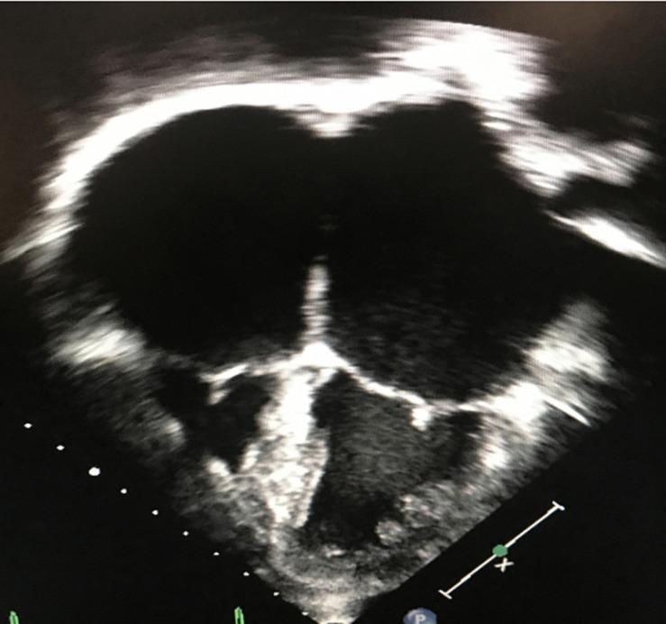
This diagnostic ultrasound image presents a 2D apical 4-chamber (A4C) view of the heart, characteristic of restrictive cardiomyopathy (RCM). The image highlights a classic 'mickey mouse' appearance due to massive biatrial enlargement. Both the left and right atria are severely dilated, occupying the majority of the cardiac silhouette. In stark contrast, the left and right ventricles appear disproportionately small, blunted, and restricted in volume. The ventricular walls exhibit a typical non-dilated morphology with preserved thickness, suggesting the restrictive physiology that leads to impaired diastolic filling and elevated filling pressures. This hallmark discrepancy between the diminutive ventricular chambers and the extremely enlarged atria serves as a primary educational example for differentiating restrictive patterns from dilated or hypertrophic cardiomyopathies. Key features visible include the central interventricular and interatrial septa and the atrioventricular valves, which separate the expansive atria from the small ventricular apex.
cardiomyopathy classification management
Cardiomyopathy - Complete Guide
Classification Overview
| Feature | Dilated (DCM) | Hypertrophic (HCM) | Restrictive (RCM) |
|---|---|---|---|
| LV cavity | Dilated | Normal / reduced | Normal or small |
| Wall thickness | Normal or thin | Markedly increased | Variable |
| Systolic function | Impaired (EF <40%) | Preserved (EF 50-80%) | Usually preserved |
| Primary dysfunction | Systolic | Diastolic | Diastolic |
| Atria | Enlarged (secondary) | Enlarged | Markedly enlarged |
1. Dilated Cardiomyopathy (DCM)
Pathophysiology
Causes (Mnemonic: ABCDE VIPER)
- A - Alcohol (acetaldehyde toxic to myocytes), doxorubicin
- B - B1 deficiency (beri-beri)
- C - Coxsackie B / Chagas / viral myocarditis (parvovirus B19, HHV-6)
- D - Dystrophin mutations (X-linked Duchenne/Becker)
- E - Endocrine (thyroid, acromegaly)
- V - Viral myocarditis progressing to DCM
- I - Idiopathic (20-50% hereditary - titin truncation mutations most common, autosomal dominant)
- P - Peripartum cardiomyopathy
- E - Election-Hemochromatosis, iron deposition
- R - Radiation, sarcoidosis
Morphology
- 4-chamber dilation with hypertrophy
- Mural thrombi common (especially LV apex) - risk of embolism
- Interstitial fibrosis on histology (non-specific)
- Floppy, flabby myocardium
Clinical Features
- Progressive HF (exertional dyspnea, orthopnea, PND)
- Dilated LV on echo
- Mitral regurgitation (annular dilation)
- Systemic emboli from mural thrombi
- Ventricular arrhythmias / sudden cardiac death
2. Hypertrophic Cardiomyopathy (HCM)
Pathophysiology
Genetics
- Autosomal dominant, gain-of-function mutations in sarcomeric proteins
- Most common: β-myosin heavy chain (most frequent) > myosin-binding protein C > troponin T
- These 3 genes account for 70-80% of all HCM cases
-
400 causative mutations identified across 9 genes
- Note: same genes that cause HCM when mutated as gain-of-function cause DCM when lost as loss-of-function - Robbins & Kumar Basic Pathology
Morphology
- Asymmetric septal hypertrophy (90% of cases) - septum disproportionately thicker than LV free wall
- Concentric hypertrophy (10%)
- On histology: myofiber disarray - haphazard arrangement of myocytes, interstitial fibrosis
Clinical Features
- Exertional dyspnea, angina, syncope (classic triad)
- Sudden cardiac death - #1 cardiovascular cause of SCD in young athletes (accounts for 1/3 of such events) - risk ~1%/year
- Harsh systolic murmur that increases with Valsalva/standing and decreases with squatting
- S4 gallop (stiff LV)
Management
- Avoid strenuous competitive sports
- Beta-blockers or calcium channel blockers (verapamil, diltiazem) for symptoms
- Disopyramide + beta-blocker if LVOTO symptoms persist
- ICD indications: prior cardiac arrest, sustained VT, FH of SCD, syncope, LV wall thickness ≥30mm, abnormal BP response to exercise
- Septal myectomy (surgical) or alcohol septal ablation for refractory LVOTO
3. Restrictive Cardiomyopathy (RCM)
Pathophysiology
Causes
| Cause | Mechanism |
|---|---|
| Cardiac amyloidosis | AL-amyloid (myeloma) or ATTR (transthyretin - wild-type in elderly, Val122Ile mutation in 4% of African Americans) |
| Sarcoidosis | Granulomatous infiltration |
| Endomyocardial fibrosis | Tropical/African; diffuse subendocardial fibrosis; associated with helminthic infections |
| Loeffler endomyocarditis | Hypereosinophilia; major basic protein damages endocardium → thrombus → fibrosis |
| Hemochromatosis | Iron deposition |
| Radiation fibrosis | Post-radiotherapy |
| Storage diseases | Mucopolysaccharidoses, sphingolipidoses |
| Idiopathic | - |
Key Features
- Normal-sized ventricles + massively enlarged atria
- Diastolic dysfunction with preserved (or near-preserved) EF
- "Square root sign" on hemodynamic tracing
- Endomyocardial biopsy - can identify specific etiology (amyloid, sarcoid)
- Hardest to distinguish from constrictive pericarditis (requires CMR/hemodynamics)
4. Arrhythmogenic Right Ventricular Cardiomyopathy (ARVC / ACM)
Pathophysiology
Genetics
- Desmosomal protein mutations most common (desmoglein, desmocollin, plakophilin-2, desmoplakin, junctional plakoglobin)
- Naxos disease - autosomal recessive ARVC + woolly hair + palmoplantar keratoderma (Greek island Naxos)
- Non-desmosomal: lamin, desmoplakin mutations
Diagnosis (Task Force Criteria)
- Structural/functional RV abnormalities
- Fibrofatty infiltration on endomyocardial biopsy
- ECG: Epsilon wave (after S in V1-V3), T-wave inversions V1-V3, prolonged terminal QRS >55ms in V1-V3
- Arrhythmias: VT with LBBB morphology (originating from RV)
Management
- Avoid strenuous endurance exercise (worsens progression)
- Beta-blockers, sotalol, amiodarone, or flecainide
- ICD: Prior SCA / sustained VT / LVEF ≤35% - then risk stratification for primary prevention
- Catheter ablation for refractory arrhythmias
5. Other/Special Forms
Peripartum Cardiomyopathy
- DCM developing in the last month of pregnancy or within 5 months of delivery
- Associated with prolactin cleavage products toxic to myocardium, anti-angiogenic factors (sFlt1)
- Risk factors: multiparity, older maternal age, African descent, pre-eclampsia
- ~50% recover LV function; recurrence risk in subsequent pregnancies
Stress (Takotsubo) Cardiomyopathy
- Acute, reversible LV dysfunction triggered by emotional/physical stress
- "Apical ballooning" on echo - apex akinetic, base hypercontractile
- Mimics ACS but no obstructive CAD
- Almost always reversible; managed supportively
Left Ventricular Non-Compaction (LVNC)
- Failure of normal myocardial compaction during embryogenesis
- Deep trabeculations and recesses in the LV
- Presents with HF, arrhythmias, thromboembolic events
- CMR is gold standard (Panel B in image below shows characteristic hyper-trabeculation)
Key Distinguishing Points (High-Yield for Exams)
| Feature | DCM | HCM | RCM | ARVC |
|---|---|---|---|---|
| Genetics | Loss-of-function (titin, dystrophin) | Gain-of-function (β-MHC) | Variable (TTR, desmin) | Desmosomal proteins |
| Primary pathology | Contractile failure | Diastolic failure | Diastolic failure | Fibrofatty RV replacement |
| Histology | Myocyte hypertrophy, interstitial fibrosis | Myofiber disarray | Interstitial fibrosis | Fibrofatty infiltration |
| SCD risk | Moderate (dilated ventricle, reentry) | High - #1 cause in athletes | Low-moderate | High - #2 cause in athletes |
| Echo hallmark | 4-chamber dilation, low EF | Asymmetric septal hypertrophy, SAM | Biatrial dilation, normal/small ventricles | RV dilation, aneurysms |
| Murmur | MR murmur (functional) | Harsh systolic, Valsalva ↑ | Usually none | None specific |
Myocarditis (Precursor to DCM)
- Most common cause: Coxsackie B and enteroviruses (in US); increasingly parvovirus B19 and HHV-6
- Also: HIV, CMV, diphtheria, Chagas (T. cruzi - most common worldwide)
- Autoimmune (SLE, rheumatic fever, drug hypersensitivity)
- Mechanism: direct viral cytopathic damage + molecular mimicry (immune-mediated)
- Clinical spectrum: asymptomatic → chest pain + fever → fulminant HF → DCM
- Diagnosis: Dallas criteria on endomyocardial biopsy (lymphocytic infiltrate + myocyte necrosis)
- Treatment: supportive; immunosuppression in giant-cell myocarditis; avoid NSAIDs acutely