
difference between dose limits and constraints
dose limits vs dose constraints radiation protection ICRP difference
Dose Limits vs. Dose Constraints
Dose Limits
| Feature | Detail |
|---|---|
| Nature | Legally binding regulatory values |
| Purpose | Set a hard ceiling - doses above this are NOT permitted |
| Set by | National regulatory bodies (based on ICRP recommendations) |
| Applies to | Occupational and public exposures in planned situations only |
| Does NOT apply to | Medical exposures of patients (diagnosis/treatment) |
| Consequence of exceeding | Regulatory violation - unlawful |
| Scope | Cumulative dose from ALL sources combined |
- Workers: 20 mSv/year averaged over 5 years (max 50 mSv in any single year), effective dose
- Public: 1 mSv/year effective dose
- Lens of the eye (workers): 20 mSv/year (updated ICRP 118)
- Skin/extremities (workers): 500 mSv/year equivalent dose
Dose Constraints
| Feature | Detail |
|---|---|
| Nature | Prospective, non-binding planning tool |
| Purpose | Upper bound used during optimization (ALARA/ALARP) for a single specific source |
| Set by | Operator, employer, or regulatory body - source-specific |
| Applies to | Planned, emergency, and existing exposure situations |
| Significance in medical exposure | Particularly important here since dose limits don't apply to patients |
| Consequence of exceeding | Triggers a review/follow-up action - NOT a legal violation |
| Scope | Single source only (not cumulative from all sources) |
Key Conceptual Differences
| Dimension | Dose Limit | Dose Constraint |
|---|---|---|
| Binding? | Yes - regulatory/legal | No - operational/planning tool |
| When applied? | Retrospectively checked for compliance | Prospectively used in design/optimization |
| Source scope | All sources combined | Per-source (one source at a time) |
| Role in ICRP framework | 3rd principle: Dose Limitation | 2nd principle: Optimization of Protection |
| Exceeding triggers... | Non-compliance/legal breach | Review and corrective action |
| Applies in emergencies? | No (reference levels used instead) | Yes (adapted form) |
| Applies to patients? | No | Yes (in absence of dose limits) |
Practical Example
- A dose constraint of 10 mSv/year set by the operator for a specific maintenance task (source-specific, optimization tool)
- A dose limit of 20 mSv/year (averaged) set by the regulator
Why the Distinction Matters
i dont know how to report fdg brain pet ct in case of autoimmune encephalitis. give me a detailed list of what i need to look for and wat are the different types of AIE and how to interpret each
FDG PET brain reporting autoimmune encephalitis anti-NMDA LGI1 GABA patterns hypometabolism hypermetabolism
https://pmc.ncbi.nlm.nih.gov/articles/PMC9996532
FDG PET brain autoimmune encephalitis hypermetabolism hypometabolism pattern
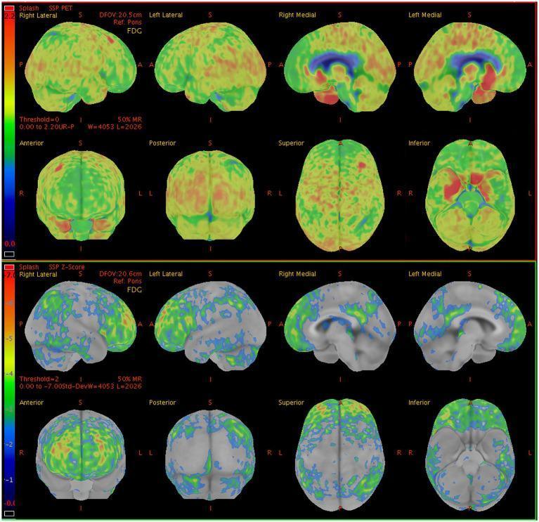
This diagnostic image displays 18F-FDG PET (fluorodeoxyglucose positron emission tomography) brain scans from a 60-year-old male presenting with seizures and memory impairment. The figure includes two rows of multi-planar reconstructions: the top row shows absolute metabolic uptake maps (SSP PET), and the bottom row displays corresponding semi-quantitative Z-score maps (SSP Z-Score) comparing the patient to a normative database. Visualized planes include lateral and medial surface views, as well as anterior, posterior, superior, and inferior projections. The Z-score maps utilize a colorimetric scale where cool colors (blue/green) represent hypometabolism and warm colors (red/orange) represent hypermetabolism. Significant bilateral hypometabolism is noted in the lateral and medial prefrontal lobes, posterior cingulate gyrus, precuneus, inferior parietal lobes, and lateral occipital regions, including the primary visual cortex. In contrast, prominent focal hypermetabolism is demonstrated in the bilateral medial temporal lobes. This metabolic pattern—diffuse cortical hypometabolism with localized medial temporal hypermetabolism—is clinically relevant in the evaluation of autoimmune encephalitides, such as LGI1-antibody encephalitis, where PET can detect functional abnormalities even when structural MRI is unremarkable.
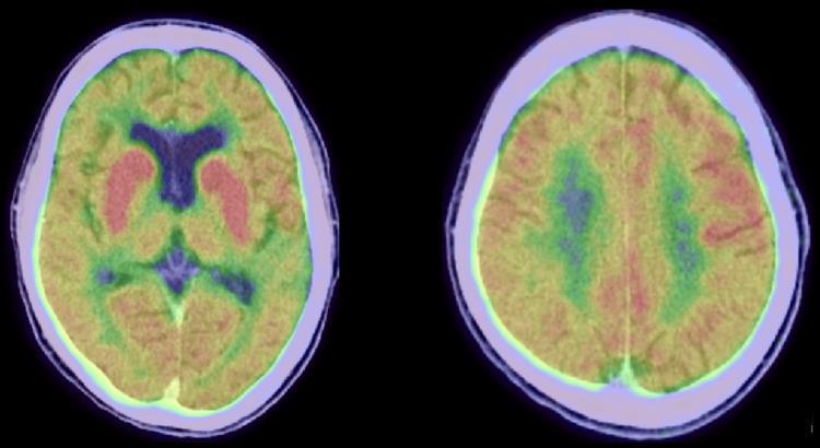
Two axial slices of an 18-Fluorodeoxyglucose Positron Emission Tomography (FDG-PET) scan of the brain demonstrating a characteristic metabolic pattern. The primary visual finding is strong, focal hypermetabolism in the bilateral basal ganglia (caudate and putamen), represented by intense red and orange color mapping. In contrast, the surrounding cerebral cortex exhibits diffuse hypometabolism, visualized through a spectrum of yellow, green, and purple shades, indicating significantly lower glucose uptake compared to the basal ganglia. These metabolic findings are indicative of neurological or autoimmune processes, such as anti-GAD antibody-associated encephalitis or chorea-related conditions, where deep gray matter structures show disproportionately high activity against a background of cortical suppression.
FDG Brain PET/CT in Autoimmune Encephalitis - Reporting Guide
Why FDG PET is Important in AIE
- PET detects abnormalities in 78-100% of AIE cases
- MRI detects abnormalities in only 42-63% of cases
- PET is particularly more sensitive than MRI in anti-NMDAR encephalitis
- PET can be positive even when antibody results are pending or negative
- FDG PET can be used for treatment response monitoring - abnormalities normalize with successful immunotherapy
Part 1: Systematic Approach - What to Look For
Step 1 - Pre-Report Checklist
- Patient's age and sex (strongly guides differential; anti-NMDAR predominantly young women, LGI1 predominantly older men)
- Clinical presentation: psychiatric symptoms, seizures, memory impairment, movement disorder, autonomic dysfunction, sleep disturbance
- Serum sodium (hyponatremia is a red flag for LGI1)
- Known antibody status if available (guides pattern recognition)
- Time from symptom onset to PET (patterns are most distinct in the first 1-2 weeks; later imaging may show mixed or less specific patterns)
Step 2 - Technical Assessment
- Patient preparation: Was the patient adequately fasted (>6h)? Blood glucose should ideally be <150-180 mg/dL; hyperglycemia causes FDG redistribution and artefactual cortical hypometabolism
- Seizure activity: Was there recent seizure activity? Peri-ictal scans show local hypermetabolism that can mimic or exaggerate AIE patterns - check EEG timing
- Sedation: Some patients with AIE are sedated/intubated; this can cause diffuse cortical hypometabolism and confound interpretation
- CT attenuation correction: Check for metal artefacts affecting PET quantification
Step 3 - Brain Regions to Systematically Evaluate
| Region | What to Look For |
|---|---|
| Medial temporal lobes (hippocampus, amygdala) | Hypermetabolism (most common finding in AIE) - often asymmetric |
| Basal ganglia (caudate, putamen) | Hypermetabolism - very characteristic, especially in LGI1 |
| Frontal lobes (prefrontal cortex) | Hypometabolism in anti-NMDAR; hypermetabolism in some |
| Parietal lobes (inferior parietal, precuneus) | Hypometabolism, especially in anti-NMDAR |
| Occipital lobes | Hypometabolism, especially "wedge-shaped" in anti-NMDAR |
| Posterior cingulate / precuneus | Hypometabolism (can mimic Alzheimer's pattern - deafferentation from temporal pathology) |
| Cerebellum | Hypermetabolism OR hypometabolism - present in ~70% of AIE cases |
| Thalamus/brainstem | Hypermetabolism can occur, note asymmetry |
| Insular cortex | Occasional involvement in limbic encephalitis |
Step 4 - Pattern Recognition
- Isolated hypermetabolism (~41% of cases) - typically medial temporal + basal ganglia
- Isolated hypometabolism (~41% of cases) - typically cortical, frontal/parietal/occipital
- Combined (~18%) - hypermetabolic limbic regions with hypometabolic cortex
Part 2: Specific Antibody Subtypes and Their FDG PET Patterns
1. Anti-NMDAR Encephalitis
- Frontotemporal hypermetabolism (early/active phase)
- Parietal and occipital hypometabolism - this is the signature finding
- "Wedge-shaped" occipital hypometabolism - characteristic and more severe/frequent than other AIE subtypes
- Diffuse cortical hypometabolism - affects both prefrontal AND posterior associative cortices simultaneously (the only subtype to do this in a majority of patients)
- "Anteroposterior gradient" - relatively higher frontal vs. posterior metabolism
- Findings are often heterogeneous and don't always fit one neat pattern
- MRI is frequently normal - PET is especially valuable here
- Patterns resolve completely with successful treatment, even after prolonged illness
2. Anti-LGI1 Encephalitis
- Basal ganglia (striatal) hypermetabolism - present in virtually ALL cases (87-100%)
- Medial temporal lobe (MTL) hypermetabolism - present in ~75-82% of cases, often bilateral
- Cerebellar hypermetabolism
- Frontal hypometabolism
- Findings are often asymmetric (differential expression of LGI1 receptor)
- Striatal hypermetabolism may correspond to the substrate for FBDS
3. Anti-CASPR2 Encephalitis
- MTL hypermetabolism - most frequent, present in virtually all (100%) CASPR2 cases
- Basal ganglia hypermetabolism (100%)
- Bilateral occipital hypometabolism (75%) - more frequent than in LGI1 or NMDAR
- "Anteroposterior gradient" similar to anti-NMDAR
- Cerebellar hypometabolism can occur
4. Anti-GABA-B Receptor Encephalitis
- Bilateral hippocampal/amygdala hypermetabolism - significantly elevated (higher SUVs than other AIE subtypes)
- Temporal lobe hypermetabolism
- Left supramarginal gyrus and right parietal lobe hypometabolism
- Pattern overlaps with other limbic encephalitides but hippocampal uptake is characteristically intense
5. Anti-GABA-A Receptor Encephalitis
- Variable, often multifocal hypermetabolism correlating with seizure foci
- Peri-ictal scans can be misleading - confirm with EEG timing
- Cortical and subcortical hypermetabolism may be widespread
6. Anti-AMPA Receptor Encephalitis
- Limbic (MTL) hypermetabolism
- Pattern similar to other limbic encephalitides
- Limited specific data due to rarity
7. Anti-GAD65 Encephalitis
- MTL hypermetabolism (54% of cases - lower than LGI1/CASPR2)
- Striatal hypermetabolism (~10%)
- Cortical hypometabolism (~35%)
- Cerebellar hypometabolism - particularly in GAD-associated cerebellar ataxia
- Temporal hypometabolism can occur
- Generally fewer abnormalities on PET than LGI1 or NMDAR
8. Paraneoplastic Limbic Encephalitis (Anti-Hu, Anti-Ma2 - Intracellular antibodies)
- Prominent, often striking bilateral mesiotemporal hypermetabolism - more consistently present than with surface antibody AIE
- Striatal and cerebellar hypermetabolism possible
- Association cortex hypometabolism (posterior cingulate, precuneus, frontal)
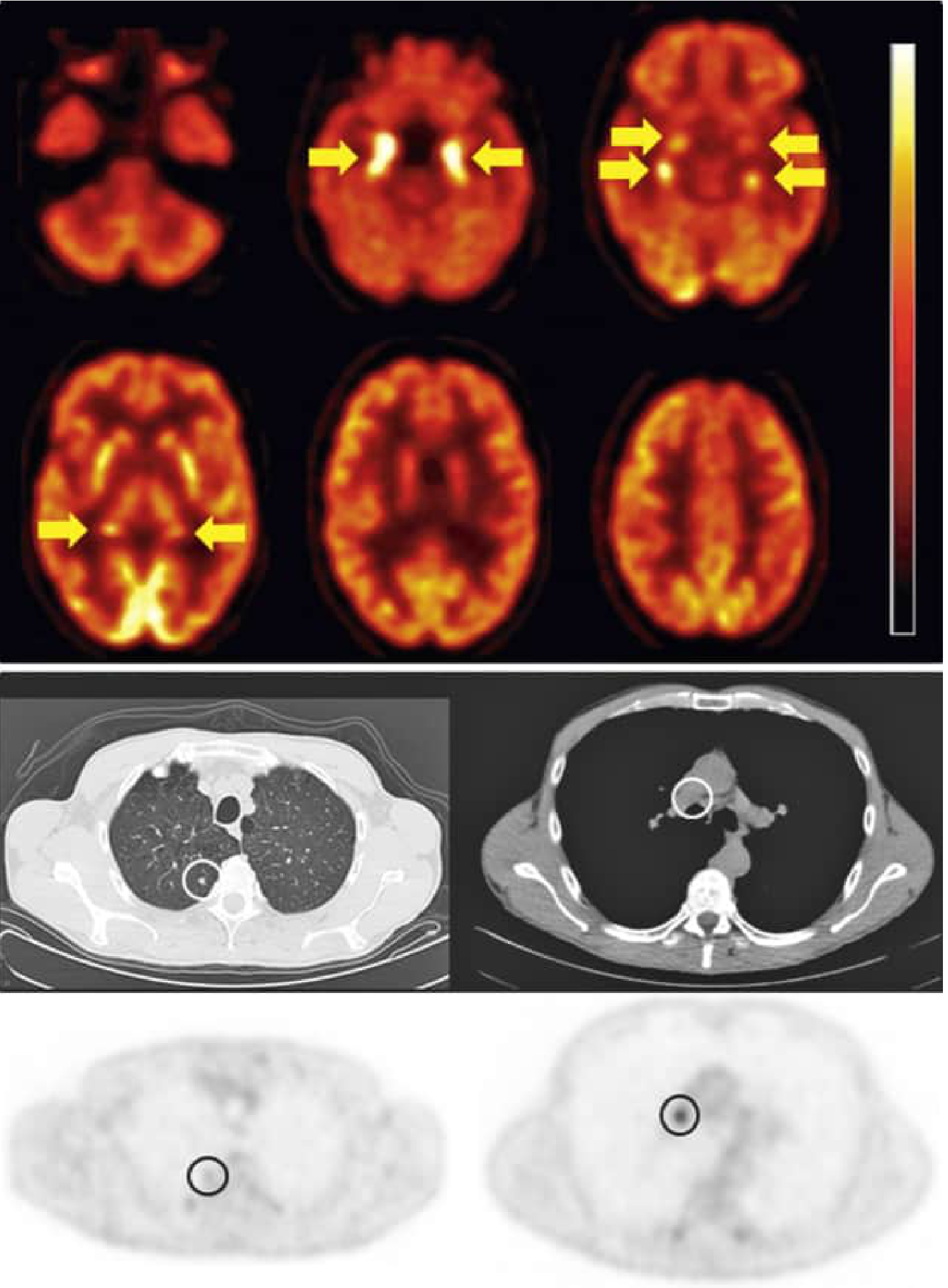
- This is the pattern shown in the textbook figure below:
Part 3: Quick Reference Summary Table
| AIE Subtype | Hypermetabolic | Hypometabolic | Key Feature |
|---|---|---|---|
| Anti-NMDAR | Frontotemporal (variable) | Parietal, occipital (diffuse), posterior cingulate | Diffuse cortical hypometabolism, "wedge-shaped" occipital |
| Anti-LGI1 | Basal ganglia, MTL (asymmetric) | Frontal | BG hypermetabolism is hallmark; asymmetry |
| Anti-CASPR2 | MTL, basal ganglia | Bilateral occipital | MTL + BG + occipital hypometabolism |
| Anti-GABA-B | Bilateral hippocampus (intense), temporal | Parietal | High hippocampal SUV |
| Anti-GABA-A | Multifocal (seizure-related) | Variable | Correlate with EEG |
| Anti-GAD65 | MTL (mild-moderate) | Cerebellar, temporal | Fewer findings; cerebellar involvement |
| Paraneoplastic (Hu/Ma2) | Bilateral MTL (intense), striatum | Association cortex | Most consistent MTL hypermetabolism |
Part 4: The CT Component
- Low-dose CT for attenuation correction - check for major structural abnormalities (mass, stroke, atrophy)
- Diagnostic CT: Look for evidence of primary malignancy (especially SCLC in older patients) - pulmonary nodule, mediastinal nodes
- Intracranial CT is often normal in AIE
- Atrophy may develop in chronic or untreated cases
Part 5: Reporting Template
- Medial temporal lobes (L/R): hypermetabolic / hypometabolic / normal
- Basal ganglia (caudate/putamen): hypermetabolic / normal
- Frontal lobes: hypometabolic / normal
- Parietal lobes: hypometabolic / normal
- Occipital lobes: hypometabolic / normal
- Posterior cingulate/precuneus: hypometabolic / normal
- Cerebellum: normal / hypermetabolic / hypometabolic
- Thalamus/brainstem: normal / abnormal
Part 6: Pitfalls and Tips
- Peri-ictal scans: Seizure activity causes local hypermetabolism - always check EEG and clinical timing. If patient had a seizure within hours of scan, interpret hypermetabolic foci cautiously
- "Recovering" phase: Mesiotemporal hypermetabolism may convert to hypometabolism after the acute phase, indicating irreversible post-inflammatory damage - not treatment failure
- Seronegative AIE: PET is still frequently abnormal (93% in one series) and the pattern (frontal/temporal hypermetabolism) can aid diagnosis when antibodies are negative
- Posterior cingulate hypometabolism: Can mimic Alzheimer's disease due to deafferentation from temporal lobe pathology - always correlate with clinical context
- Voxel-based analysis (SSP/SPM): More sensitive than visual reading alone, especially for detecting extra-limbic hypermetabolism in LGI1 and CASPR2; use when available
- Follow-up PET: Normalization of PET correlates with clinical improvement (mRS improvement) and can confirm treatment response
what is meant by anterior posterior gradient?
FDG PET anteroposterior gradient anti-NMDA encephalitis frontal hypermetabolism occipital hypometabolism
Anteroposterior Gradient on FDG PET
FRONT (Frontal) --> BACK (Parietal/Occipital)
Higher metabolism Lower metabolism
Relatively "hot" Relatively "cold"
Why It Happens in Anti-NMDAR Encephalitis
- Posterior cortex (parietal, occipital, posterior cingulate, precuneus) loses its normal resting activity → hypometabolism
- Frontal lobes are relatively spared or may even show increased activity due to disinhibition of frontal circuits → relatively hypermetabolic or normal-appearing
- Frontal lobes appear warm/preserved
- Parietal and occipital lobes appear cold/suppressed
- There is a visible step-change from front to back - this is the "gradient"
How to Recognise It on a Scan
- The anterior cortex (frontal) will appear orange/red relative to the posterior cortex
- The parietal and occipital cortex will appear green/blue (hypometabolic)
- On SSP surface projections it is very clear - the posterior surfaces are washed out compared to the anterior surfaces
Which AIE Subtypes Show This Pattern?
| Subtype | Anteroposterior Gradient |
|---|---|
| Anti-NMDAR | Yes - classic, most prominent and diffuse |
| Anti-CASPR2 | Yes - described in all 4 patients in one series |
| Anti-LGI1 | Less prominent; pattern is more about basal ganglia + MTL hypermetabolism |
| GAD65 / GABA-B | Not a defining feature |
Clinical Significance
- It is not pathognomonic of anti-NMDAR encephalitis alone - CASPR2 can show it too
- The severity of the gradient correlates with clinical severity - more widespread posterior hypometabolism = more severe disease
- It resolves with successful treatment, even in prolonged cases - making it a useful treatment response marker
- Do not confuse it with Alzheimer's disease (AD also shows posterior hypometabolism, but in AD the frontal lobes are usually also eventually involved, and the clinical context is completely different - gradual memory loss vs acute neuropsychiatric presentation)
Simple Way to Remember It
In anti-NMDAR encephalitis, the brain is "front-heavy" on FDG PET - the frontal lobe lights up relatively more, while the parietal and occipital lobes go quiet. Front is hot, back is cold - that slope from front to back is the anteroposterior gradient.
what is limbic encephalitis?
limbic encephalitis definition causes clinical features diagnosis treatment 2024
limbic encephalitis MRI hippocampus T2 FLAIR hyperintensity medial temporal
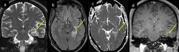
This diagnostic image set consists of four MRI brain sequences—(a) Coronal T2, (b) Axial T2-FLAIR, (c) Apparent Diffusion Coefficient (ADC) map, and (d) Coronal T1 post-contrast—demonstrating characteristic findings of medial temporal lobe encephalitis. Yellow arrows across all sequences highlight pathology in the left hippocampus and medial temporal lobe. The Coronal T2 and Axial FLAIR images show abnormal high signal intensity and mild swelling of the hippocampus and parahippocampal gyrus. The ADC map reveals facilitated diffusion (hyperintensity) in the corresponding region, indicating vasogenic edema rather than acute infarction. The T1 post-contrast image displays patchy parenchymal enhancement within the affected medial temporal structures, suggesting blood-brain barrier breakdown or inflammation. These findings are clinically significant for diagnosing encephalitis patterns, such as those associated with viral infections (including SARS-CoV-2) or autoimmune etiologies, targeting the limbic system.
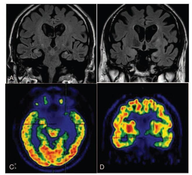
This composite diagnostic image displays neuroimaging findings in a patient with anti-LGI1 limbic encephalitis. Panels A and B are coronal T2-weighted Fluid-Attenuated Inversion Recovery (T2W FLAIR) MRI scans. Panel A demonstrates abnormal hyperintensities within the left medial temporal lobe and hippocampus, while Panel B reveals moderate hippocampal atrophy, classified as Medial Temporal Lobe Atrophy (MTA) score 3. Panels C and D provide functional assessment via 18F-FDG PET/CT. Panel C (axial view) shows hypometabolism, indicated by a partial absence of radioactivity (cool blue tones) in the left medial temporal cortex and hippocampus compared to the contralateral side. Panel D (coronal view) highlights markedly reduced tracer uptake in the head of the left caudate nucleus. Collectively, these images illustrate the correlation between structural MRI changes (hyperintensity and atrophy) and functional PET deficits (hypometabolism) characteristic of autoimmune limbic encephalitis involving the medial temporal lobes and basal ganglia.
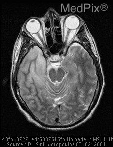
**Imaging Modality:** Magnetic Resonance Imaging (MRI), axial section.
Limbic Encephalitis
What Is It?
Memory loss + Psychiatric/behavioural change + Seizures - developing subacutely over days to weeks
- Hippocampus - memory consolidation
- Amygdala - emotion and fear responses
- Parahippocampal gyrus and entorhinal cortex
- Cingulate gyrus (especially anterior cingulate)
- Hypothalamus (in some subtypes)
- Insula
The Limbic System - Why These Symptoms?
| Structure Inflamed | Symptom Produced |
|---|---|
| Hippocampus | Short-term memory loss, anterograde amnesia |
| Amygdala | Fear, anxiety, agitation, personality change |
| Parahippocampal cortex | Spatial disorientation, complex partial seizures |
| Cingulate gyrus | Behavioural disturbance, emotional dysregulation |
| Hypothalamus | Hyperthermia, autonomic instability, hypersomnia |
Causes - Two Main Categories
1. Paraneoplastic Limbic Encephalitis (antibodies against INTRACELLULAR antigens)
| Antibody | Associated Tumor | Key Feature |
|---|---|---|
| Anti-Hu | SCLC (most common) | Often part of widespread encephalomyelitis |
| Anti-Ma2 | Testicular germ cell tumor (young men) | Upper brainstem involvement, vertical gaze palsy |
| Anti-CV2/CRMP5 | SCLC, thymoma | Also causes chorea, cerebellar ataxia, optic neuritis |
| Anti-amphiphysin | Breast cancer, SCLC | Can coexist with stiff-person syndrome |
- These are T-cell mediated (intracellular targets are not accessible to antibodies)
- Poor response to immunotherapy - treatment is directed at the tumor
- Irreversible neuronal damage is common
- Only ~30% of Ma2-positive patients improve with treatment
2. Autoimmune Limbic Encephalitis (antibodies against CELL SURFACE antigens)
| Antibody | Key Features |
|---|---|
| Anti-LGI1 | Older men, FBDS (faciobrachial dystonic seizures), hyponatremia |
| Anti-CASPR2 | Morvan syndrome, autonomic dysfunction, neuromyotonia |
| Anti-GABA-B | Refractory seizures, SCLC association |
| Anti-AMPA | Prominent psychiatric features, lung/breast cancer |
| Anti-NMDAR | Young women, ovarian teratoma, psychiatric onset (technically wider than just limbic) |
| Anti-GAD65 | Non-paraneoplastic, associated with T1DM/autoimmune disorders |
Clinical Features
Core Triad
- Subacute onset (days to ~12 weeks) of:
- Memory impairment - short-term memory loss is hallmark; long-term memory relatively preserved initially
- Neuropsychiatric symptoms - anxiety, depression, agitation, personality change, psychosis, confusion
-
- Seizures - usually complex partial (temporal lobe type); may be the presenting feature (especially in LGI1)
Additional Features (vary by subtype)
- Sleep disturbance (insomnia, hypersomnia, REM behaviour disorder)
- Autonomic dysfunction (tachycardia, blood pressure swings, hyperhidrosis)
- Movement disorders (dyskinesias, faciobrachial dystonic seizures in LGI1)
- Decreased level of consciousness in severe cases
- Hyponatremia (particularly in LGI1)
Diagnostic Criteria (Graus et al., Lancet Neurol 2016)
- Subacute onset (<3 months) of working memory deficits, seizures, or psychiatric symptoms suggesting limbic involvement
- Bilateral brain abnormalities on T2-weighted MRI, highly restricted to medial temporal lobes
- At least one of:
- CSF pleocytosis (>5 WBC/mm³)
- EEG with epileptic or slow-wave activity in the temporal lobes
- Reasonable exclusion of alternative causes
Investigations
MRI Brain
- T2/FLAIR: Bilateral (or unilateral) medial temporal hyperintensity and swelling - hippocampus is the epicenter
- T1: May show hypointensity in the medial temporal lobes
- DWI: Facilitated diffusion (vasogenic edema) - helps distinguish from HSV encephalitis which shows restricted diffusion
- Contrast: Patchy enhancement may occur (blood-brain barrier breakdown)
- Normal MRI does NOT exclude LE - especially early or in anti-NMDAR encephalitis
FDG PET
- Abnormal in 78-100% of cases (vs 42-63% for MRI)
- Medial temporal hypermetabolism is the hallmark in acute LE
- Striatal hypermetabolism, cortical hypometabolism (pattern varies by antibody - see previous answer)
CSF Analysis
- Lymphocytic pleocytosis (usually mild, 10-100 cells/mm³)
- Mildly elevated protein
- Normal glucose
- Oligoclonal bands may be present
Antibody Testing
- Serum AND CSF - some antibodies are detected better in CSF (e.g., anti-NMDAR)
- Panel should include: NMDAR, LGI1, CASPR2, GABA-B, AMPA, GABA-A, DPPX, GAD65
- Paraneoplastic panel: Hu, Ma2, CV2/CRMP5, Ri, amphiphysin, Yo
EEG
- Temporal lobe epileptiform discharges
- Slowing over temporal regions
- Helps localise and characterise seizure activity
Whole-Body FDG PET/CT
- Mandatory in ALL cases - screen for occult malignancy
- Especially important in Hu/Ma2 positive cases and GABA-B (high tumor frequency)
Key Differential Diagnoses
| Condition | How to Differentiate |
|---|---|
| HSV encephalitis | Often unilateral, involves insula + orbitofrontal cortex, restricted diffusion on DWI, fever, PCR positive |
| Creutzfeldt-Jakob disease | Rapidly progressive, "cortical ribboning" on DWI, EEG triphasic waves |
| Alzheimer's disease | Gradual onset over years, not subacute; posterior cortical hypometabolism on PET |
| Psychiatric illness (first-break psychosis) | Normal MRI, EEG, CSF - always exclude LE in new-onset psychosis |
| Status epilepticus | Peri-ictal MRI changes in temporal lobes can mimic LE - correlate with EEG |
Treatment
Immunotherapy (for autoimmune/surface-antibody LE)
- IV methylprednisolone (1g/day x 5 days)
- IV immunoglobulin (IVIG, 2g/kg over 5 days)
- Plasma exchange (PLEX)
- Rituximab
- Mycophenolate mofetil
- Azathioprine
- Treat the underlying tumor (first priority)
- Immunotherapy as adjunct
- Response is poor compared to surface-antibody LE
Symptomatic
- Anti-seizure medications (note: FBDS in LGI1 respond poorly to ASMs but well to immunotherapy)
- Psychiatric support/behavioural management
Prognosis
- Surface antibody LE (LGI1, CASPR2, GABA-B, NMDAR): Good prognosis with early immunotherapy; reversible in most cases; MRI and PET changes normalize with treatment
- Intracellular antibody paraneoplastic LE (Hu, Ma2): Poor neurological prognosis; irreversible damage; depends on tumor treatment
- Chronic hippocampal atrophy and memory impairment can remain even after treatment
What is the difference between paraneoplastic vs autoimmune limbic encephalitis?
Paraneoplastic vs. Autoimmune Limbic Encephalitis
The Fundamental Mechanism - Visualised
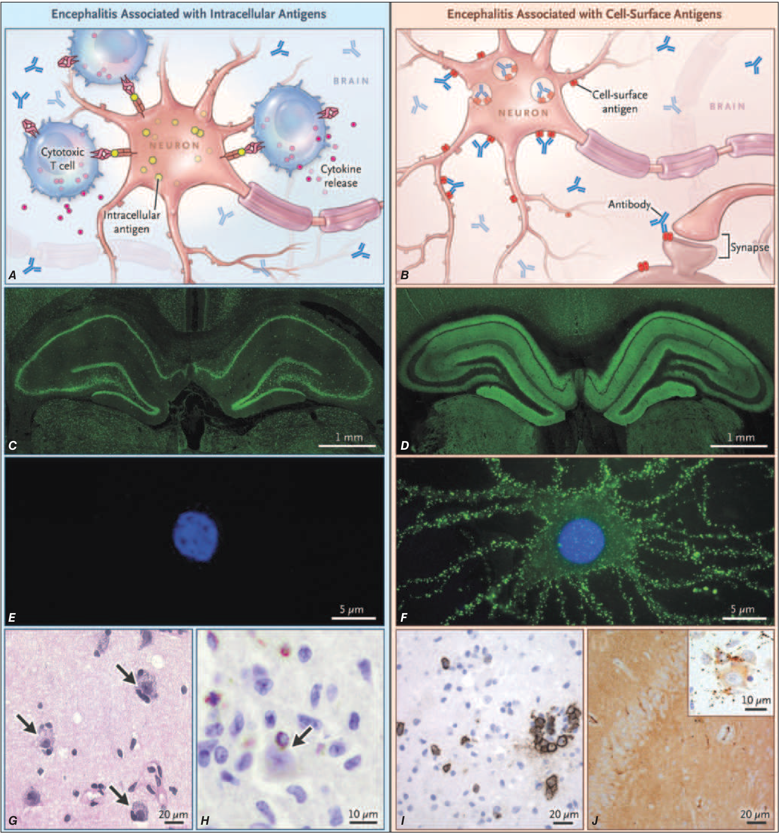
Side-by-Side Comparison
| Feature | Paraneoplastic LE | Autoimmune LE |
|---|---|---|
| Antibody target location | Intracellular (inside the neuron - nucleus, cytoplasm) | Cell surface / synaptic (on the neuronal membrane, accessible) |
| Example antibodies | Anti-Hu, Anti-Ma2, Anti-CV2/CRMP5, Anti-amphiphysin, Anti-Yo | Anti-NMDAR, Anti-LGI1, Anti-CASPR2, Anti-GABA-B, Anti-AMPA, Anti-GAD65 |
| Tumor association | Always - by definition, the tumor triggers the immune response | Variable - may or may not have a tumor |
| Common tumors | SCLC (Hu), Testicular germ cell (Ma2), Thymoma (CV2), Ovarian teratoma | SCLC (GABA-B ~50%), Thymoma (LGI1 <20%), Ovarian teratoma (NMDAR ~50% females) |
| Primary immune mechanism | Cytotoxic T-cell mediated - T cells directly kill neurons; antibodies are biomarkers, NOT the effectors | Antibody-mediated - antibodies directly bind, crosslink, internalize, or block receptors → receptor dysfunction |
| Can antibodies cause damage? | No - the intracellular antigen is inaccessible to circulating antibodies | Yes - antibodies directly alter receptor structure and function |
| Neuropathology | Neuronal loss + dense T-cell infiltrates (perforin/granzyme mechanisms) | Moderate inflammation, plasma cells, IgG deposits, minimal neuronal loss |
| Reversibility | Largely irreversible - neurons are destroyed by T cells; structural damage persists | Largely reversible - antibody removal restores receptor function; neurons mostly intact |
| Response to immunotherapy | Poor - removing antibodies doesn't stop T-cell damage; ~1/3 improve with tumor treatment | Good - steroids, IVIG, plasma exchange, rituximab are effective |
| Treatment priority | Treat the tumor first - this is the definitive treatment | Immunotherapy first - tumor treatment secondary if applicable |
| Prognosis | Generally poor; significant residual neurological disability common | Generally good with early treatment; full recovery possible |
| Relapse | Less common (tumor drives the immune response; treat the tumor, stop the trigger) | Can relapse, especially if no tumor found or maintenance immunotherapy stopped |
Why Antibodies Are Markers vs. Effectors
- Tumor expresses neuronal proteins (e.g., Hu protein in SCLC)
- Immune system mounts a T-cell response against the tumor
- Those same T cells cross-react with neurons expressing the same protein
- T cells kill neurons directly (perforin/granzyme pathway)
- The antibodies (Anti-Hu etc.) are produced alongside but cannot reach the intracellular epitope - they are diagnostic biomarkers, not the weapon
- Antibodies are produced against a surface antigen (often a receptor)
- Antibodies cross the blood-brain barrier and bind directly to the receptor
- The receptor is internalized, blocked, or structurally altered
- Synaptic transmission is disrupted → symptoms
- Remove the antibodies (plasma exchange, IVIG) → receptor function recovers → symptoms improve
The "Gray Zone" - Overlap and Complexity
- Surface antibody LE CAN have a tumor - GABA-B has SCLC in ~50%, anti-NMDAR has ovarian teratoma in up to 50% of young women. So "autoimmune LE" is not always tumor-free - the key distinction is the antibody target location and mechanism, not strictly whether a tumor is present.
- GAD65 is technically an intracellular antibody (GAD65 is inside the vesicle) but behaves more like surface-antibody disease - partially responds to immunotherapy, lower tumor association. It is a borderline case.
- Immune checkpoint inhibitor drugs (anti-PD1, anti-CTLA4) used in cancer treatment can unmask or trigger both types - classical paraneoplastic antibodies (Hu) and cell-surface antibodies can emerge after immunotherapy.
MRI Comparison
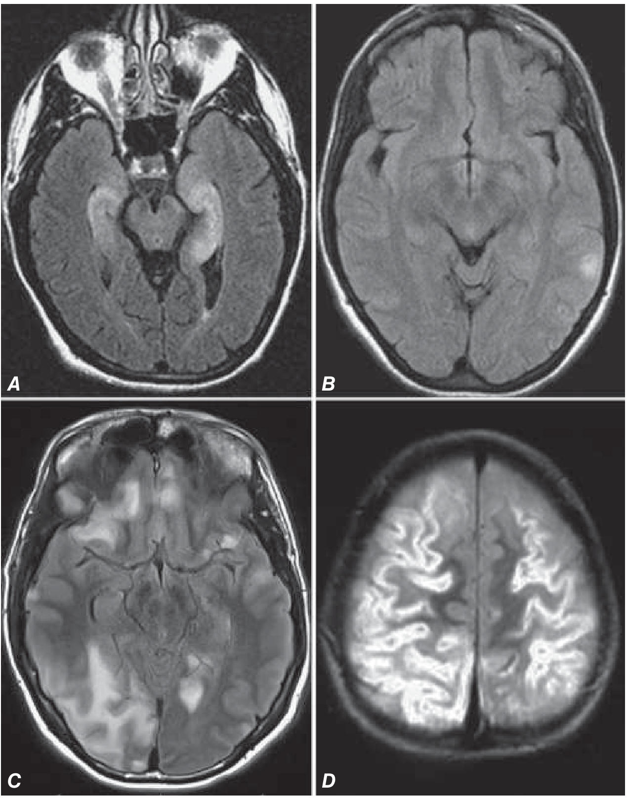
Practical Clinical Clues to Tell Them Apart at the Bedside
| Clue | Suggests Paraneoplastic | Suggests Autoimmune |
|---|---|---|
| Age | Older (Hu, CV2 - middle-aged to elderly) | Younger (NMDAR - young women; LGI1 - older men but responds) |
| Known cancer / smoking history | Strong pointer | Less likely |
| Rapid deterioration, not responding to immunotherapy | Paraneoplastic (T-cell damage ongoing) | Autoimmune would usually show some response |
| Multifocal neurological deficits (cerebellar + limbic + PNS) | Paraneoplastic encephalomyelitis (Hu) | Less typical |
| Prominent psychiatric onset | Less typical | Anti-NMDAR (very common) |
| Faciobrachial dystonic seizures | Not typical | Anti-LGI1 (pathognomonic) |
| Autonomic dysfunction + neuromyotonia | Less typical | Anti-CASPR2 (Morvan) |
| Hyponatremia | Not specific | LGI1 (common) |
| Whole-body PET shows avid primary tumor | Strong pointer | May be incidental |
| CSF: heavy pleocytosis, T-cell dominant | Paraneoplastic | Autoimmune usually milder pleocytosis |
Summary in One Sentence Each
show all pet images of AIE
FDG PET LGI1 CASPR2 limbic encephalitis basal ganglia medial temporal hypermetabolism
PET scan encephalitis striatum caudate putamen hypermetabolism cortical hypometabolism
paraneoplastic limbic encephalitis FDG PET mesiotemporal hypermetabolism whole body tumor
FDG PET brain encephalitis surface projection SSP Z-score map temporal frontal occipital metabolism
FDG PET Images in Autoimmune Encephalitis
Image 1 - Paraneoplastic Limbic Encephalitis (Anti-Hu / SCLC)
- Top panel (brain PET): Striking bilateral mesiotemporal (hippocampal) hypermetabolism - the bright white/yellow foci marked by arrows. Mild left association cortex hypometabolism in the background.
- Bottom panel (whole-body PET/CT): The primary SCLC - a 5mm spiculated right upper lobe nodule (low metabolic activity, circled) and a strongly FDG-avid mediastinal lymph node.
- Subtype: Anti-Hu positive, 68-year-old male with paraneoplastic LE
- Key point: Intracellular antibody subtypes show the most consistent and dramatic mesiotemporal hypermetabolism
Image 2 - LGI1 Encephalitis: SSP Surface Projection + Z-Score Map
- Top row (SSP PET): Raw metabolic map. Red/orange = high uptake. The inferior surfaces (temporal) show warm activity; the medial surfaces (posterior cingulate/precuneus) show a blue dropout.
- Bottom row (SSP Z-Score): Comparison to age-matched normals. Blue = significantly hypometabolic (Z < -2). Warm colors = hypermetabolic (Z > +2).
- Bilateral medial temporal and inferior temporal hypermetabolism (warm, seen on the inferior and medial projections)
- Prefrontal and posterior cortical hypometabolism (blue on lateral and medial surface views)
- Subtype: LGI1 encephalitis in a 60-year-old male with seizures and memory impairment
- Key point: SSP/Z-score maps are far more sensitive than visual axial slice reading alone
Image 3 - Axial FDG PET/CT: Basal Ganglia Hypermetabolism + Cortical Hypometabolism
- Left slice (basal ganglia level): Intense bilateral caudate and putamen hypermetabolism - the deep red/pink foci in the center of the brain. Surrounding cortex is relatively cold (green/blue = hypometabolic).
- Right slice (cortical level): Background cortical hypometabolism with a single area of relative left-sided preserved activity.
- Subtype: Anti-GAD antibody encephalitis / autoimmune encephalitis with chorea-related presentation
- Key point: Disproportionate deep grey matter (basal ganglia) hypermetabolism against suppressed cortex is a hallmark of both LGI1 and autoimmune chorea-encephalitis subtypes
Image 4 - Anti-NMDAR Encephalitis: Longitudinal FDG PET + MRI Comparison (3 Patients, 3 Time Points)
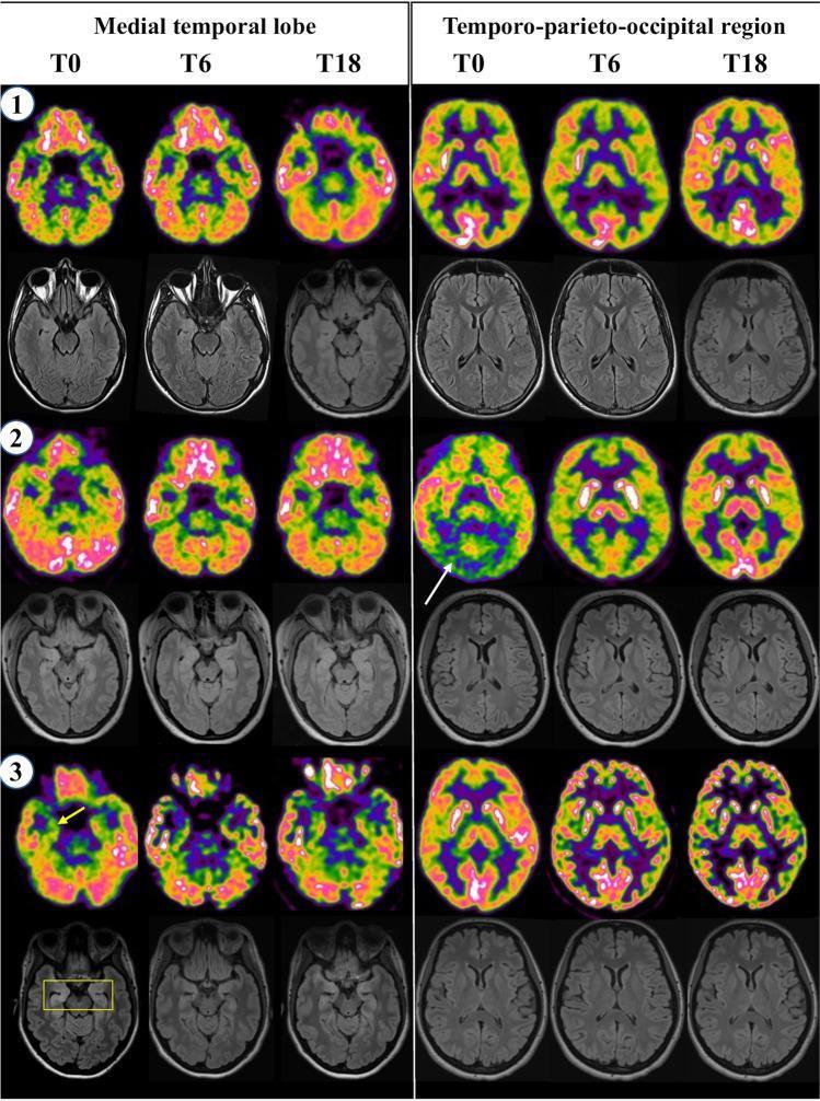
- Layout: 3 patients (rows 1, 2, 3). Each row shows PET (top) and MRI FLAIR (bottom) at T0 (onset), T6 (6 months), T18 (18 months).
- Left 3 columns = medial temporal lobe level; Right 3 columns = temporo-parieto-occipital level
- Patient 1: Bilateral lateral temporal and posterior parietal hypometabolism at onset (T0), peaking at T6, resolving by T18 - MRI is normal throughout
- Patient 2: Bilateral occipital hypometabolism at T0 (white arrow) - the anteroposterior gradient in action; basal ganglia relative hypermetabolism visible against cortical hypometabolism at T6
- Patient 3: Right medial temporal hypometabolism on PET (yellow arrow) corresponding exactly to the FLAIR hyperintensity on MRI (yellow rectangle) at T0; both resolve by T18
- Key points:
- Anti-NMDAR patterns are heterogeneous - some show posterior hypometabolism, some temporal, some mixed
- PET changes resolve with successful treatment - confirming the reversibility of surface-antibody LE
- MRI may be normal even when PET is clearly abnormal
Summary Reference: Which Pattern Goes with Which Subtype
| Image | Subtype | Dominant PET Finding |
|---|---|---|
| Image 1 | Paraneoplastic (Anti-Hu) | Bilateral MTL hypermetabolism |
| Image 2 | Anti-LGI1 | MTL + frontal hyper; posterior cortex hypo |
| Image 3 | Anti-GAD / chorea-type AIE | Basal ganglia hyper + cortical hypo |
| Image 4 | Anti-NMDAR | Occipital/parietal hypo + variable temporal; resolves with Rx |
Create a quick reference PET pattern checklist for reporting each AIE subtype
ls -la /tmp/workspace/ 2>/dev/null || mkdir -p /tmp/workspace/aie-checklist
mkdir -p /tmp/workspace/aie-checklist && cat > /tmp/workspace/aie-checklist/generate_checklist.py << 'PYEOF' from reportlab.lib.pagesizes import A4 from reportlab.lib import colors from reportlab.lib.units import cm, mm from reportlab.platypus import (SimpleDocTemplate, Table, TableStyle, Paragraph, Spacer, HRFlowable) from reportlab.lib.styles import getSampleStyleSheet, ParagraphStyle from reportlab.lib.enums import TA_CENTER, TA_LEFT, TA_RIGHT from reportlab.platypus import KeepTogether from reportlab.graphics.shapes import Drawing, Rect, String, Line from reportlab.graphics import renderPDF W, H = A4 # 595.27 x 841.89 pts # ── Color palette ────────────────────────────────────────────────────────────── NAVY = colors.HexColor('#1A2E4A') STEEL = colors.HexColor('#2C5F8A') TEAL = colors.HexColor('#1A7A7A') ORANGE = colors.HexColor('#E05A00') PURPLE = colors.HexColor('#5B2D8E') GREEN = colors.HexColor('#1A7A3A') CRIMSON = colors.HexColor('#8B0000') GOLD = colors.HexColor('#B8860B') SLATE = colors.HexColor('#4A5568') LIGHT_BG = colors.HexColor('#F0F4F8') WHITE = colors.white PALE_GOLD = colors.HexColor('#FFF8E7') PALE_TEAL = colors.HexColor('#E6F4F4') PALE_PURP = colors.HexColor('#F3EEFF') PALE_ORAN = colors.HexColor('#FFF0E6') PALE_CRIM = colors.HexColor('#FFF0F0') PALE_GREE = colors.HexColor('#EFF8F0') PALE_BLUE = colors.HexColor('#EEF4FF') PALE_NAVY = colors.HexColor('#E8EDF4') DIVIDER = colors.HexColor('#CBD5E0') styles = getSampleStyleSheet() def S(name, **kw): """Quick paragraph style factory""" base = styles['Normal'] ps = ParagraphStyle(name, parent=base, **kw) return ps title_style = S('Title', fontSize=16, fontName='Helvetica-Bold', textColor=NAVY, spaceAfter=2, leading=20) subtitle_style = S('Subtitle', fontSize=9, fontName='Helvetica', textColor=SLATE, spaceAfter=6, leading=12) section_hdr = S('SecHdr', fontSize=8.5, fontName='Helvetica-Bold', textColor=WHITE, spaceAfter=0, leading=11, alignment=TA_LEFT) body_sm = S('BodySm', fontSize=7.5, fontName='Helvetica', textColor=colors.black, leading=10) body_bold = S('BodyBold', fontSize=7.5, fontName='Helvetica-Bold', textColor=colors.black, leading=10) caption = S('Cap', fontSize=7, fontName='Helvetica-Oblique', textColor=SLATE, leading=9) check_style = S('Chk', fontSize=7.5, fontName='Helvetica', leading=10) footer_style = S('Ftr', fontSize=6.5, fontName='Helvetica-Oblique', textColor=SLATE, alignment=TA_CENTER) label_style = S('Lbl', fontSize=7, fontName='Helvetica-Bold', textColor=WHITE, leading=9, alignment=TA_CENTER) CHECKBOX = "☐" HYPER = "▲ HYPER" HYPO = "▼ HYPO" NORMAL = "– NORMAL" VAR = "~ VARIABLE" def cb(text, bold=False): st = body_bold if bold else body_sm return Paragraph(f"{CHECKBOX} {text}", st) def region_row(region, finding, note=""): icon = "" if "HYPER" in finding: icon = "🔴" elif "HYPO" in finding: icon = "🔵" elif "VARIABLE" in finding: icon = "🟡" else: icon = "⚪" note_txt = f" <i><font color='#666666' size='6.5'>{note}</font></i>" if note else "" return [Paragraph(f"{CHECKBOX} {region}", body_sm), Paragraph(f"<b>{finding}</b>{note_txt}", body_sm)] def make_subtype_table(title, color, pale, rows, clinical_note, abbrev): """Build one subtype block as a table""" # Header row header = Table( [[Paragraph(f" {abbrev}", label_style), Paragraph(f" {title}", section_hdr), Paragraph(f" {clinical_note}", caption)]], colWidths=[1.4*cm, 8.0*cm, 7.4*cm] ) header.setStyle(TableStyle([ ('BACKGROUND', (0,0), (-1,0), color), ('VALIGN', (0,0), (-1,0), 'MIDDLE'), ('TOPPADDING', (0,0), (-1,0), 4), ('BOTTOMPADDING', (0,0), (-1,0), 4), ('LEFTPADDING', (0,0), (-1,0), 6), ('GRID', (0,0),(-1,0), 0.3, colors.white), ])) # Data rows data = [] for r in rows: data.append(r) tbl = Table(data, colWidths=[8.5*cm, 8.3*cm]) tbl.setStyle(TableStyle([ ('BACKGROUND', (0,0), (-1,-1), pale), ('TOPPADDING', (0,0),(-1,-1), 3), ('BOTTOMPADDING', (0,0),(-1,-1), 3), ('LEFTPADDING', (0,0),(-1,-1), 8), ('RIGHTPADDING', (0,0),(-1,-1), 6), ('ROWBACKGROUNDS',(0,0),(-1,-1), [pale, colors.white]), ('GRID', (0,0),(-1,-1), 0.2, DIVIDER), ('VALIGN',(0,0),(-1,-1),'MIDDLE'), ])) return KeepTogether([header, tbl, Spacer(1, 4)]) # ── Build document ───────────────────────────────────────────────────────────── doc = SimpleDocTemplate( '/tmp/workspace/aie-checklist/AIE_FDG_PET_Checklist.pdf', pagesize=A4, leftMargin=1.5*cm, rightMargin=1.5*cm, topMargin=1.5*cm, bottomMargin=1.5*cm, title="FDG PET/CT Brain – AIE Reporting Checklist", author="Orris Clinical AI" ) story = [] # ── TITLE BLOCK ──────────────────────────────────────────────────────────────── title_tbl = Table( [[Paragraph("FDG Brain PET/CT", title_style), Paragraph("Autoimmune Encephalitis – Reporting Checklist", subtitle_style)]], colWidths=[7*cm, 9.8*cm] ) title_tbl.setStyle(TableStyle([ ('VALIGN',(0,0),(-1,-1),'BOTTOM'), ('LEFTPADDING',(0,0),(-1,-1),0), ('BOTTOMPADDING',(0,0),(-1,-1),0), ])) story.append(title_tbl) story.append(HRFlowable(width='100%', thickness=2, color=NAVY, spaceAfter=6)) # ── PRE-REPORT CHECKLIST ─────────────────────────────────────────────────────── pre_hdr = Table([[Paragraph(" PRE-REPORT CHECKLIST", section_hdr)]], colWidths=[16.8*cm]) pre_hdr.setStyle(TableStyle([ ('BACKGROUND',(0,0),(-1,-1), NAVY), ('TOPPADDING',(0,0),(-1,-1), 4), ('BOTTOMPADDING',(0,0),(-1,-1), 4), ('LEFTPADDING',(0,0),(-1,-1), 6), ])) pre_data = [ [cb("Patient age & sex noted (guides differential)"), cb("Blood glucose at injection < 150–180 mg/dL")], [cb("Subacute clinical onset confirmed (days–weeks)"), cb("Recent seizure activity checked (EEG timing)")], [cb("Known antibody status recorded if available"), cb("Sedation / intubation noted (can cause diffuse hypometabolism)")], [cb("Time from symptom onset to scan noted"), cb("CT component reviewed for occult malignancy / structural lesion")], ] pre_tbl = Table(pre_data, colWidths=[8.4*cm, 8.4*cm]) pre_tbl.setStyle(TableStyle([ ('BACKGROUND',(0,0),(-1,-1), PALE_NAVY), ('TOPPADDING',(0,0),(-1,-1), 3), ('BOTTOMPADDING',(0,0),(-1,-1), 3), ('LEFTPADDING',(0,0),(-1,-1), 6), ('GRID',(0,0),(-1,-1), 0.2, DIVIDER), ('VALIGN',(0,0),(-1,-1),'MIDDLE'), ('ROWBACKGROUNDS',(0,0),(-1,-1),[PALE_NAVY, colors.white]), ])) story.append(KeepTogether([pre_hdr, pre_tbl, Spacer(1,5)])) # ── LEGEND ───────────────────────────────────────────────────────────────────── legend_data = [[ Paragraph("🔴 ▲ HYPER = Hypermetabolism (increased FDG uptake vs background)", body_sm), Paragraph("🔵 ▼ HYPO = Hypometabolism (decreased FDG uptake vs background)", body_sm), Paragraph("🟡 ~ VARIABLE", body_sm), Paragraph("⚪ – NORMAL / not a defining feature", body_sm), ]] legend_tbl = Table(legend_data, colWidths=[5.2*cm, 5.2*cm, 2.8*cm, 3.6*cm]) legend_tbl.setStyle(TableStyle([ ('BACKGROUND',(0,0),(-1,-1), colors.HexColor('#F7FAFC')), ('BOX',(0,0),(-1,-1), 0.5, DIVIDER), ('TOPPADDING',(0,0),(-1,-1), 3), ('BOTTOMPADDING',(0,0),(-1,-1), 3), ('LEFTPADDING',(0,0),(-1,-1), 5), ('VALIGN',(0,0),(-1,-1),'MIDDLE'), ])) story.append(legend_tbl) story.append(Spacer(1, 5)) # ══════════════════════════════════════════════════════════════════════════════ # SUBTYPE BLOCKS # ══════════════════════════════════════════════════════════════════════════════ # 1. Anti-NMDAR story.append(make_subtype_table( title = "Anti-NMDAR Encephalitis", color = STEEL, pale = PALE_BLUE, abbrev = "NMDAR", clinical_note = "Young women | Ovarian teratoma | Psychiatric onset → seizures → autonomic dysfunction", rows = [ region_row("Occipital lobes", HYPO, '"Wedge-shaped" bilateral hypometabolism – MOST characteristic'), region_row("Parietal lobes / Precuneus", HYPO, "Diffuse; often simultaneous with occipital dropout"), region_row("Posterior cingulate cortex", HYPO, "Deafferentation pattern – can mimic Alzheimer's"), region_row("Frontal lobes", VAR, "Relatively preserved or mildly hyper → creates anteroposterior gradient"), region_row("Temporal lobes (lateral)", HYPO, "Variable; less consistent than occipital"), region_row("Medial temporal lobes (MTL)", VAR, "Less prominent than in LGI1/CASPR2; may be hypo or normal"), region_row("Basal ganglia", VAR, "May show relative hypermetabolism against cortical background"), region_row("Cerebellum", NORMAL, "Usually spared"), [Paragraph("☑ Overall pattern", body_bold), Paragraph("<b>ANTEROPOSTERIOR GRADIENT</b> – front warm, back cold. Diffuse cortical hypometabolism " "is the defining feature. MRI often NORMAL – PET is especially valuable here.", body_sm)], ] )) # 2. Anti-LGI1 story.append(make_subtype_table( title = "Anti-LGI1 Encephalitis", color = TEAL, pale = PALE_TEAL, abbrev = "LGI1", clinical_note = "Older men (~57y) | Faciobrachial dystonic seizures (FBDS) | Hyponatremia | Thymoma <20%", rows = [ region_row("Basal ganglia (caudate + putamen)", HYPER, "HALLMARK – present in 87–100%; often bilateral"), region_row("Medial temporal lobes (hippocampus)", HYPER, "75–82%; ASYMMETRIC in many cases"), region_row("Temporal lobes (lateral)", HYPER, "Common accompaniment"), region_row("Cerebellum", HYPER, "Frequent; may be hypermetabolic"), region_row("Frontal lobes", HYPO, "Frontal hypometabolism as counterpart to limbic hypermetabolism"), region_row("Posterior cingulate / Parietal", HYPO, "Variable, less prominent than in NMDAR"), [Paragraph("☑ Overall pattern", body_bold), Paragraph("<b>Bilateral (asymmetric) MTL + Basal Ganglia HYPER</b> with frontal HYPO. " "Most 'classic' AIE pattern. Asymmetry is a useful clue.", body_sm)], ] )) # 3. Anti-CASPR2 story.append(make_subtype_table( title = "Anti-CASPR2 Encephalitis", color = PURPLE, pale = PALE_PURP, abbrev = "CASPR2", clinical_note = "Middle-aged men | Morvan syndrome | Autonomic dysfunction | Neuromyotonia | Thymoma", rows = [ region_row("Medial temporal lobes", HYPER, "Most frequent – present in ~100% of cases"), region_row("Basal ganglia", HYPER, "100%; similar to LGI1 pattern"), region_row("Bilateral occipital lobes", HYPO, "75% – MORE prominent than in LGI1"), region_row("Frontal lobes", HYPO, "Variable"), region_row("Cerebellum", HYPO, "Can occur – cerebellar hypometabolism"), [Paragraph("☑ Overall pattern", body_bold), Paragraph("<b>MTL + BG HYPER</b> (like LGI1) <b>BUT with prominent bilateral occipital HYPO</b>. " "Anteroposterior gradient also seen. Consider CASPR2 when occipital dropout is striking.", body_sm)], ] )) # 4. Anti-GABA-B story.append(make_subtype_table( title = "Anti-GABA-B Receptor Encephalitis", color = ORANGE, pale = PALE_ORAN, abbrev = "GABA-B", clinical_note = "SCLC in ~50% | Refractory seizures as dominant feature | Middle-aged to elderly", rows = [ region_row("Bilateral hippocampus / amygdala", HYPER, "INTENSE – higher SUVs than other AIE subtypes"), region_row("Temporal lobes", HYPER, "Medial + lateral temporal hypermetabolism"), region_row("Left supramarginal gyrus", HYPO, "Statistically significant vs other subtypes"), region_row("Right parietal lobe", HYPO, "Significant in GABA-B vs anti-Hu group"), region_row("Basal ganglia", NORMAL, "Less prominent than LGI1"), [Paragraph("☑ Overall pattern", body_bold), Paragraph("<b>Intense bilateral hippocampal HYPER</b> (highest SUV of all AIE subtypes) " "+ parietal HYPO. Think GABA-B when seizures are the dominant symptom " "and hippocampal uptake is strikingly bright.", body_sm)], ] )) # 5. Anti-GAD65 story.append(make_subtype_table( title = "Anti-GAD65 Encephalitis", color = GREEN, pale = PALE_GREE, abbrev = "GAD65", clinical_note = "Non-paraneoplastic | Young women | Associated T1DM / autoimmune | Stiff-person / cerebellar ataxia", rows = [ region_row("Medial temporal lobes", HYPER, "54% – less consistent than LGI1/CASPR2"), region_row("Basal ganglia", HYPER, "~10% – less prominent"), region_row("Cerebellum", HYPO, "CHARACTERISTIC when cerebellar ataxia is present"), region_row("Temporal lobes", HYPO, "Temporal hypometabolism can occur"), region_row("Cortex (general)", HYPO, "~35% – mild and less dramatic than NMDAR"), [Paragraph("☑ Overall pattern", body_bold), Paragraph("<b>Fewest PET abnormalities of all AIE subtypes.</b> " "MTL hypermetabolism present but mild. Cerebellar hypometabolism is " "the key differentiating feature when present.", body_sm)], ] )) # 6. Paraneoplastic (Anti-Hu / Anti-Ma2) story.append(make_subtype_table( title = "Paraneoplastic LE – Anti-Hu / Anti-Ma2 (Intracellular Antibodies)", color = CRIMSON, pale = PALE_CRIM, abbrev = "PLE", clinical_note = "Anti-Hu: SCLC | Anti-Ma2: Testicular GCT (young men) | T-cell mediated | Poor Rx response", rows = [ region_row("Bilateral medial temporal lobes", HYPER, "MOST CONSISTENT & INTENSE of all LE types; often striking"), region_row("Striatum / basal ganglia", HYPER, "Common accompaniment"), region_row("Cerebellum", HYPER, "Can be hypermetabolic"), region_row("Brainstem", HYPER, "Especially anti-Ma2; upper brainstem"), region_row("Association cortices", HYPO, "Posterior cingulate, precuneus, frontal – variable"), region_row("Whole-body PET/CT", "⚠ MANDATORY", "Screen for primary tumor in ALL cases"), [Paragraph("☑ Overall pattern", body_bold), Paragraph("<b>Bilateral MTL HYPER is most intense and consistent here vs surface-Ab LE.</b> " "Key distinction: intracellular Ab = T-cell mediated = irreversible. " "Always search for primary tumor on whole-body scan.", body_sm)], ] )) # ── GENERAL PITFALLS BOX ──────────────────────────────────────────────────────── pit_hdr = Table([[Paragraph(" ⚠ PITFALLS & IMPORTANT NOTES", section_hdr)]], colWidths=[16.8*cm]) pit_hdr.setStyle(TableStyle([ ('BACKGROUND',(0,0),(-1,-1), GOLD), ('TOPPADDING',(0,0),(-1,-1), 4), ('BOTTOMPADDING',(0,0),(-1,-1), 4), ('LEFTPADDING',(0,0),(-1,-1), 6), ])) pitfalls = [ [cb("Peri-ictal scan: Seizure within hours → focal HYPERMETABOLISM at seizure focus – always check EEG timing before calling it AIE"), cb("Recovery phase: Mesiotemporal HYPERMETABOLISM may convert to HYPOMETABOLISM = irreversible post-inflammatory damage (NOT treatment failure)")], [cb("MRI often normal (especially anti-NMDAR): Negative MRI does NOT exclude AIE – PET detects abnormality in 78–100% vs MRI 42–63%"), cb("Posterior cingulate HYPO: Can mimic Alzheimer's disease due to deafferentation – always correlate with clinical context and age of onset")], [cb("Seronegative AIE: PET still abnormal in ~93%; frontal/temporal hypermetabolism supports diagnosis even without antibody confirmation"), cb("Blood glucose: Hyperglycaemia redistributes FDG away from brain → artefactual cortical hypometabolism – note glucose at injection")], [cb("Sedation/ventilation: Diffuse cortical hypometabolism – state of consciousness must be documented"), cb("Use SSP / Z-score maps: Voxel-based analysis detects extra-limbic hypermetabolism missed on visual axial reading alone")], ] pit_tbl = Table(pitfalls, colWidths=[8.4*cm, 8.4*cm]) pit_tbl.setStyle(TableStyle([ ('BACKGROUND',(0,0),(-1,-1), PALE_GOLD), ('TOPPADDING',(0,0),(-1,-1), 3), ('BOTTOMPADDING',(0,0),(-1,-1), 3), ('LEFTPADDING',(0,0),(-1,-1), 6), ('GRID',(0,0),(-1,-1), 0.2, DIVIDER), ('VALIGN',(0,0),(-1,-1),'MIDDLE'), ('ROWBACKGROUNDS',(0,0),(-1,-1),[PALE_GOLD, colors.white]), ])) story.append(KeepTogether([pit_hdr, pit_tbl, Spacer(1,5)])) # ── QUICK REFERENCE SUMMARY TABLE ────────────────────────────────────────────── sum_hdr = Table([[Paragraph(" QUICK REFERENCE SUMMARY", section_hdr)]], colWidths=[16.8*cm]) sum_hdr.setStyle(TableStyle([ ('BACKGROUND',(0,0),(-1,-1), SLATE), ('TOPPADDING',(0,0),(-1,-1), 4), ('BOTTOMPADDING',(0,0),(-1,-1), 4), ('LEFTPADDING',(0,0),(-1,-1), 6), ])) sum_col_hdr = [ Paragraph("<b>Subtype</b>", body_bold), Paragraph("<b>Hypermetabolic</b>", body_bold), Paragraph("<b>Hypometabolic</b>", body_bold), Paragraph("<b>Key Differentiator</b>", body_bold), ] sum_rows = [ sum_col_hdr, [Paragraph("Anti-NMDAR", S('X', parent=body_sm, textColor=STEEL, fontName='Helvetica-Bold')), Paragraph("Frontal (relative)", body_sm), Paragraph("Occipital, Parietal, Post.Cing.", body_sm), Paragraph("Diffuse cortical HYPO; wedge occipital; MRI often normal", caption)], [Paragraph("Anti-LGI1", S('X', parent=body_sm, textColor=TEAL, fontName='Helvetica-Bold')), Paragraph("Basal ganglia ✦, MTL (asymmetric)", body_sm), Paragraph("Frontal lobes", body_sm), Paragraph("BG hypermetabolism virtually universal; asymmetry is key", caption)], [Paragraph("Anti-CASPR2", S('X', parent=body_sm, textColor=PURPLE, fontName='Helvetica-Bold')), Paragraph("MTL, Basal ganglia", body_sm), Paragraph("Bilateral occipital ✦, Cerebellar", body_sm), Paragraph("Like LGI1 but prominent occipital HYPO; A-P gradient", caption)], [Paragraph("Anti-GABA-B", S('X', parent=body_sm, textColor=ORANGE, fontName='Helvetica-Bold')), Paragraph("Hippocampus (intense ✦)", body_sm), Paragraph("Parietal, Supramarginal gyrus", body_sm), Paragraph("Highest hippocampal SUV; refractory seizures; look for SCLC", caption)], [Paragraph("Anti-GAD65", S('X', parent=body_sm, textColor=GREEN, fontName='Helvetica-Bold')), Paragraph("MTL (mild)", body_sm), Paragraph("Cerebellum ✦, Temporal", body_sm), Paragraph("Fewest findings; cerebellar HYPO when ataxia present", caption)], [Paragraph("Paraneoplastic\n(Hu/Ma2)", S('X', parent=body_sm, textColor=CRIMSON, fontName='Helvetica-Bold')), Paragraph("Bilateral MTL (most intense ✦)", body_sm), Paragraph("Association cortices", body_sm), Paragraph("Most striking MTL HYPER; whole-body PET mandatory for tumor", caption)], ] sum_tbl = Table(sum_rows, colWidths=[2.6*cm, 3.6*cm, 4.2*cm, 6.4*cm]) sum_tbl.setStyle(TableStyle([ ('BACKGROUND',(0,0),(-1,0), colors.HexColor('#4A5568')), ('TEXTCOLOR',(0,0),(-1,0), WHITE), ('ROWBACKGROUNDS',(0,1),(-1,-1),[colors.HexColor('#F7FAFC'), WHITE]), ('TOPPADDING',(0,0),(-1,-1), 3), ('BOTTOMPADDING',(0,0),(-1,-1), 3), ('LEFTPADDING',(0,0),(-1,-1), 5), ('GRID',(0,0),(-1,-1), 0.3, DIVIDER), ('VALIGN',(0,0),(-1,-1),'MIDDLE'), ('LINEBELOW',(0,0),(-1,0), 1, SLATE), ])) story.append(KeepTogether([sum_hdr, sum_tbl, Spacer(1,5)])) # ── FOOTER ───────────────────────────────────────────────────────────────────── story.append(HRFlowable(width='100%', thickness=0.5, color=DIVIDER, spaceBefore=4, spaceAfter=3)) story.append(Paragraph( "Sources: Bradley & Daroff's Neurology in Clinical Practice; Harrison's Principles of Internal Medicine 22e; " "Solnes et al. J Nucl Med 2017; Baumgartner et al. 2013; Brain Metabolic Alterations in AE (MDPI 2023) | " "Generated by Orris Clinical AI – For educational/reporting reference use only", footer_style )) doc.build(story) print("PDF created successfully.") PYEOF python3 /tmp/workspace/aie-checklist/generate_checklist.py
~/aie-checklist/generate_checklist.py
Successfully replaced 1 instance(s) of the string in '/tmp/workspace/aie-checklist/generate_checklist.py'
python3 /tmp/workspace/aie-checklist/generate_checklist.py
AIE FDG PET Checklist
PDF Document · PDF
- Brain region-by-region checklist with expected finding (HYPER / HYPO / VARIABLE)
- Inline notes explaining the significance
- Summary "overall pattern" row at the bottom
| Subtype | Colour |
|---|---|
| Anti-NMDAR | Blue |
| Anti-LGI1 | Teal |
| Anti-CASPR2 | Purple |
| Anti-GABA-B | Orange |
| Anti-GAD65 | Green |
| Paraneoplastic (Hu/Ma2) | Crimson |
1-5 questions answers please
What topic should the Q&A cover? · What format do you want?
previous questions which were asked to select before these questions
- Paraneoplastic (anti-Hu, anti-Ma2): Antibody targets are intracellular - antibodies cannot access them. Damage is caused by cytotoxic T-cells directly killing neurons → irreversible neuronal loss. Treatment is directed at the underlying tumor; immunotherapy response is poor (~1/3 improve).
- Autoimmune (anti-LGI1, anti-NMDAR etc.): Antibody targets are on the cell surface - antibodies directly bind, internalize, and functionally disrupt receptors → reversible dysfunction (neurons remain intact). Responds well to steroids, IVIG, plasma exchange, and rituximab.
- Mesiotemporal hypermetabolism = active inflammation, ongoing metabolic activity in affected hippocampus - seen in the acute phase
- Conversion to hypometabolism on follow-up = irreversible post-inflammatory damage - neurons have been lost and the hippocampus is now metabolically silent (atrophic/scarred)