
Adductor canal block
Reading File
Reading File
Searching the Web
adductor canal block technique indications complications ultrasound guidance authoritative anesthesia
Searching PubMed
"adductor canal block" AND analgesia
Finding Sources
Finding Sources
Reading Article
PMID: 38561898
Reading File
Reading File
Reading File
Reading File
Reading Article
PMID: 41533004
Adductor canal block (ACB)
An adductor canal block is an ultrasound-guided regional anesthetic technique used mainly for analgesia after knee surgery, especially total knee arthroplasty, ACL reconstruction, and selected medial leg or foot procedures. It primarily blocks the saphenous nerve, with variable spread to the nerve to vastus medialis and sometimes obturator contributions.
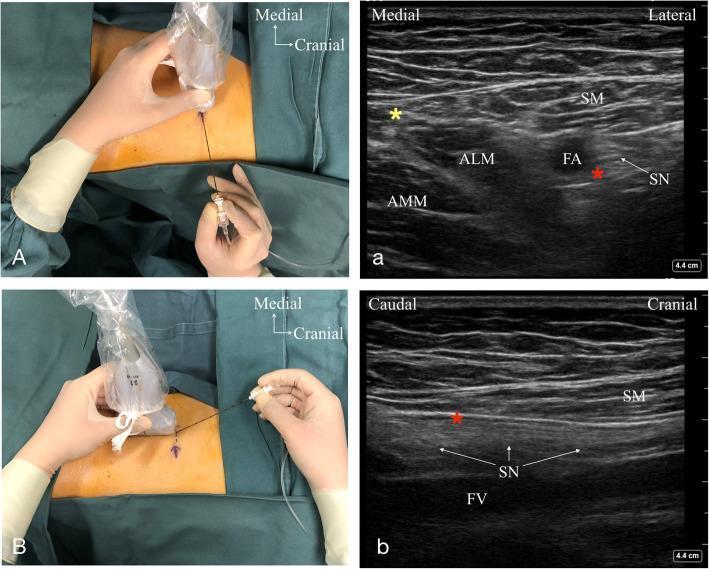
Why it is used
- Provides analgesia to the anteromedial knee and medial leg.
- Causes less quadriceps weakness than a femoral nerve block, so it is generally more compatible with early postoperative mobilization.
- Does not reliably cover posterior knee pain. For knee arthroplasty, it is commonly one component of multimodal analgesia, potentially combined with local infiltration analgesia and, in appropriate cases, an iPACK block.
Relevant anatomy
The adductor canal is bounded by:
- Sartorius anteriorly/medially
- Vastus medialis laterally/anteriorly
- Adductor muscles posteriorly
Its key ultrasound landmark is the femoral artery, deep to sartorius. The saphenous nerve is usually adjacent to the artery, often anterior or lateral to it. The intended injection is a periarterial local-anesthetic deposit, deep to sartorius.
Typical ultrasound-guided technique
Performed by an appropriately trained anesthesia clinician using full monitoring, sterile technique, aspiration, and local-anesthetic toxicity precautions.
- Supine position, leg slightly externally rotated.
- Place a high-frequency linear ultrasound probe transversely on the mid-to-distal anteromedial thigh.
- Identify sartorius superficially and the femoral artery beneath it.
- Advance a needle in-plane toward the fascial plane deep to sartorius, generally lateral/anterior to the artery.
- After negative aspiration and visual confirmation of spread, inject local anesthetic incrementally.
Common reference volumes are approximately 10-20 mL. Larger volumes or proximal spread can increase the chance of motor involvement, including quadriceps weakness. Miller's Anesthesia, 10e, pp. 6270-6272; Morgan and Mikhail's Clinical Anesthesiology, 7e, pp. 1926-1927.
Benefits versus femoral nerve block
| Feature | Adductor canal block | Femoral nerve block |
|---|---|---|
| Main target | Predominantly sensory branches | Femoral nerve, sensory and motor |
| Quadriceps weakness | Usually less | More frequent/marked |
| Early walking | Often better preserved | May be limited by weakness |
| Analgesia | Useful for anteromedial knee | Often stronger but less motor-sparing |
ACB is not completely motor-free. Unexpected thigh weakness can occur, particularly with injection location, volume, or proximal spread. Fall precautions still apply.
Complications and cautions
- Local anesthetic systemic toxicity
- Vascular puncture, hematoma, rare arterial injury/pseudoaneurysm
- Infection
- Nerve injury or persistent paresthesia
- Inadequate block or incomplete posterior-knee analgesia
- Quadriceps weakness and falls
- Rare intramuscular injection-related myonecrosis
Avoid or carefully individualize the block in patients with local infection, significant allergy to the planned local anesthetic, or bleeding risk that makes deep regional procedures unsafe.
Evidence update
A 2026 systematic review of 26 randomized trials (2,400 patients) found that ACB plus local infiltration analgesia reduced pain at 24 and 48 hours after total knee arthroplasty compared with infiltration alone, with lower opioid use and better 24-hour range of motion in the combined group. Certainty varied from moderate to very low because of study bias and heterogeneity. A 2024 network meta-analysis found the mobility advantage of ACB versus femoral blockade, while indicating that single-shot ACB may provide less analgesia than some continuous femoral-block strategies. See the 2026 meta-analysis and 2024 network meta-analysis.
This is an educational overview, not a guide for self-administration. An ACB should be performed by a trained clinician with resuscitation capability and local-anesthetic toxicity management available.
Explain the ultrasound anatomy and needle approach step by step
Reading File
Finding Sources
Searching Images
adductor canal block ultrasound anatomy sartorius femoral artery saphenous nerve in plane needle approach labeled
Searching PubMed
"adductor canal block" AND ultrasound
Searching the Web
adductor canal block ultrasound anatomy needle approach ultrasound guide ASRA
Ultrasound anatomy and needle approach for an adductor canal block
This is a clinician-performed, ultrasound-guided block. The key is to identify the femoral artery under sartorius, then place the needle tip in the subsartorial fascial plane rather than in muscle or a vessel.
1. Position the patient and probe
- Patient supine.
- Operative leg slightly externally rotated, with the medial thigh exposed.
- Use a high-frequency linear transducer.
- Begin with the probe transverse across the anteromedial mid-thigh, approximately halfway between the anterior superior iliac spine and superior pole of the patella.
2. Find the femoral artery first
On the transverse ultrasound image, identify:
- Femur: bright curved line with posterior acoustic shadow, deep in the image.
- Femoral artery: round pulsatile anechoic structure, usually superficial/medial to the femur.
- Femoral vein: usually adjacent to the artery and compressible. Use color Doppler if uncertain.
The artery is the dependable landmark. Do not depend on seeing the saphenous nerve, since it can be small or indistinct.
3. Identify the three walls of the canal
At the intended level, recognize the characteristic subsartorial configuration:
| Structure | Ultrasound position |
|---|---|
| Sartorius muscle | Superficial, forming a roof over the artery; often triangular or strap-like |
| Vastus medialis | Lateral/anterior to the artery |
| Adductor longus or adductor magnus | Posterior or posteromedial to the artery |
| Femoral artery | Deep to sartorius, near the center/medial portion of the image |
| Saphenous nerve | Small hyperechoic oval or fascicular structure near the anterolateral/anterior aspect of artery; may not be visible |
The “true” adductor canal is identified where the medial border of sartorius converges with the medial border of adductor longus, and the vastoadductor membrane forms a deep fascial boundary.
4. Confirm you are at the appropriate level
There are two related targets:
- Proximal adductor canal or distal femoral triangle level: often selected for knee analgesia, because spread may include the saphenous nerve and nerve to vastus medialis.
- More distal adductor canal: tends to be more selectively saphenous and may reduce motor involvement, but can provide less coverage of knee joint pain.
For post-total-knee-arthroplasty analgesia, many clinicians target the mid-thigh subsartorial region to achieve coverage of both the saphenous nerve and relevant articular branches. Miller's Anesthesia, 10e, pp. 6270-6272.
5. Plan the needle path
The standard approach is in-plane, lateral-to-medial:
- The needle enters from the lateral edge of the probe footprint.
- It advances under continuous visualization through the lateral soft tissues toward the artery.
- Keep the entire shaft and, especially, the needle tip visible.
- The desired endpoint is the fascial plane deep to sartorius and immediately anterior or anterolateral to the femoral artery.
A lateral-to-medial path makes it easier to see the needle and keeps the target relationship to the artery clear. Avoid aiming directly at a visible saphenous nerve. The purpose is fascial-plane spread, not intraneural injection.
6. Advance to the target plane
Advance incrementally, adjusting the probe and needle together as needed.
Your target is:
- Deep to sartorius
- Adjacent to, but not within, the femoral artery
- Outside the vastus medialis and adductor muscles
- In the fascial plane containing or immediately adjacent to the saphenous nerve
Do not mistake a superficial injection that only lifts sartorius for correct canal spread. Proper injectate spread should dissect the subsartorial space around the anteromedial aspect of the artery.
7. Confirm position with a small test injection
After negative aspiration, inject a small incremental test dose while watching ultrasound.
Correct spread
- Hypoechoic fluid opens the plane deep to sartorius.
- Fluid spreads around the anterior/lateral aspect of the artery.
- The artery may be displaced slightly deeper.
- The local anesthetic creates a crescent or semilunar spread around the artery and nearby saphenous nerve.
Incorrect spread
- Fluid remains inside vastus medialis or an adductor muscle: reposition.
- Fluid merely elevates sartorius without tracking around the artery: the tip is likely too superficial.
- Resistance, severe pain, paresthesia, or nerve swelling: stop injection and reassess.
The ASRA ultrasound guidance discussion similarly describes desired semilunar periarterial spread and warns that extensive lifting of sartorius suggests an incorrect plane.
8. Inject incrementally with continuous observation
Inject only after repeated aspiration and under real-time visualization of spread. Volume and drug choice should follow the clinician’s institutional protocol, surgical indication, patient size, concurrent local-anesthetic exposure, and toxicity-risk assessment. Higher volumes and proximal spread may lead to quadriceps weakness.
9. Re-scan before finishing
Before removing the needle, confirm:
- No intravascular injection or hematoma
- Local anesthetic has spread in the intended subsartorial plane
- No obvious intramuscular collection
- The patient has no concerning neurologic symptoms
Key practical errors to avoid
- Too proximal: may effectively become a femoral triangle block and increase motor involvement.
- Too distal: may miss nerve-to-vastus-medialis contributions and give weaker knee analgesia.
- Injecting into muscle: produces poor spread and may cause pain or tissue injury.
- Direct nerve injection: not required, and increases risk of neural injury.
- Assuming motor sparing means no fall risk: quadriceps weakness can still occur. Maintain fall precautions.
The standard textbook description places the needle in-plane in the triangular subsartorial space, anterior/lateral to the artery, with ultrasound visualization of local-anesthetic spread. Morgan and Mikhail's Clinical Anesthesiology, 7e, pp. 1926-1927.
This is a shared conversation. Sign in to Orris to start your own chat.