
Patellar dislocation
Reading File
Searching sources...
Finding Sources
Searching PubMed
"patellar dislocation" AND management
Good results. Now let me read the key textbook sections in parallel.
Reading File
Reading File
Reading File
Let me get the rest of the Roberts & Hedges reduction technique section and the Rheumatology recurrent dislocation section.
Reading File
Reading File
Now let me fetch the key images to include visually.
Loading Image
Loading Image
Excellent - I have comprehensive content from multiple textbooks plus images. Here is the full structured answer:
Patellar Dislocation
Overview
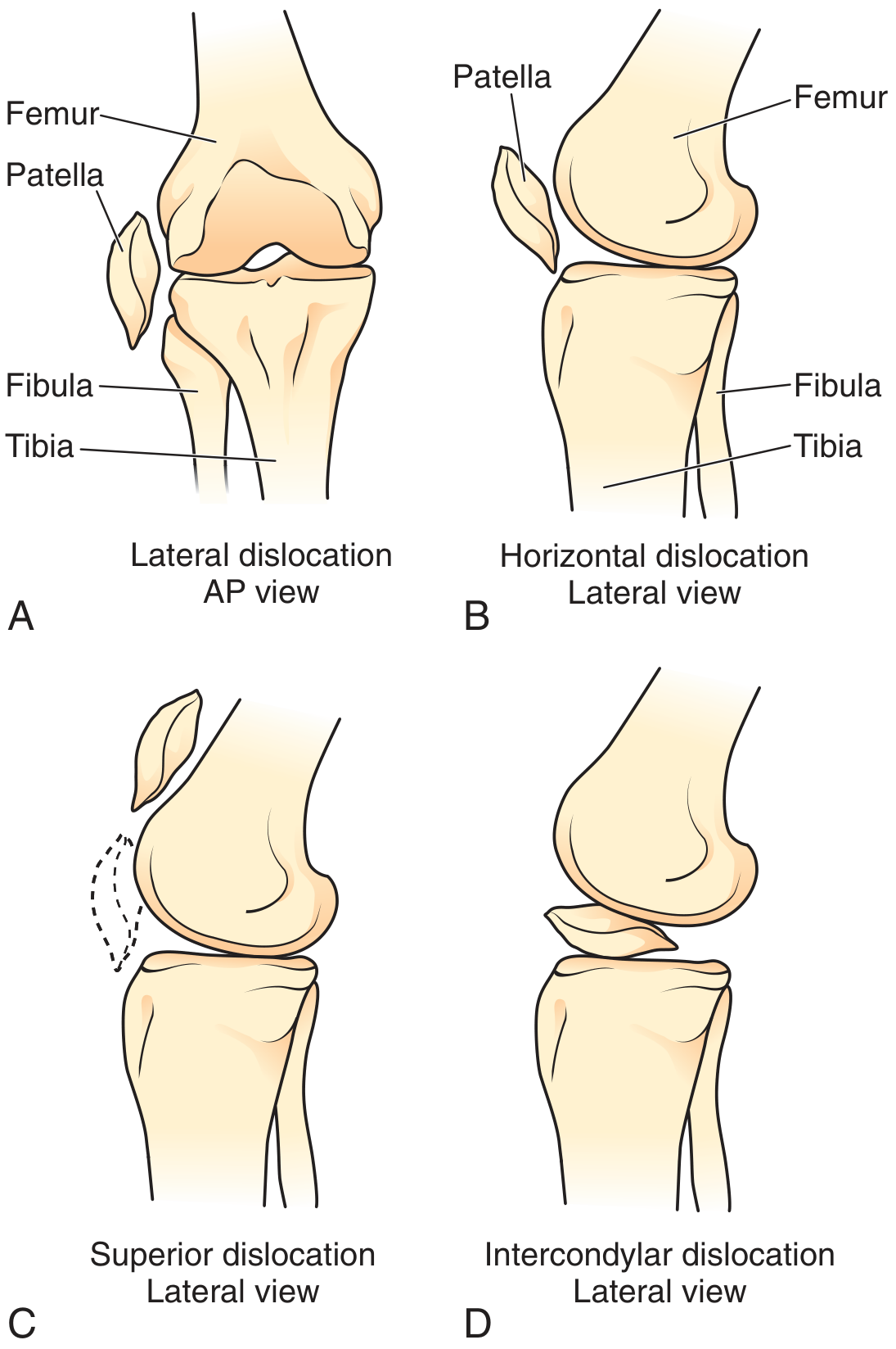
Patellar dislocation is a common musculoskeletal injury, especially in adolescents and young athletes. The vast majority are lateral dislocations - the patella displaces over the lateral femoral condyle. Other subtypes (superior, horizontal/medial, intercondylar) are rare.
Mechanism
Two classic mechanisms:
- Quadriceps contraction + valgus/external rotation - e.g., cutting movements in sports or dancing
- Direct blow to the anterior or medial patella with the knee flexed
The resulting forces tear the medial retinaculum (and medial patellofemoral ligament - MPFL), allowing the patella to displace laterally. Subluxation (incomplete displacement) typically implies a stretched, rather than torn, medial retinaculum.
Predisposing factors:
- Genu valgum
- Femoral anteversion
- Patella alta
- Trochlear dysplasia
- Ligamentous laxity / joint hypermobility
- Prior dislocation history
Clinical Features
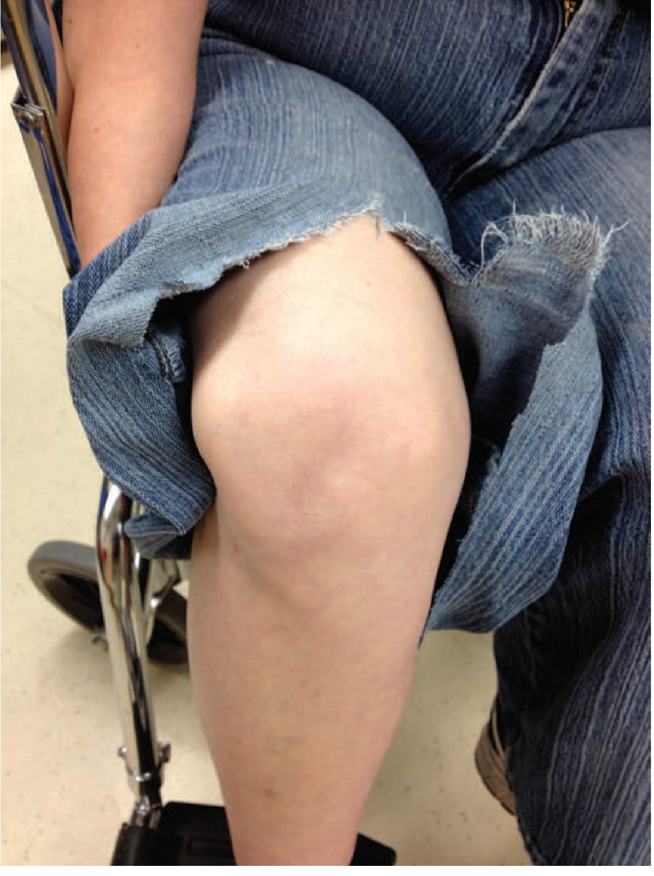
The diagnosis is usually clinically obvious for unreduced dislocations:
- Acute pain and inability to bear weight or flex the knee
- Knee held in flexion
- Patella visible and palpable on the lateral aspect of the knee
- Tenting of overlying skin unless significant swelling
Spontaneous reduction in the field is common - the patient may simply describe the knee "going out." Clues to a spontaneously reduced patella:
- Knee effusion (may develop hours after injury)
- Medial patellar edge tenderness (MPFL injury)
- Fairbanks'/Apprehension sign - patient grabs the knee when the patella is pushed laterally, reflecting fear of re-dislocation
Diagnostic Testing
X-rays (AP + lateral views) are standard. Expected findings:
-
Empty trochlear groove
-
Laterally displaced patella
-
Joint effusion
-
Possible osteochondral avulsion fracture
-
Sunrise (Merchant) view: difficult to obtain pre-reduction due to pain; obtain post-reduction
-
Lipohemarthrosis on x-ray warrants careful search for fracture fragments
-
Osteochondral fractures occur in ~50% of cases; many are only visible on arthroscopy
-
CT or MRI may be needed to fully characterize associated injuries
Note: The clinical diagnosis in older patients should be made with caution - patellar dislocations are primarily injuries of the young.
Reduction Technique
Closed reduction is the standard approach. It is usually straightforward and often does not require sedation.
Steps:
- Reassure the patient verbally - premedication (analgesia/anxiolysis) as needed
- Flex the hip to relax the quadriceps
- Gently extend the knee (white arrow in procedure diagrams)
- Apply medial pressure on the lateral face of the patella (black arrow), lifting the most lateral edge over the femoral condyle
This produces immediate pain relief. A neurovascular exam should follow every reduction.
If reduction is difficult: gentle downward pressure on the lateral patella may help open the medial patellar facet.
Irreducible dislocation requires orthopaedic consultation and surgical reduction. Clues to irreducibility include:
- Older patient age
- Pre-existing patellofemoral arthritis
- Knee flexion < 45°
- Anterolateral (not purely lateral) patellar position
- Internal rotation of patellar axis
Non-lateral dislocations (intracondylar, superior, vertical/dorsal fin) are rare and almost always require operative reduction.
Post-Reduction Care
| Step | Detail |
|---|---|
| Immobilisation | Knee immobilizer in full extension |
| Weight-bearing | Partial weight-bearing; provide crutches |
| Exercises | Straight leg raises to strengthen quadriceps |
| Follow-up | Orthopaedics within 1-2 weeks |
| Post-reduction films | AP + lateral + sunrise view to confirm reduction and check for osteochondral fragments |
Complications
- Osteochondral fracture - seen in ~50%; may require arthroscopy for fragment removal
- Hemarthrosis - more pronounced with associated fractures
- Medial retinaculum / MPFL tear - near universal in true dislocations
- Recurrent dislocation - 15-71% recurrence rate (widely cited as 15% overall, up to 71% in high-risk populations)
Recurrent Patellar Dislocation
Recurrence is a major concern after first dislocation. The pathogenesis is multifactorial with a genetic component.
Conservative management:
- Quadriceps/VMO strengthening physiotherapy
- Bracing / taping
- Weight loss if applicable
- Adequate analgesia
In patients with joint hypermobility, surgery should not be viewed as the first-line approach - joint laxity is a prognostic indicator of poor operative outcomes.
Surgical options (for recurrent instability or associated osteochondral fracture):
- MPFL reconstruction - the most common modern approach; evidence supports reconstruction with or without tibial tubercle osteotomy
- Tibial tubercle osteotomy (Fulkerson, Maquet, Elmslie-Trillat) - for abnormal TT-TG distance
- Trochleoplasty - for severe trochlear dysplasia
Recent Evidence
A 2025 meta-analysis (Recker et al., Arthroscopy 2025 - PMID 39983797) found that operative treatment has greater expected value than nonoperative treatment for first-time patellar dislocations using decision analysis - a shift from the traditional view that both approaches yield similar long-term outcomes. A 2024 systematic review (Su et al., BMC Musculoskelet Disord 2024 - PMID 39143601) found that MPFL reconstruction with and without tibial tubercle osteotomy both achieve good outcomes in patellar instability, with selection based on anatomy.
Sources: Rosen's Emergency Medicine, 9th Ed. | Tintinalli's Emergency Medicine, 9th Ed. | Roberts & Hedges' Clinical Procedures in Emergency Medicine, 7th Ed. | Rheumatology 2-Vol Set (2022, Elsevier)
Different index
I want to make sure I give you exactly what you need. Could you clarify what you mean by "different index"?
Asking for Preferences
What do you mean by 'different index' in the context of patellar dislocation?
Searching sources...
Finding Sources
Finding Sources
Campbell's Operative Orthopaedics and Miller's Review have dedicated sections. Let me read both in parallel.
Reading File
Reading File
Reading File
The Campbell's table is excellent and comprehensive. Let me also get the Miller's dedicated patellofemoral section and the Rheumatology content.
Reading File
Reading File
Excellent - I have a detailed diagram description. Let me fetch the key patellar height index diagram.
Loading Image
Loading Image
I now have everything needed for a thorough, well-illustrated answer on patellar height indices.
Patellar Height Indices
Patellar height (position) is measured on a lateral radiograph with the knee flexed at 30 degrees. Patella alta (high-riding patella) is a key predisposing factor for patellar dislocation and instability; patella baja (low patella) is a common surgical complication.
1. Insall-Salvati Index
View: Lateral radiograph, knee flexed 30°
Measurement:
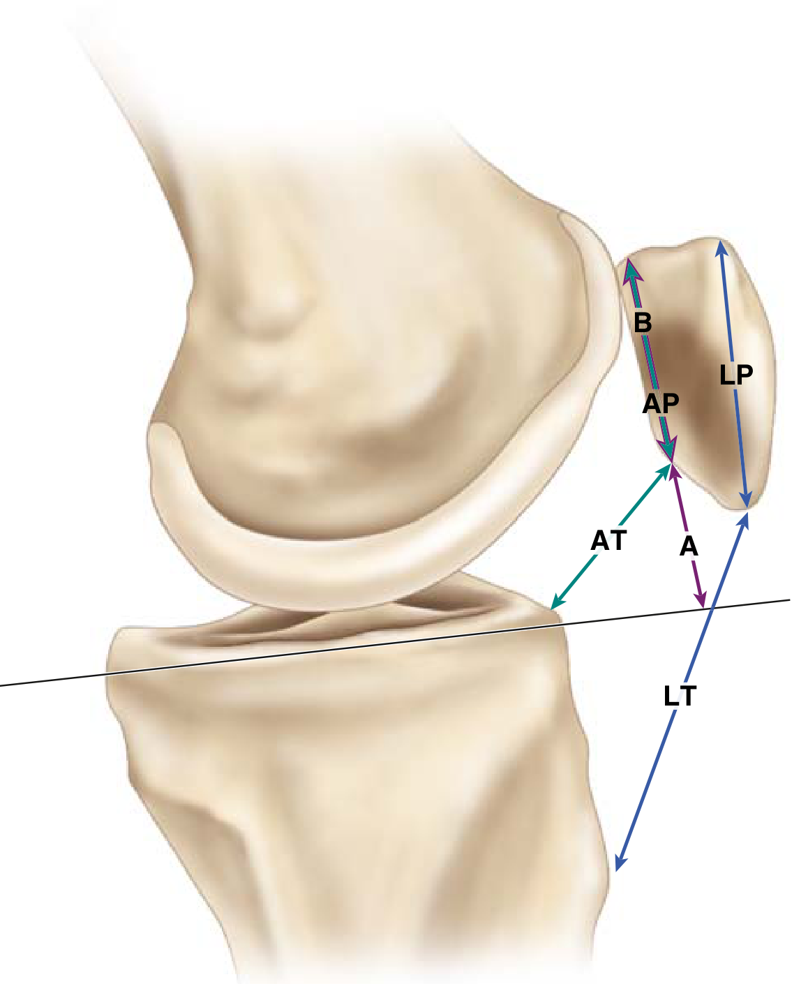
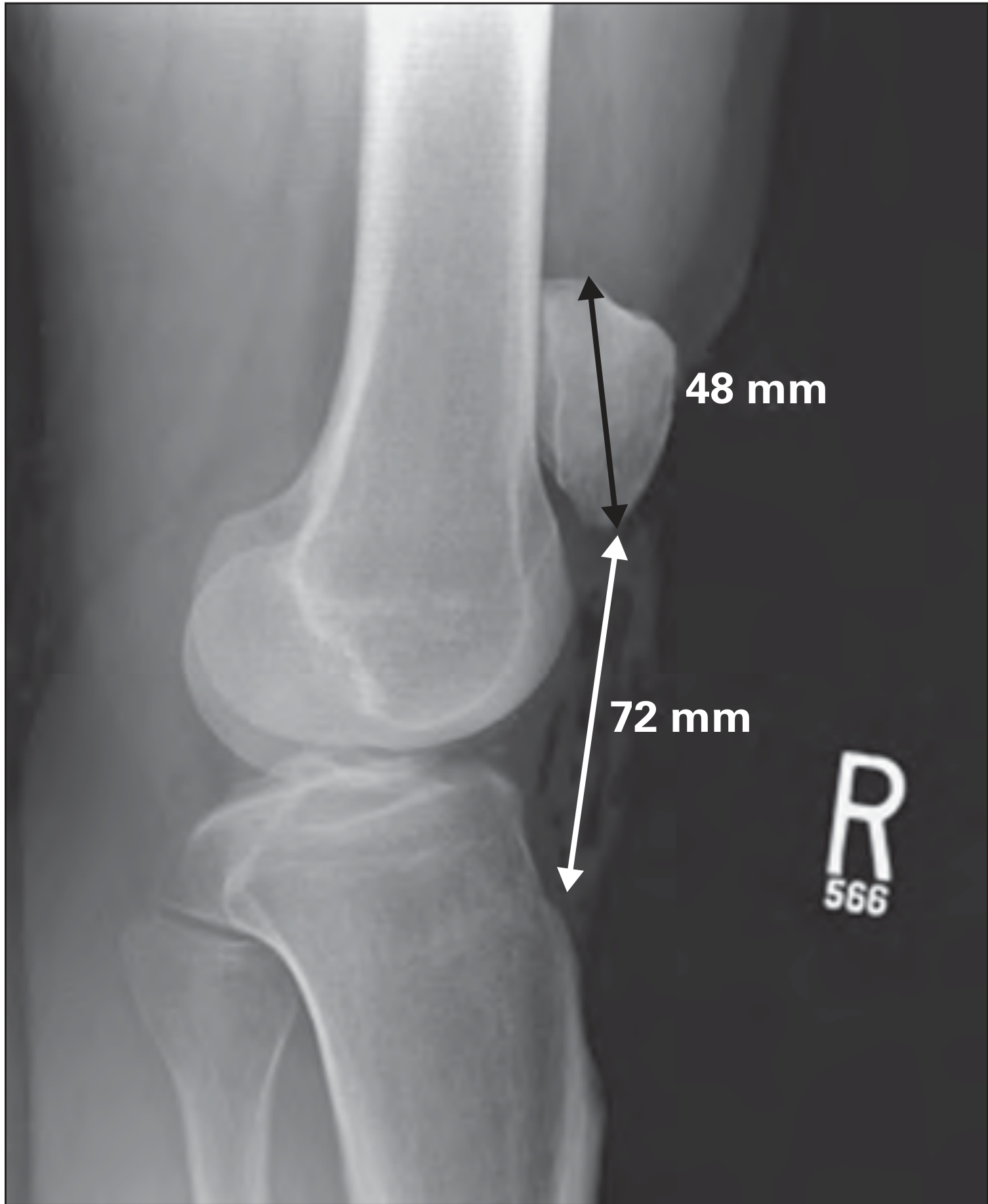
LT / LP = Patellar tendon length (LT) ÷ Longest sagittal diameter of patella (LP)
| Value | Interpretation |
|---|---|
| ~1.0 | Normal |
| > 1.2 | Patella alta |
| < 0.8 | Patella baja |
Key points:
- The most widely known and historically used index
- Measures the patellar tendon from the lower pole of the patella to the tibial tuberosity
- Limitation: LP (patellar length) can vary with fracture, surgery, or bone resorption, making it less reliable in post-operative knees
2. Caton-Deschamps Index
View: Lateral radiograph, knee flexed 30°
Measurement:
AT / AP = Distance from lower edge of patellar articular surface to anterosuperior tibial angle (AT) ÷ Length of patellar articular surface (AP)
| Value | Interpretation |
|---|---|
| 0.6 - 1.3 | Normal |
| > 1.3 | Patella alta |
| < 0.6 | Patella infera (baja) |
Key points:
- Uses articular surface landmarks rather than total patellar length
- Less affected by patellar morphology changes (e.g. after fracture or inferior pole excision)
- Widely considered the most reliable index, though this is debated
- Preferred for evaluating patellar instability and post-operative height assessment
3. Blackburne-Peel Index
View: Lateral radiograph, knee flexed 30°
Measurement:
A / B = Perpendicular height from tibial plateau tangent to inferior pole of patellar articular surface (A) ÷ Length of patellar articular surface (B)
| Value | Interpretation |
|---|---|
| 0.54 - 1.06 | Normal |
| > 1.06 | Patella alta |
| < 0.54 | Patella baja |
Key points:
- Uses the tibial plateau as the reference line (rather than the tibial tuberosity)
- Not affected by tibial tuberosity position or tibial slope
- Useful when tibial anatomy is abnormal (e.g. post-osteotomy)
Quick Comparison Table
| Index | Ratio | Normal | Alta | Baja | Key Reference Point |
|---|---|---|---|---|---|
| Insall-Salvati | LT / LP | ~1.0 | >1.2 | <0.8 | Tibial tuberosity + total patella length |
| Caton-Deschamps | AT / AP | 0.6-1.3 | >1.3 | <0.6 | Anterosuperior tibial angle + articular surface |
| Blackburne-Peel | A / B | 0.54-1.06 | >1.06 | <0.54 | Tibial plateau tangent + articular surface |
Other Related Radiographic Indices (Patellofemoral)
Beyond patellar height, these indices assess alignment and trochlear morphology relevant to instability:
| Index | Method | Normal / Threshold |
|---|---|---|
| Blumensaat line | Lateral x-ray at 30° - lower patellar pole should approximate this line through intercondylar notch | Patella proximal to line = alta |
| TT-TG distance | Axial CT/radiograph - distance between tibial tuberosity and trochlear groove (two lines perpendicular to posterior bicondylar axis) | >20 mm = malalignment; surgical relevance for tubercle osteotomy |
| Patellar tilt | CT at 20° flexion - angle between posterior condyle tangent and major patellar axis | Normal <20°; >20° = dysplasia |
| Trochlear depth | Lateral x-ray (Dejour) - measured 1 cm from top of groove | Normal ≥5 mm |
| Crossing sign | Lateral x-ray - anterior condylar cortex intersects trochlear outline | Present = dysplastic sulcus |
| Trochlear bump | Lateral x-ray - trochlear line extends anterior to femoral cortex | Present = dysplastic sulcus |
Clinical Relevance in Patellar Dislocation
- Patella alta (high-riding patella) reduces patellofemoral contact area and engagement in the trochlear groove at low flexion angles, predisposing to lateral subluxation and dislocation
- TT-TG > 20 mm is the threshold commonly used to select patients for tibial tubercle anteromedialization (e.g. Fulkerson osteotomy)
- Caton-Deschamps > 1.2-1.3 may prompt distal tubercle transfer as part of surgical correction of instability
- The Caton-Deschamps method is generally preferred in surgical planning for patellar instability because it is not distorted by changes to the inferior patellar pole
Sources: Campbell's Operative Orthopaedics, 15th Ed (2026) | Miller's Review of Orthopaedics, 9th Ed | Rheumatology 2-Vol Set, Elsevier 2022
This is a shared conversation. Sign in to Orris to start your own chat.