

photo-1785937065070.jpg
Explain this table specially pathogenesis in detain
Reading File
Finding Sources
Finding Sources
Reading File
Reading File
Loading Image
Reading File
Here is a detailed explanation of the table comparing Dystrophic vs. Metastatic Calcification, with special focus on pathogenesis:
Dystrophic vs. Metastatic Calcification
Quick Overview of the Table
| Feature | Dystrophic | Metastatic |
|---|---|---|
| Tissue | Dead/dying tissues | Normal tissues |
| Calcium metabolism | Normal | Deranged (abnormal) |
| Calcium in blood | Normal serum levels | Hypercalcemia |
| Reversibility | Irreversible | Reversible if metabolic disorder corrected |
| Pathogenesis | Phosphates bind to necrotic tissue -> calcium phosphate precipitates | Hypercalcemia causes calcium-phosphate precipitation at specific sites |
Pathogenesis - Detailed Explanation
1. Dystrophic Calcification - Pathogenesis
This occurs in dead and dying tissue despite completely normal serum calcium levels. The mechanism is local and tissue-driven:
Step-by-step mechanism:
a) Cell injury leads to calcium influx
- Normally, living cells actively pump calcium OUT of the cytoplasm (intracellular Ca²+ is kept very low ~0.1 µmol).
- When a cell is injured or dies (necrosis - coagulative, caseous, or liquefactive), the cell membrane loses integrity and the calcium pump fails.
- Calcium floods into the dying cell from the extracellular fluid (where Ca²+ concentration is ~1.3 mmol - over 10,000x higher).
b) Phosphate release from dead cells
- Necrotic and degenerative cells release phosphate-containing compounds from their damaged organelles (especially mitochondria and cell membranes containing phospholipids).
- These intracellular phosphates accumulate locally at the site of cell death.
c) Calcium-phosphate precipitation
- The locally released phosphates have an increased affinity/binding to the dead and necrotic tissue debris.
- Phosphate binds to calcium and forms calcium phosphate precipitates (primarily hydroxyapatite crystals, the same mineral in bone).
- Single necrotic cells can act as "seed crystals" that get encrusted with mineral, sometimes forming psammoma bodies (concentric lamellated calcifications).
d) Progressive mineralization
- The initial micro-deposits act as a nidus, and more calcium-phosphate layers are deposited over time.
- The result is visible as white, gritty, chalky deposits at the necrotic site.
Key examples of dystrophic calcification:
- Calcific aortic stenosis (damaged valve cusps develop chalk-white calcium deposits - as shown in the Robbins image above)
- Atherosclerotic plaques
- Caseous necrosis of tuberculosis (lymph nodes can turn literally "to stone")
- Areas of fat necrosis (e.g., post-pancreatitis)
Serum calcium is NORMAL - the problem is purely local at the site of tissue injury.
2. Metastatic Calcification - Pathogenesis
This occurs in normal, living tissues and is driven entirely by systemic hypercalcemia (elevated blood calcium).
Step-by-step mechanism:
a) Hypercalcemia develops systemically
The four main causes of hypercalcemia driving metastatic calcification are (per Robbins):
- Hyperparathyroidism - excess PTH causes bone resorption -> calcium released into blood. Also seen with PTH-related protein secreted by malignant tumors.
- Bone destruction - from primary bone marrow tumors (multiple myeloma), diffuse skeletal metastases (e.g., breast cancer), Paget disease, or immobilization.
- Vitamin D disorders - vitamin D intoxication, sarcoidosis (macrophages activate vitamin D precursor), Williams syndrome.
- Renal failure - phosphate retention leads to secondary hyperparathyroidism.
b) Solubility product exceeded at specific alkaline sites
- When serum calcium rises, the calcium x phosphate solubility product is exceeded.
- Calcium phosphate precipitates out at tissues that have an alkaline (high pH) microenvironment - because calcium phosphate is LESS soluble in alkaline conditions.
- Tissues that excrete acid (like gastric mucosa, renal tubules) have an alkaline compartment on their interstitial/serosal side, predisposing them to calcium deposition.
c) Preferred deposition sites (the classic 4 + 1):
- Lungs (alveolar septa, vessel walls) - very commonly affected; can cause respiratory compromise
- Stomach (gastric mucosa)
- Blood vessels (systemic arteries, pulmonary veins)
- Cornea (band keratopathy)
- Kidneys (nephrocalcinosis - can cause renal failure over time)
All these sites have an alkaline internal compartment due to their acid-excreting function.
d) Morphology
- Calcium salts appear identical to dystrophic calcification microscopically: basophilic, amorphous or crystalline (hydroxyapatite) deposits.
- The key difference is that the surrounding tissue is NORMAL.
Reversibility: Unlike dystrophic calcification, metastatic calcification can regress if the underlying hypercalcemia is corrected (e.g., treating hyperparathyroidism, stopping vitamin D excess).
Memory Hook
| Dystrophic | Metastatic | |
|---|---|---|
| Calcium in blood? | Normal | High (hypercalcemia) |
| Tissue condition? | Dead/necrotic | Normal/alive |
| Driver | Local phosphate release from dying cells | Systemic calcium overload precipitating at alkaline sites |
| Reversible? | No | Yes (treat the cause) |
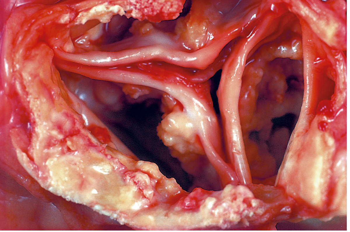
Gross specimen showing dystrophic calcification of the aortic valve - irregular yellowish-white calcium masses on the fibrotic valve cusps, with normal serum calcium. (Robbins, Cotran & Kumar - Pathologic Basis of Disease)
Source: Robbins, Cotran & Kumar - Pathologic Basis of Disease, p. 76
What are the cardinal pillars of ethics in MBBS AETCOM answer
The four cardinal pillars of medical ethics are Autonomy, Beneficence, Non-maleficence, and Justice - a framework by Beauchamp & Childress widely used in AETCOM. Let me search the medical library for the exact AETCOM curriculum content.
Searching sources...
Finding Sources
Finding Sources
Finding Sources
Finding Sources
Searching the Web
AETCOM MBBS cardinal pillars of ethics four principles India NMC
Reading File
Reading Web Page
https://dokumen.pub/aetcom-attitude-ethics-and-communication…
Reading File
Here is a complete AETCOM-aligned answer on the Four Cardinal Pillars of Medical Ethics:
The Four Cardinal Pillars of Ethics (AETCOM - Module 2.2)
As per the NMC AETCOM Manual (Module 2.2: Foundations of Bioethics), the four cardinal pillars of bioethics are:
Autonomy | Beneficence | Non-maleficence | Justice
These four prima facie principles were propounded by Beauchamp and Childress (Principalism) and form the foundation of ethical medical practice. They are "binding" moral responsibilities in healthcare delivery, irrespective of an individual doctor's personal ideology.
1. AUTONOMY
Meaning: Respect for the patient's right to self-determination - the right to make informed decisions about their own health.
Key points:
- The word comes from Greek: autos (self) + nomos (rule/law) = self-rule
- The patient has the right to accept or refuse any treatment, even life-saving treatment, if they have decision-making capacity
- This is the basis of informed consent - consent must be:
- Informed (patient knows diagnosis, planned treatment, risks, benefits, alternatives, and prognosis)
- Voluntary (freely given, not under pressure)
- Competent (given by a person with capacity to decide)
- Autonomy also includes shared decision-making - doctor and patient together deciding the best course
- Limits of autonomy: A patient cannot demand treatment that is harmful or unethical; autonomy may be restricted in emergencies or when the patient lacks capacity
Clinical example: A Jehovah's Witness refusing a blood transfusion must be respected, even if the doctor believes it is life-saving.
2. BENEFICENCE
Meaning: The duty to "do good" - acting in the patient's best interest.
- From Latin: bene (good) + facere (to do)
- It is an act of charity, mercy, and kindness and the doctor's moral obligation to safeguard the patient's health
- Includes:
- Providing competent, up-to-date treatment
- Actively promoting the patient's welfare
- Preventing harm where possible
- Balancing benefits against risks of treatment
- Paternalism (doing good without patient consent) conflicts with autonomy - modern ethics requires balancing both
Clinical example: A surgeon who delays emergency surgery to explain the risks to the patient is balancing beneficence with autonomy.
3. NON-MALEFICENCE
Meaning: The duty to "do no harm" (Primum non nocere - First, do no harm).
- The oldest principle in medicine, rooted in the Hippocratic Oath
- Every medical intervention carries some risk; non-maleficence does NOT mean avoiding all risk, but ensuring that:
- Benefits outweigh risks
- No unnecessary harm is inflicted
- Proportionality is maintained
- Applies to:
- Avoiding unnecessary investigations or procedures
- Not prescribing harmful drugs or dosages
- Not prolonging futile treatment (causing suffering without benefit)
- Maintaining competence - an incompetent doctor harms patients
Distinction from Beneficence:
| Beneficence | Non-maleficence |
|---|---|
| Actively do good | Avoid causing harm |
| Positive duty | Negative duty |
| "Help the patient" | "Don't hurt the patient" |
Clinical example: Prescribing antibiotics unnecessarily (antibiotic resistance, side effects) violates non-maleficence.
4. JUSTICE
Meaning: Fair, equitable, and appropriate treatment for all patients; equal distribution of health resources.
-
Justice in healthcare operates at two levels:
- Individual justice - treating each patient fairly, without discrimination based on caste, religion, gender, economic status
- Distributive justice - fair allocation of scarce healthcare resources across society
-
As per the AETCOM Manual, justice in distribution of health care resources means every individual must receive an equal share of health resources as per:
- Their need
- Their effort and contribution
- Their merit
- Free-market exchanges
-
Includes equal time for examination of each patient (though not always possible in every clinical situation)
-
Current Government of India initiative under justice: Ayushman Bharat / PM-JAY scheme providing health insurance to the poor
Clinical example: Giving preferential treatment to a VIP patient while other patients with equal or greater need are waiting violates the principle of justice.
Summary Table (Exam-Ready)
| Pillar | Meaning | Key Concept | Mnemonic |
|---|---|---|---|
| Autonomy | Patient's right to decide | Informed consent | "Self-rule" |
| Beneficence | Do good for patient | Act in patient's best interest | "Be nice" |
| Non-maleficence | Do no harm | Primum non nocere | "No harm" |
| Justice | Fairness & equity | Equal distribution of care | "Just treat all equally" |
Mnemonic: A-B-N-J → "A Brave Nurse Jumps" or simply "JAB-N" (Justice, Autonomy, Beneficence, Non-maleficence)
Conflicts Between Pillars (Important for SAQs/Viva)
These pillars do not always align - ethical dilemmas arise when they conflict:
- Autonomy vs. Beneficence: Patient refuses treatment the doctor knows is beneficial
- Beneficence vs. Non-maleficence: A chemotherapy drug that cures but causes suffering
- Autonomy vs. Justice: A patient demands expensive treatment at the cost of resources others need
- In such situations, the doctor must do a "balancing act" considering all principles together for a workable solution
Source: NMC AETCOM Manual, Module 2.2 - "The Foundations of Bioethics" (Professional Year 2); Miller's Anesthesia 10e, p. 602
This is a shared conversation. Sign in to Orris to start your own chat.