
Tell me about pres
Asking for Preferences
What do you mean by 'pres'?
PRES (Posterior Reversible Encephalopathy Syndrome) is a clinico-radiological syndrome characterized by headache, seizures, visual disturbances, and altered consciousness, typically associated with hypertensive emergencies, eclampsia, immunosuppressive drugs, or renal failure. MRI shows characteristic posterior white matter edema (vasogenic), which is usually reversible with treatment.
Searching sources...
Reading File
Finding Sources
Searching PubMed
"posterior reversible encephalopathy syndrome" PRES
Finding Sources
Reading Article
PMID: 40818477
Reading File
Reading File
Reading File
Reading File
Loading Image
Reading File
Loading Image
Here is a detailed overview of Posterior Reversible Encephalopathy Syndrome (PRES):
Posterior Reversible Encephalopathy Syndrome (PRES)
Also known as Reversible Posterior Leukoencephalopathy Syndrome (RPLS), PRES is a clinicoradiological syndrome characterized by acute neurological symptoms in the setting of endothelial dysfunction and vasogenic edema, predominantly affecting the posterior cerebral white matter.
Definition
PRES results from vascular endothelial dysfunction leading to blood-brain barrier (BBB) breakdown and vasogenic edema. It is seen in malignant hypertension, eclampsia, immunosuppressive/cytotoxic drug use, and autoimmune disease. - Kanski's Clinical Ophthalmology, 10th Ed.
Causes / Precipitating Factors
- Severe hypertension (systolic >195 mmHg) - most common; occurs in essential hypertension, renal artery stenosis, acute glomerulonephritis, pheochromocytoma, Cushing syndrome, cocaine use
- Eclampsia and HELLP syndrome (can occur at lower blood pressures due to rapid rise)
- Immunosuppressive/cytotoxic drugs: tacrolimus, cyclosporine (ciclosporin), FK-506, cyclophosphamide, interferons, tyrosine kinase inhibitors, CAR-T cell therapies
- Renal failure / hemodialysis
- Autoimmune disease (e.g., systemic lupus erythematosus)
- Sympathomimetic and serotonergic drugs
- Adams and Victor's Principles of Neurology, 12th Ed.
Clinical Features
The neurological syndrome is dominated by symptoms referable to the occipital and adjacent parietal regions:
| Feature | Details |
|---|---|
| Headache | Often the first symptom |
| Seizures | May be focal or generalized; can be more marked on one side |
| Visual disturbances | Visual field deficits, hallucinations, cortical blindness, Balint syndrome |
| Encephalopathy | Confusion, altered consciousness, stupor, coma in advanced cases |
| Focal neurological signs | Hemiparesis, aphasia (from clustering of microinfarcts) |
| Papilledema | Retinal hemorrhages may occur (but not required for diagnosis) |
Pathophysiology
Two main theories exist:
- Failure of cerebrovascular autoregulation: Severe hypertension overwhelms the adaptive capacity of arterioles, causing overdistention, endothelial damage, and passive extravasation of fluid into the brain parenchyma - producing vasogenic edema.
- Vasospasm-mediated ischemia: Some evidence for segmental vasoconstriction contributing to tissue injury.
The net result is endothelial dysfunction with damage to capillary endothelial junctions, vasodilatation, fluid extravasation, and vasogenic edema. Inflammatory cytokines (IL-6, IL-10, VEGF) are increasingly recognized as contributors. Neuropathologically: fibrinoid necrosis of arteriole/capillary walls, fibrin thrombi, microinfarcts, and zones of cerebral edema. - Adams and Victor's Principles of Neurology, 12th Ed.; Bradley and Daroff's Neurology, 8th Ed.
Imaging
MRI is the investigation of choice:
- T2/FLAIR: Bilateral hyperintense signal in the posterior white matter (parietooccipital lobes), representing vasogenic edema
- Edema affects cortex AND subcortical white matter
- Little or no mass effect (distinguishes from tumor or stroke)
- Does NOT typically spread along white matter tracts (e.g., corpus callosum)
- Scattered cortical lesions may occur in a watershed distribution
- In severe cases: hemorrhage and heterogeneous infarction
- DWI: Typically shows restricted diffusion is absent (vasogenic not cytotoxic edema) - an important distinguishing feature
- Changes are usually reversible over weeks with treatment
The image below (from Adams and Victor's Principles of Neurology) shows typical PRES: symmetric T2-FLAIR hyperintensity in the parietooccipital lobes, involving cortex and subcortical white matter:
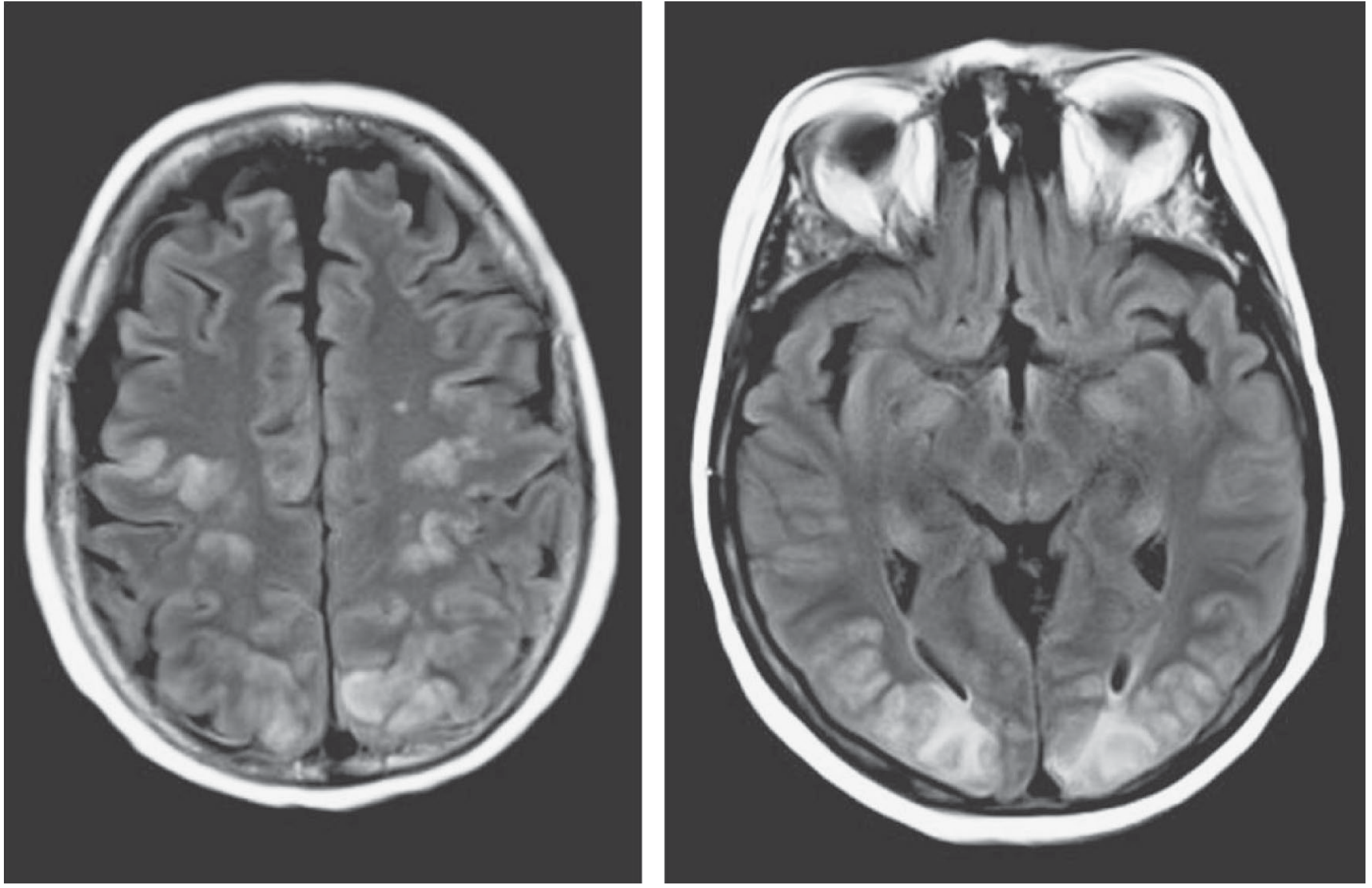
Hypertensive encephalopathy with PRES. Symmetric T2-FLAIR hyperintensity in the parietooccipital lobes, affecting cortex and subcortical white matter, with minimal mass effect.
This second image (from Bradley and Daroff's Neurology in Clinical Practice) shows unilateral-predominant posterior white matter hyperintensity in a patient with hypertension, visual changes, altered consciousness, and seizure:
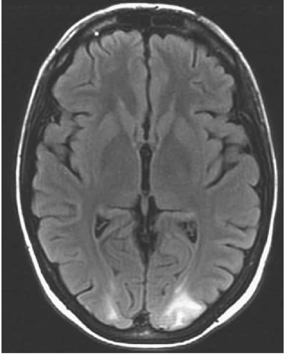
Axial FLAIR: hyperintense signal in the occipital subcortical white matter bilaterally (left > right) in a patient presenting with elevated BP, visual changes, altered mental status, and seizure.
CT: Shows corresponding reduced density (hypodensity) in posterior white matter. Useful in emergency settings.
CSF: Elevated pressure and protein (>100 mg/dL) in many cases, no cellular reaction; LP not required for diagnosis.
Treatment
Treatment is focused on removing the precipitating cause and controlling blood pressure:
- Blood pressure reduction: Target ~150/100 mmHg or a 20% reduction in mean arterial pressure (avoid excessive reduction, which may cause watershed ischemia)
- Acute: IV sodium nitroprusside, IV labetalol, IV nicardipine, or oral nifedipine
- Sustained: ACE inhibitors or calcium channel blockers
- Eclampsia: Magnesium sulfate
- Drug-induced PRES: Discontinue or reduce the offending agent
- Seizure management: Standard antiepileptic treatment acutely; long-term AED therapy usually not needed if the underlying cause is resolved
- Cerebral edema/raised ICP: Dexamethasone 4-6 mg every 6 hours may be used, though evidence is limited; hyperosmolar therapy has also been used with unclear benefit
With treatment, neurological deficits and imaging changes typically resolve over days to weeks - hence "reversible." - Adams and Victor's Principles of Neurology, 12th Ed.
Prognosis
- Generally favorable when the underlying cause is treated promptly
- Incomplete resolution or recurrence can occur
- Complications include epilepsy, ischemic stroke, and rarely death
- Severe or drug-induced PRES (e.g., chemotherapy-related) may have a more complex course
Key Distinguishing Points
| Feature | PRES | Ischemic Stroke | Vasogenic Tumor Edema |
|---|---|---|---|
| Distribution | Posterior, bilateral | Vascular territory | Perilesional |
| DWI restriction | Usually absent | Present | Absent |
| Mass effect | Minimal | Variable | Often present |
| Reversibility | Yes (usually) | No | Depends |
| Precipitant | HTN, eclampsia, drugs | Embolism, thrombosis | Tumor |
Recent Evidence (2025)
A 2025 review in Lancet Neurology by Fugate, Hawkes, and Rabinstein highlights that novel immunotherapies (tyrosine kinase inhibitors, CAR-T cell therapies) have expanded the spectrum of PRES triggers, and that emerging biomarkers (IL-6, IL-10, VEGF) offer promise for improved diagnosis and prognosis. Notably, evidence from randomized trials remains absent - all management is based on observational data and expert consensus. [PMID: 40818477]
This is a shared conversation. Sign in to Orris to start your own chat.