
BASIC RADIATION: LONG QUESTION: Discuss in detail about the effect of ionizing radiation on Biologic molecules. Write a note on Osteoradionecrosis. (2018) Describe the measures of protection from radiation hazards. (16) 7. Define electromagnetic radiation? Describe structure and function of x ray tube. (12) Biological effects of radiation? (14,23), NS. Describe x-ray tube with diagram? (13) 6. Discuss in detail about the parts of X-ray tube with diagram. Write in short about production of x-ray. (19) What are the protection measures for the patient and the operator against the adverse effect of radiation? (12) What is x-ray? How x-ray is produced? What are the radiation hazards? (11) Describe in details about the hazards of radiation. Describe in detail about various methods of radiation protection. (20s) 10. Draw a labeled diagram of x-ray head and explain in detail about the working mechanism of x-ray tube head. (20) 11. Discuss the methods of radiation safety and protection of operator, patient and public. Write a short note on Acute radiation syndrome. (20) SHORT NOTE: Radiation Mucositis. (2018) Characteristics Radiation. (2018) Osteoradionecrosis. (16,13) TLD Badge. (16) Properties of x-ray. (12,13) Photoelectric effect. (12,14,16) Collimation (20s,17,12,14,22) Breaking Radiation. (15,23) 9. Compton scatters. (12) 10.- Thermo luminescent dose meters. (12) 11. Radiation monitoring badge. (12) Filtration. 9,) N.'13. Radiolysis of water 19 14. Write in brief about dosimetry. (20) 15. Production of X-ray. (22) 16 Radiation effects on oral cavity. (22)
ionizing radiation effects on DNA biological molecules
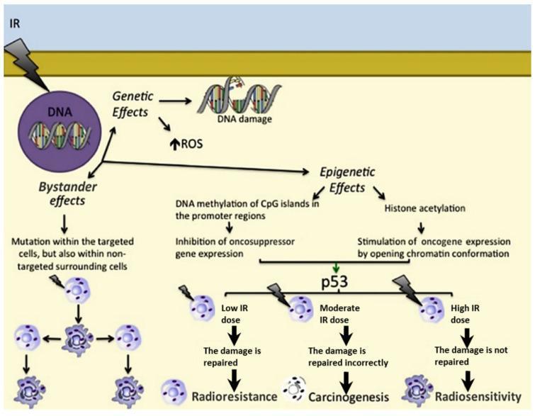
This pathophysiology diagram illustrates the biological responses of human cells to ionizing radiation (IR), categorized into three primary pathways: Genetic, Epigenetic, and Bystander effects. The Genetic pathway highlights direct DNA damage and increased reactive oxygen species (ROS). The Epigenetic pathway details mechanisms such as DNA methylation of CpG islands (leading to inhibition of oncosuppressor genes) and histone acetylation (leading to oncogene stimulation through chromatin opening). These pathways converge on the p53 tumor suppressor protein, which mediates cellular outcomes based on radiation intensity. A 'Low IR dose' results in effective DNA repair and radioresistance; a 'Moderate IR dose' causes incorrect repair, leading to carcinogenesis; and a 'High IR dose' prevents repair entirely, resulting in radiosensitivity and cell death. The Bystander effects pathway demonstrates how radiation induces mutations not only in targeted cells but also in non-targeted surrounding cells, contributing to genomic instability. This diagram is designed for intermediate to advanced medical education regarding radiobiology, molecular oncology, and clinical radiation therapy principles.
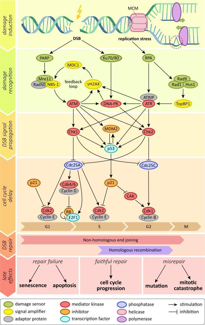
This pathophysiology diagram illustrates the comprehensive molecular signaling network of the human DNA damage response (DDR). The schematic is organized into temporal and functional stages: damage induction, damage recognition, DSB signal propagation, cell cycle delay, DSB repair, and late effects. The initial phase identifies two primary triggers: ionizing radiation causing double-strand breaks (DSBs) and replication stress involving stalled replication forks with MCM complexes. The signaling cascade categorizes molecules by color-coded functions, including damage sensors (e.g., PARP, Ku70/80, MRN complex), signal amplifiers (e.g., γH2AX, MDC1, TopBP1), and central mediator kinases (ATM, ATR, DNA-PK). These kinases activate downstream effectors Chk1 and Chk2, which regulate cell cycle checkpoints (G1, S, G2) by inhibiting Cdc25 phosphatases and activating p53-mediated p21 inhibition of Cyclin/Cdk complexes. The diagram concludes with repair pathways—Non-homologous end joining (NHEJ) and Homologous recombination (HR)—and their potential biological outcomes: faithful repair leading to cell cycle progression, repair failure leading to senescence or apoptosis, and misrepair leading to mutations or mitotic catastrophe.
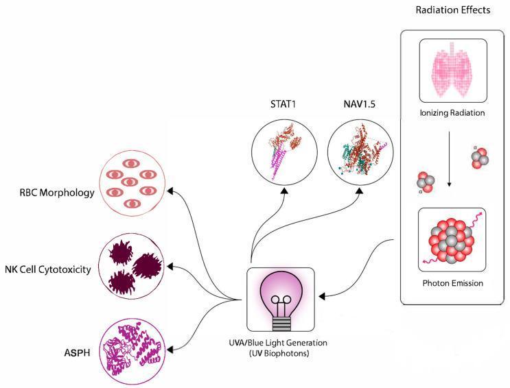
A pathophysiology diagram illustrating the hypothetical pathways linking ionizing radiation exposure to systemic biological effects. The process is divided into two main sections. On the right, a 'Radiation Effects' vertical flow depicts initial ionizing radiation exposure (represented by a lung schematic) leading to photon emission. This is linked to the central node, 'UVA/Blue Light Generation (UV Biophotons)'. From this central node, several signaling pathways diverge to the left, targeting specific molecular and cellular components. These include protein markers STAT1 and NAV1.5 (a voltage-gated sodium channel), and enzymes like ASPH (Aspartate ̠-Hydroxylase), shown as complex protein structures. Further downstream effects are indicated on the far left, highlighting changes in Red Blood Cell (RBC) morphology and Natural Killer (NK) cell cytotoxicity. The diagram uses stylized icons and molecular modeling to explain how low-dose radiation may induce bystander effects or chronic fatigue through biophotonic signaling and subsequent cellular dysfunction.
x-ray tube diagram cathode anode filament tungsten target
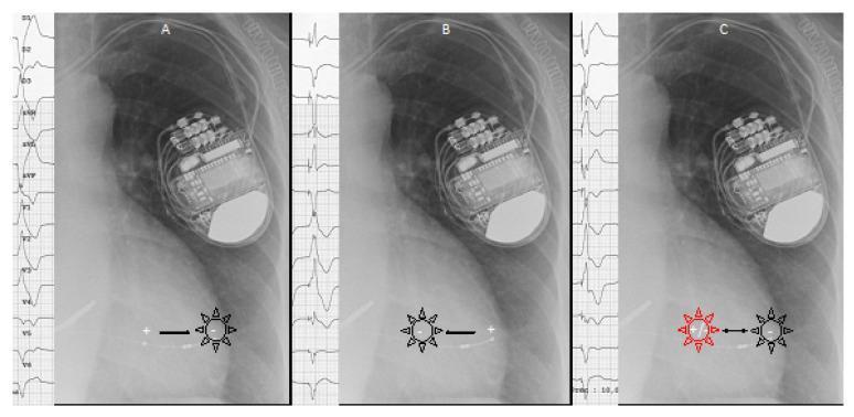
This composite educational graphic consists of three panels (A, B, and C) illustrating cardiac pacing concepts using chest X-rays, ECG tracings, and superimposed diagrams. Each panel features a chest X-ray showing an implanted cardiac device (pacemaker or ICD) in the left infraclavicular region with leads extending toward the heart. Panel A and B demonstrate conventional bipolar pacing where a depolarization wavefront (indicated by a black sunburst icon) originates solely from the cathode (-). Panel A shows the cathode on the right and anode (+) on the left, while Panel B reverses this orientation. Panel C illustrates 'pseudo-multisite pacing' (pseudo-MPP) via intentional anodal capture. It shows depolarization wavefronts originating from both the cathode and the anode (indicated by red and black sunburst icons), occurring when high-density current is applied. Adjacent to each X-ray is a 12-lead ECG strip demonstrating the resulting QRS complex morphologies. The diagram highlights how anodal capture in bipolar pacing creates a fusion QRS complex, relevant for optimizing Cardiac Resynchronization Therapy (CRT).
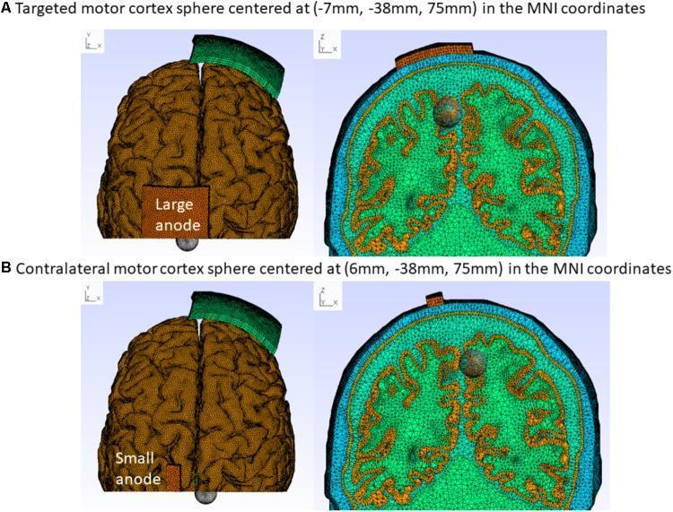
Educational diagram illustrating finite element method (FEM) head models for transcranial Direct Current Stimulation (tDCS) research. The figure shows the Colin27 head model used to analyze electric field distribution in the motor cortex. It is organized into two rows: (A) demonstrates a 'Large-anode' montage (5 cm x 7 cm) and (B) demonstrates a 'Small-anode' montage (3.5 cm x 1 cm). The left panels provide a superior (top) view of the cortical surface mesh, highlighting the anode placement (orange rectangle) relative to a cathode (green band) and a target grey sphere representing a 1 cm radius cortical tissue cluster. Row A targets the motor cortex at MNI coordinates (-7mm, -38mm, 75mm), while row B focuses on the contralateral leg motor cortex at (6mm, -38mm, 75mm). The right panels present coronal cross-sections of the FEM mesh, visualizing the internal brain geometry, sulci, and gyri using a triangular mesh discretization. This visual resource is used to compare how different electrode sizes and spatial coordinates affect targeted neurostimulation in clinical neurology and biomedical engineering applications.
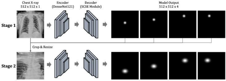
A technical diagram illustrating a two-stage deep learning architecture for key point detection in chest radiography, specifically designed to evaluate endotracheal tube (ETT) positioning. Stage 1 takes a full-field chest X-ray (512x512x1) as input, passing it through a DenseNet121 encoder and a decoder with a Spatial and Channel Squeeze & Excitation (SCSE) module. The output is a set of four heatmaps representing the initial localization of four clinical key points: the tracheal tube end, the carina, and the left and right clavicular heads. In Stage 2, the process is refined by cropping and resizing the region of interest based on Stage 1 results, passing it through identical encoder-decoder blocks to produce high-resolution probability distributions of the target coordinates. The model facilitates clinical assessment by calculating distances between these anatomical landmarks to determine if the ETT position is appropriate (typically 20–70 mm from the carina). This tool is intended for medical AI research and radiology computer-aided diagnosis training.
BASIC RADIATION - Complete Study Notes
LONG QUESTIONS
1. Effects of Ionizing Radiation on Biologic Molecules (2018, 14, 23)
Definition of Ionizing Radiation
Mechanisms of Action on Biologic Molecules
A. Direct Effect
B. Indirect Effect (Radiolysis of Water)
H₂O + radiation → H₂O⁺ + e⁻ (ion pair) H₂O⁺ → H⁺ + OH• (hydroxyl radical) e⁻ + H₂O → H₂O⁻ → OH⁻ + H• H• + H• → H₂ OH• + OH• → H₂O₂ (hydrogen peroxide)
Effects on Specific Biologic Molecules
1. DNA (Primary Target)
- Single-strand breaks (SSBs): One strand cut - usually repaired accurately using the complementary strand as template. Requires a dose of ~1 Gy to produce 1000 SSBs per cell.
- Double-strand breaks (DSBs): Both strands broken close together - far more lethal and mutagenic. DSBs occur at ~1/20th the frequency of SSBs but are the primary lethal lesion. They are repaired by:
- Non-Homologous End Joining (NHEJ) - fast but error-prone
- Homologous Recombination (HR) - accurate but only in S/G2 phase
- Base damage: Alteration or loss of purine/pyrimidine bases
- Cross-links: DNA-DNA and DNA-protein cross-links
- Chromosomal aberrations: Deletions, translocations, dicentrics, ring chromosomes
- Low dose: Effective DNA repair → radioresistance
- Moderate dose: Misrepair → mutations → carcinogenesis
- High dose: Repair failure → cell death/apoptosis
2. Proteins
- Ionization of amino acid side chains - especially sulfhydryl groups (cysteine, methionine)
- Cross-linking and aggregation of protein molecules
- Enzyme inactivation - loss of catalytic activity
- Structural proteins disrupted
- At diagnostic X-ray doses, protein damage is less significant than DNA damage
3. Lipids
- Lipid peroxidation: OH• attacks polyunsaturated fatty acids in cell membranes
- Chain reaction propagation via lipid radicals
- Membrane disruption → altered permeability, loss of ion gradients
- Especially affects mitochondrial and plasma membranes
4. Carbohydrates
- Less radiosensitive than DNA/proteins
- Hydroxyl radical attacks glycosidic bonds
- Depolymerization of polysaccharides
- Clinical significance: glycosaminoglycans in connective tissue affected → fibrosis
5. Nucleic Acids (RNA)
- Similar mechanisms to DNA
- mRNA damage → impaired protein synthesis
- rRNA damage → ribosomal dysfunction
- tRNA damage → translation errors
The 4 R's of Radiobiology (Factors modifying radiation effects)
- Repair - sublethal damage repair occurs between fractions
- Redistribution - cells cycle into more sensitive phases
- Repopulation - surviving cells proliferate between fractions
- Reoxygenation - hypoxic cells (3x more radioresistant) become oxygenated
Radiosensitivity (Law of Bergonie and Tribondeau)
- Divide rapidly
- Have high mitotic index
- Are undifferentiated
- Most sensitive: lymphocytes, erythroblasts, spermatogonia, intestinal crypt cells
- Least sensitive: neurons, muscle cells, chondrocytes
2. Osteoradionecrosis (ORN) (2018, 16, 13)
Definition
Pathogenesis
- Inhibition of mitosis and reduction in tissue repair capacity
- Vasculitis leading to progressive obliteration of blood vessels
- Avascular necrosis (Marx's "3H tissue" - Hypoxic, Hypocellular, Hypovascular)
- Fibroatrophic mechanism - damage to fibroblasts and osteoblasts
- Histologically: death of osteocytes and osteoblasts → empty lacunae, preponderance of osteoclasts
Incidence
- Mandibular ORN: 5-10% of patients treated with conventional RT or HDR brachytherapy
- Severe ORN in ~2% of cases
- Higher incidence with hyperfractionated regimens (23% vs 9% with conventional RT in one study)
Predisposing Factors
- Dental extractions in irradiated regions (post-RT extraction carries higher risk)
- Total radiation dose (>60 Gy significantly increases risk)
- Microvascular disease (diabetes, atherosclerosis)
- Poor oral hygiene/dental disease
- Brachytherapy with high local doses
- Trauma post-irradiation
Clinical Features
- Pain, bad breath (halitosis)
- Dysgeusia (taste disturbance), dysesthesia or anesthesia
- Trismus, difficulty with mastication, deglutition, and/or speech
- Fistula formation
- Pathologic fracture
- Exposed necrotic bone in the mouth
- Local, spreading, or systemic infection
Management
| Stage | Treatment |
|---|---|
| Mild ORN | Conservative debridement, antibiotics, occasional ultrasound |
| Moderate | Sequestrectomy, wound irrigation, hyperbaric oxygen (HBO) therapy |
| Severe / Extensive | Radical resection of mandible + immediate microvascular reconstruction |
Prevention
- Reduce mandibular volumes receiving high doses (IMRT planning)
- Improve salivary flow rates and oral health
- Uniform prophylactic dental care before RT
- Repair/extract diseased teeth before RT (not healthy/restorable teeth)
- Use megavoltage (vs kilovoltage) X-rays - reduces bone dose
- Multiple radiation fields; IMRT; Hadron therapy (avoids exit dose)
3. Radiation Mucositis (2018)
Definition
Pathogenesis
- Radiation kills rapidly dividing mucosal basal cells (high mitotic index)
- Mucosal atrophy → erosion → ulceration
- Typically begins in Week 2-3 of RT (cumulative dose ~20 Gy)
- Confluent mucositis by Week 4-5 (~40 Gy)
Grading (RTOG Scale)
| Grade | Features |
|---|---|
| 1 | Injection/mild erythema |
| 2 | Patchy mucositis with inflammatory serosanguinous discharge |
| 3 | Confluent fibrinous mucositis; severe pain; difficulty eating |
| 4 | Ulceration, hemorrhage, necrosis |
Management
- Good oral hygiene, frequent saline rinses
- Mucosal anesthetics (viscous lidocaine)
- Analgesics (step-up: NSAIDs → opioids)
- Nutritional support (nasogastric feeds if severe)
- Antifungal for secondary Candida infection
- Amifostine (radioprotector) may reduce severity
- Resolves 2-4 weeks after completing RT
4. X-Ray Tube - Structure and Function (2012, 13, 19, 20)
Definition of Electromagnetic Radiation
X-Ray Tube - Labeled Diagram
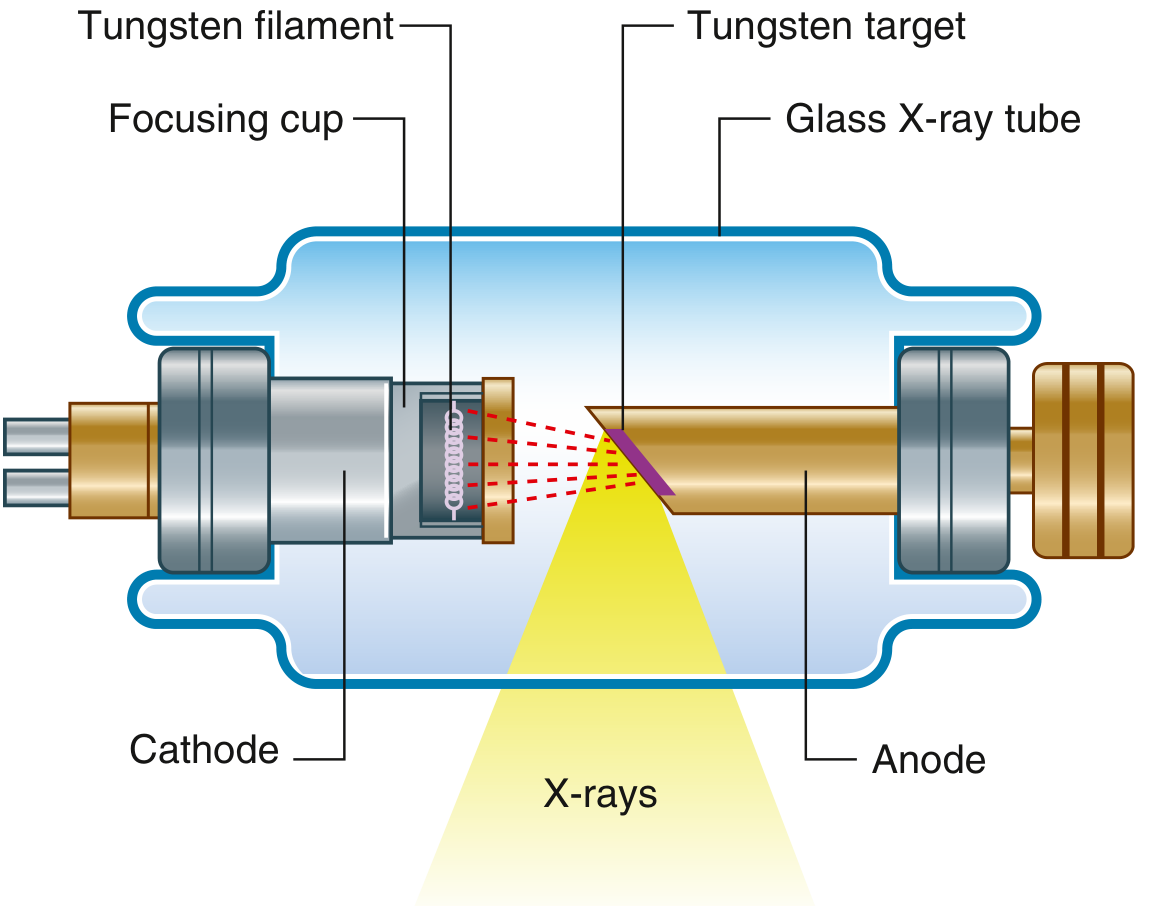
Parts of the X-Ray Tube
A. Glass Envelope (Housing)
- Hard borosilicate glass vacuum tube (evacuated to <10⁻⁶ torr)
- Vacuum is essential - prevents electron collision with gas molecules
- The entire assembly is housed in an oil-filled metal shield (tube housing) that provides:
- Radiation shielding
- Heat dissipation
- Electrical insulation
B. Cathode (Negative Electrode)
- Tungsten filament (coiled wire): Heated by low-voltage current (6-12 V, 3-5 A) → thermionic emission of electrons (electrons boil off the hot filament)
- Focusing cup (molybdenum): Negatively charged metal cup surrounding the filament that focuses the electron beam onto a small area of the anode
- The filament temperature determines tube current (mA) and thus X-ray quantity
C. Anode (Positive Electrode)
- The target material is tungsten (Z=74) - chosen because:
- High atomic number (Z) → high X-ray production efficiency
- Very high melting point (3422°C) → handles intense heat
- High thermal conductivity
- Stationary anode: Used in dental X-ray units (low power)
- Rotating anode: Used in diagnostic radiology units - the disc rotates at 3000-10,000 rpm to spread heat over a larger target area, allowing higher mA and kVp
- Anode angle (typically 10-20°): Affects the focal spot size and field coverage (line focus principle)
D. Induction Motor / Rotor
- Drives the rotating anode
- External stator coils (electromagnetic induction)
E. High-Voltage Generator
- Provides 40-150 kVp (kilovolts peak) across the tube
- Higher kVp → greater electron acceleration → higher energy X-rays (greater penetrating power)
Production of X-Rays
1. Bremsstrahlung (Braking Radiation / Characteristic of continuous spectrum)
- German for "braking radiation"
- Electrons decelerate as they pass near the nucleus of tungsten atoms
- Kinetic energy is converted to X-ray photons
- Continuous spectrum of energies - from zero up to maximum (= kVp in keV)
- Accounts for ~80-90% of the X-ray beam
- Maximum photon energy = eV (electron charge × accelerating voltage)
2. Characteristic Radiation
- Incident electron ejects an inner-shell (K-shell) electron from tungsten
- An outer-shell electron drops down to fill the vacancy
- Energy difference emitted as a characteristic X-ray photon
- Produces discrete energy lines characteristic of the anode material
- Tungsten K-alpha characteristic radiation: ~57-69 keV
- Only produced when kVp exceeds the K-shell binding energy of tungsten (69.5 keV)
Energy Efficiency
- Only ~1% of electron kinetic energy is converted to X-rays
- The remaining ~99% is released as heat at the anode
- Hence the need for rotating anodes and oil cooling
Properties of X-Rays
- Travel in straight lines at the speed of light
- No mass, no charge - cannot be deflected by magnetic fields
- Cause ionization of matter
- Cause fluorescence in certain materials (e.g., calcium tungstate)
- Affect photographic film (photochemical effect)
- Exponential attenuation as they pass through matter (Beer-Lambert law)
- Penetrate matter (penetrating power increases with kVp)
- Produce photoelectric effect
- Undergo Compton scatter
- Can cause biologic damage (mutagenic, carcinogenic)
- Can be collimated (directed) and filtered
5. Photoelectric Effect (2012, 14, 16)
Definition
Mechanism
- Incoming photon energy (Ei) must exceed the binding energy (Eb) of the electron
- Photoelectron kinetic energy = Ei - Eb
- The vacancy is filled by an outer-shell electron → characteristic radiation emitted
- The photoelectron has short range and deposits all energy locally (high dose to tissue)
Factors Governing Photoelectric Effect
- Probability ∝ Z³ (atomic number cubed) - hence bone (calcium, Z=20) attenuates far more than soft tissue
- Probability ∝ 1/E³ (inverse cube of photon energy) - more common at low kVp
- No scatter produced - image contrast is excellent
Clinical Significance
- Dominant interaction at low energies (diagnostic range: <100 kVp in bone/contrast media)
- Explains contrast between bone (Z=13 for Ca) and soft tissue (Z~7)
- Basis of contrast angiography (iodine Z=53, barium Z=56 - high Z → strong photoelectric absorption)
- Responsible for the high dose absorbed by bone at low kVp
6. Compton Scatter (2012)
Definition
Mechanism
- Incident photon → scattered photon (lower energy, changed direction) + Compton electron
- Energy and momentum are conserved
- Scattered photon angle: 0° (forward scatter) to 180° (backscatter)
Factors
- Probability proportional to electron density (number of electrons per gram × density)
- Independent of atomic number Z (hence no tissue discrimination - poor contrast)
- Dominant interaction at intermediate energies (26 keV - 30 MeV in soft tissue)
- Dominant in the diagnostic energy range for soft tissue
Clinical Significance
- Major source of scattered radiation → reduces image contrast, increases patient dose
- Scattered radiation is a major radiation hazard to operator
- Compton scatter is reduced by: collimation, grids, air gaps, low mA-high kVp techniques
7. Collimation (2020s, 17, 12, 14, 22)
Definition
Types of Collimators
- Lead diaphragm/aperture diaphragm: Simple fixed-size opening
- Cone collimator: Cone-shaped cylinder restricting beam to round field (used in dental panoramic, CT)
- Light beam diaphragm (rectangular/variable): Most common - adjustable lead shutters producing rectangular fields; light projects the beam shape onto the patient before exposure
- Multi-leaf collimator (MLC): Used in radiotherapy linacs - individual tungsten leaves shaped to conform to tumor volume
Purpose and Advantages
- Reduces patient dose - irradiates only the area of interest
- Reduces scatter radiation - smaller field = less Compton scatter = less operator exposure
- Improves image contrast - less scatter fogging the film/detector
- Radiation protection - primary means of limiting beam size
8. Filtration (Short Note)
Definition
Types
- Inherent filtration: Provided by components of the X-ray tube itself - glass envelope, tube oil, window. Equivalent to ~0.5-1 mm aluminum.
- Added filtration: Aluminum sheets (or copper for high kVp beams) placed in the beam path.
- Total filtration = Inherent + Added filtration
Requirements (NCRP/AERB)
- Below 70 kVp: ≥1.5 mm Al equivalent
- 70-150 kVp: ≥2.5 mm Al equivalent
Purpose
- Removes soft X-rays that would be absorbed by superficial tissues (skin dose reduction) without contributing to the diagnostic image
- Hardens the beam (increases mean photon energy)
- Half-Value Layer (HVL): Thickness of material needed to reduce beam intensity by half; a measure of beam quality
9. Radiation Hazards (2011, 20, 20s)
Classification of Radiation Effects
Deterministic (Non-stochastic) Effects
- Have a threshold dose; severity increases with dose above threshold
- Examples: skin erythema (threshold ~3-6 Gy), cataract (2-5 Gy), infertility (2-6 Gy), radiation sickness
- Can be predicted and prevented by keeping doses below threshold
Stochastic Effects
- No threshold dose; probability increases with dose
- Severity independent of dose once they occur
- Examples: carcinogenesis, hereditary/genetic effects
- Cannot be entirely prevented - only minimized (ALARA principle)
Specific Radiation Hazards
1. Acute Radiation Syndrome (ARS)
| Syndrome | Dose | Features |
|---|---|---|
| Hematopoietic | 1-6 Gy | Pancytopenia, infection, hemorrhage (bone marrow suppression) |
| Gastrointestinal | 6-15 Gy | Nausea, vomiting, diarrhea, mucosal stripping, electrolyte imbalance |
| Cerebrovascular | >15 Gy | Seizures, ataxia, cardiovascular collapse, death within 1-2 days |
- Prodrome (minutes-hours): Nausea, vomiting, fatigue, fever
- Latent period (hours-weeks): Apparent recovery
- Manifest illness: Syndrome-specific symptoms
- Recovery or death
2. Carcinogenesis
- Leukemia: earliest malignancy (latency 2-5 years, peaks at 5-10 years)
- Solid tumors: latency 10-40 years
- Thyroid cancer (especially in children after neck irradiation)
- Skin cancer (historical example: early radiologists' hands)
3. Genetic/Hereditary Effects
- Mutations in germ cells transmitted to offspring
- Risk is low at diagnostic doses
4. Local Tissue Effects
- Skin: Erythema, epilation, dry/wet desquamation, radionecrosis (chronic radiodermatitis)
- Bone: Osteoradionecrosis, growth retardation (in children)
- Lens: Radiation cataract (posterior subcapsular type, latency 2-10 years)
- Gonads: Temporary/permanent infertility
- Oral cavity effects (22):
- Mucositis (acute)
- Xerostomia (dry mouth) due to salivary gland damage - increases caries risk
- Radiation caries (rapid cervical caries)
- Trismus (fibrosis of masticatory muscles)
- Osteoradionecrosis (late complication)
- Taste disturbance (dysgeusia/ageusia)
- Secondary infections (Candidiasis)
10. Radiation Protection (2012, 16, 20, 20s)
Principles of Radiation Protection
- Justification - no practice shall be adopted unless it produces sufficient benefit
- Optimization (ALARA) - doses kept As Low As Reasonably Achievable
- Dose limitation - individual dose limits must not be exceeded
Dose Limits (ICRP/AERB)
| Category | Effective Dose Limit |
|---|---|
| Occupational workers | 20 mSv/year (averaged over 5 years) |
| General public | 1 mSv/year |
| Pregnant workers (abdomen) | 2 mSv for remainder of pregnancy |
| Lens of eye (workers) | 20 mSv/year |
Protection Measures
A. Protection of Patient
- Clinical justification - X-ray only when clinically indicated; no routine screening
- Collimation - restrict beam to area of interest
- Filtration - remove soft X-rays with aluminum filters
- High-speed film/digital detectors - reduce exposure needed
- Appropriate kVp/mAs - use lowest adequate technique
- Lead aprons and thyroid collars - shield radiosensitive organs
- Gonadal shielding - lead shield over gonads (especially children/reproductive age)
- Minimum repeat radiographs - proper positioning first time
- Pregnant women - avoid abdominal/pelvic X-rays; if essential, shield uterus
- Rare earth screen-film combinations / digital sensors
B. Protection of Operator/Radiographer
- Distance - Inverse square law: intensity ∝ 1/d²; doubling distance reduces dose to 1/4
- Operator should stand at minimum 2 meters from tube during exposure
- Never hold film in patient's mouth during exposure
- Shielding - Use lead barriers (0.5-1 mm Pb equivalent):
- Lead apron (0.25 mm Pb)
- Lead gloves
- Lead-lined control booth
- Lead glass viewing window
- Time - Minimize time in radiation field
- Never stand in primary beam; stand at 90°-135° to the beam
- Use remote control or long exposure switch
- Use protective barriers/walls
- Regular maintenance and calibration of equipment
C. Protection of Public
- Adequate structural shielding of X-ray rooms (lead-lined walls, floors, ceilings)
- Warning lights and signs (X-ray in progress)
- Restricted access during exposures
- Primary beam pointed toward floor or adequately shielded walls
- Regular radiation surveys of rooms
11. Radiation Monitoring / Dosimetry (2012, 16, 20)
TLD Badge / Thermoluminescent Dosimeter (TLD) (2016, 2012)
- Badge worn by radiation worker for a defined period (monthly/quarterly)
- Crystal exposed to radiation → electrons trapped
- Crystal heated to 200-400°C → trapped electrons released → light emitted (thermoluminescence)
- Light measured with photomultiplier tube → dose calculated
- Wide dose range: 0.1 mGy to 10 Gy
- Small and lightweight
- Can be reused (after reading)
- Tissue-equivalent response (LiF effective Z=8.2, soft tissue Z=7.4)
- No energy dependence issues
- Can measure mixed fields (X-rays, gamma, beta)
- Cannot be read in real-time (not immediate)
- Reading destroys the information (not rereadable)
- Requires laboratory processing
Other Radiation Monitoring Devices
Film Badge
- Contains photographic film in a plastic holder with various filters
- Filters (Cu, Al, Pb) help differentiate radiation type and energy
- Monthly replacement
- Permanent record (archivable)
- Disadvantages: sensitive to heat and humidity, not reusable after reading
Pocket Dosimeter (Direct-reading)
- Ion chamber type - immediate reading
- Used for high-dose procedures
- Less accurate for low doses
Electronic Personal Dosimeter (EPD)
- Solid-state detector with immediate digital readout
- Real-time monitoring
- Alarming capability at set dose rates
Survey Meters
- Geiger-Muller (GM) counter: Detects and counts individual radiation events; used for contamination monitoring
- Ionization chamber survey meter: Measures dose rate; used for area monitoring
12. Bremsstrahlung / Breaking Radiation (2015, 23)
- The electron does not directly collide with the nucleus
- The Coulombic interaction deflects the electron trajectory
- The loss of kinetic energy is emitted as an X-ray photon
- The photon energy can range from near-zero to the maximum electron kinetic energy (= eV where V = tube voltage in kVp)
- Produces a continuous (white) X-ray spectrum
- Probability of production increases with: higher Z target material, higher electron energy (higher kVp)
- Accounts for ~80-90% of X-ray output in diagnostic tubes
Summary Table of Short Notes
| Topic | Key Point |
|---|---|
| Bremsstrahlung | Continuous X-ray spectrum from electron braking near nucleus; 80-90% of beam |
| Characteristic radiation | Discrete energy peaks from inner-shell electron ejection; Z-specific |
| Photoelectric effect | Complete photon absorption; ∝ Z³/E³; excellent image contrast; no scatter |
| Compton scatter | Partial photon absorption; Z-independent; major scatter source; dominant at intermediate energies |
| Collimation | Limits beam size; reduces dose + scatter + improves contrast |
| Filtration | Removes soft X-rays; reduces skin dose; hardens beam |
| TLD badge | LiF crystal; reads cumulative dose by thermoluminescence; 0.1 mGy-10 Gy range |
| Osteoradionecrosis | Post-RT avascular necrosis; mandible > maxilla; hypoxic-hypocellular-hypovascular; Rx: debridement/resection ± HBO |
| Radiation mucositis | Acute RT complication; grade 1-4; manage with analgesia, hygiene, nutrition |
| Radiolysis of water | OH• radical; H₂O₂; primary indirect mechanism of radiation damage; 70% of X-ray effects |
What are the types of Intraoral films? Their size, advantage, disadvantage & uses. (2017) 2 Describe the procedure of processing of radiograph and enumerate the reasons of fatty radiograph. (15) 2. 4. Describe in detail the process of film development. (14) Define image sharpness and resolution? Describe the principles and methods for minimizing loss of image clarity. (07) 5. Factors controlling x-ray beam. (07) Discuss in detail about processing of radiograph and formation of latent image. SHORT NOTE: . Developing & Fixing Solution. (2018) 2. Dark room. (19,17,14). S. Film fog. (13) Developer solution. (20s,12) 5. X-ray film. (20s,12) рб. Composition of OPA film. (14). Factors affecting contrast of radiograph. (06) 8. Fixer solution. (19,16,08) 9. Different size of intraoral film. (08) 10. Intensitying screen. (20s,17,08,09,16,21) t. Film speed. (06) Nt2. Fixer solution in radiology. (20) 13. Classification of X-ray films(2023)
intraoral dental x-ray film sizes periapical bitewing occlusal
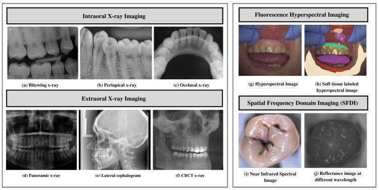
A comprehensive compilation of dental imaging modalities categorized into four sections. The first section, Intraoral X-ray Imaging, displays (a) Bitewing X-rays for interproximal caries detection, (b) Periapical X-rays showing the whole tooth and surrounding bone, and (c) Occlusal X-rays visualizing larger segments of the arch. The second section, Extraoral X-ray Imaging, includes (d) Panoramic X-rays for a broad view of the maxilla and mandible, (e) Lateral Cephalograms for orthodontic analysis of craniofacial structures, and (f) CBCT X-ray for 3D volumetric reconstruction. The third section illustrates Fluorescence Hyperspectral Imaging with (g) a standard hyperspectral image and (h) a soft tissue labeled version for tissue differentiation. The final section demonstrates Spatial Frequency Domain Imaging (SFDI) with (i) a near-infrared spectral image of an occlusal surface and (j) a reflectance image at different wavelengths. This figure serves as an educational overview of diagnostic tools used for assessing dental anatomy, pathology, and soft tissue health in dentistry and maxillofacial surgery.
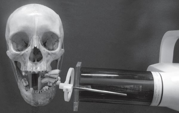
This clinical research photograph demonstrates a standardized dental radiography experimental setup using a human dry skull specimen. The image shows a frontal view of the skull with the mandible positioned to simulate a periapical or bitewing radiograph procedure. An intraoral dental film is fixed in a specialized film holder, which is attached to the posterior segment of the mandible. To replicate clinical conditions, a block of self-polymerizing acrylic resin is interposed on the buccal surface of the mandible to act as a soft tissue substitute. The x-ray machine's collimator (position-indicating device) is aligned perpendicular to the film, guided by a metal aiming rod and an external circular centering ring to maintain a fixed focus-film distance and geometric accuracy. This setup is characteristic of dental physics studies or quality assurance testing intended to evaluate the attenuation properties of different materials and their effect on radiographic image density and contrast in oral and maxillofacial radiology.
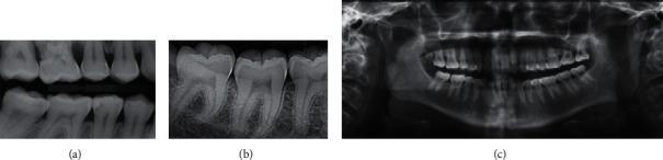
This comparative medical illustration presents three primary types of diagnostic dental radiography. (a) Bitewing X-ray: A localized intraoral view capturing the coronal portions and cervical thirds of the roots of both maxillary and mandibular teeth in a specific segment. It is primarily used to detect interproximal caries and monitor alveolar bone levels. (b) Periapical X-ray: An intraoral radiographic view focusing on a small group of teeth, showing the entire tooth structure from the crown to the apex of the root, as well as the surrounding periapical bone and periodontal ligament space. This modality is essential for assessing root morphology and apical pathology. (c) Orthopantomogram (OPG): An extraoral panoramic radiograph providing a comprehensive, curved-plane view of the entire maxillofacial region. It displays all teeth in both the maxillary and mandibular arches, the temporomandibular joints (TMJ), and the maxillary sinuses, serving as a vital tool for orthodontic planning and forensic odontology.
darkroom film processing radiograph developer fixer safelight
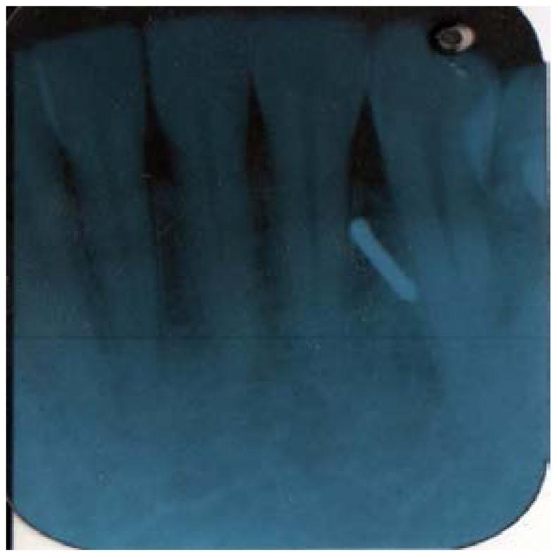
This diagnostic image is a periapical radiograph of the mandibular anterior region, displaying the lower incisors and their supporting structures. The film utilizes a blue-tone processing filter. The anatomical focus is on the dental crowns, roots, and surrounding alveolar bone. A significant clinical finding is a highly radiopaque, cylindrical foreign body located in the soft tissue near the apex of the mandibular left lateral incisor/canine region. This object, consistent with a broken dental bur, is oriented diagonally. The radiograph demonstrates clear delineation of the periodontal ligament spaces and the dense cortical nature of the foreign object relative to the surrounding trabecular bone and soft tissue. This image is used in dentistry to illustrate the detection of iatrogenic foreign bodies and the importance of radiographic follow-up when instruments fail during clinical procedures.
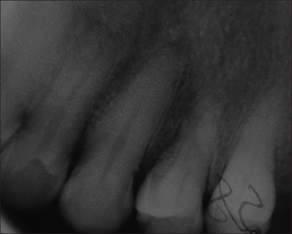
**Imaging Modality:** Intraoral periapical radiograph. **Anatomical Region:** Maxillary anterior segment, showing multiple permanent incisors and associated alveolar bone. **Observed Pathology/Diagnostic Features:** The radiograph demonstrates significant image degradation due to a "film fog" or "static electricity" artifact. Specifically, there is a prominent, radiopaque, tree-like or "lightning bolt" branching pattern superimposed over the crown of a lateral tooth. This is a characteristic static discharge artifact often caused by rapid removal of the film from its packet or low humidity environments during processing. **Additional Radiographic Findings:** - **Dental Anatomy:** The teeth show intact pulp chambers and root canals with no immediate evidence of periapical radiolucency. - **Bone Quality:** The interdental alveolar bone displays a normal trabecular pattern, though visual clarity is reduced by generalized graininess and poor contrast (radiographic noise). - **Restorations:** A radiopaque restorative material is visible on the incisal/proximal aspect of one of the central incisors. **Key Features for Retrieval:** Dental radiography, static discharge artifact, film fog, maxillary incisors, periapical view, technical processing error.
intensifying screen rare earth phosphor cassette radiography
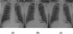
This Comparison Chart features a side-by-side display of three posteroanterior (PA) chest radiographs of a 42-year-old male, labeled (a), (b), and (c). The images serve as a technical demonstration of diagnostic imaging evolution, specifically comparing traditional screen-film (S/F) technology with Fuji Computed Radiography (FCR) systems. Visible anatomical structures across all three panels include the lung fields, cardiac silhouette, mediastinum, diaphragm, and rib cage. The lung fields are radiolucent, showing branching pulmonary vascular markings. The central radiopaque structures consist of the heart and mediastinal great vessels. Diaphragmatic contours are clearly defined above the abdominal cavity. The educational focus is on image quality metrics: panel (a) shows traditional screen-film results; panel (b) uses FCR 5000 technology; and panel (c) uses the FCR 5501D system. There is a progressive increase in edge sharpness, contrast, and signal-to-noise ratio from (a) to (c), with (c) providing the most detailed visualization of fine pulmonary markings and interstitial textures due to advanced dual-side phosphor plate scanning. This material is suitable for radiology and medical physics students studying imaging modality optimization.
![This diagnostic image displays a series of phosphor screen autoradiography experiments comparing [18F]-AV-1451 binding in brain tissue from Alzheimer’s Disease (AD) and Chronic Traumatic Encephalopathy (CTE). The layout organizes tissue slices from the parietal, temporal, and occipital cortex across four CTE cases and one AD positive control, categorized by washing protocol (Ethanol vs. No Ethanol). In the AD sample, a strong, dark tracer signal is visible in the cortical ribbons, which is markedly reduced in the corresponding 'Self-block' control, indicating specific binding to neurofibrillary tangles. In contrast, the CTE cases (Cases #1-#4) show negligible [18F]-AV-1451 signal across all cortical regions and washing conditions, appearing nearly identical to their self-block counterparts. Rare, localized focal spots in CTE cases represent off-target binding to leptomeningeal melanocytes rather than tau aggregates. The comparison demonstrates the high sensitivity of [18F]-AV-1451 for AD-type tau pathology and its relative lack of binding to the distinct tau filaments found in CTE, regardless of tissue processing techniques.](https://cdn.orris.care/cdss_images/pmc_clinical_VQA_b2223fbc9b7ec01c8fd1fdcdb71d2c9a9c8031a35626926e3d3f826c23858532.jpg)
This diagnostic image displays a series of phosphor screen autoradiography experiments comparing [18F]-AV-1451 binding in brain tissue from Alzheimer’s Disease (AD) and Chronic Traumatic Encephalopathy (CTE). The layout organizes tissue slices from the parietal, temporal, and occipital cortex across four CTE cases and one AD positive control, categorized by washing protocol (Ethanol vs. No Ethanol). In the AD sample, a strong, dark tracer signal is visible in the cortical ribbons, which is markedly reduced in the corresponding 'Self-block' control, indicating specific binding to neurofibrillary tangles. In contrast, the CTE cases (Cases #1-#4) show negligible [18F]-AV-1451 signal across all cortical regions and washing conditions, appearing nearly identical to their self-block counterparts. Rare, localized focal spots in CTE cases represent off-target binding to leptomeningeal melanocytes rather than tau aggregates. The comparison demonstrates the high sensitivity of [18F]-AV-1451 for AD-type tau pathology and its relative lack of binding to the distinct tau filaments found in CTE, regardless of tissue processing techniques.
DENTAL RADIOLOGY: FILMS, PROCESSING & IMAGE QUALITY - Complete Study Notes
LONG QUESTIONS
1. Intraoral Films - Types, Sizes, Advantages, Disadvantages & Uses (2017)
Classification of X-Ray Films (2023)
- Intraoral films (placed inside the mouth)
- Extraoral films (placed outside the mouth)
| Speed Group | Relative Speed | Exposure Needed |
|---|---|---|
| Group A | Slowest | Highest dose |
| Group B | - | - |
| Group C | Moderate | - |
| Group D (Standard) | Moderate-fast | Widely used historically |
| Group E | Fast | ~50% less dose than D |
| Group F (Insight) | Fastest | ~60-70% less dose than D |
Types of Intraoral Films
Type 1: Periapical (PA) Film
| Size No. | Dimensions | Use |
|---|---|---|
| Size 0 | 22 × 35 mm | Children (primary teeth, narrow arches) |
| Size 1 | 24 × 40 mm | Anterior teeth (narrower arch), children |
| Size 2 | 31 × 41 mm | Posterior adult teeth (most common - standard) |
| Size 3 | 27 × 54 mm | Long narrow - lower anterior bitewing/occlusal |
| Size 4 | 57 × 76 mm | Occlusal films |
- Shows entire root length and periapical region
- Reveals periapical pathology (abscess, cysts, granuloma)
- Assesses root morphology, root canal length
- Evaluates bone level and alveolar socket
- Shows furcation involvement
- Limited area of coverage per film
- Difficult placement in patients with shallow palate, gag reflex, restricted opening
- Geometric distortion if angulation incorrect
- Multiple films needed for full-mouth survey (FMX = 14-18 films)
- Endodontic working length determination
- Periapical pathology diagnosis
- Evaluation of root fractures
- Pre/post-extraction assessment
- Implant evaluation
- Detection of root resorption
Type 2: Bitewing (Interproximal) Film
| Size | Dimensions | Use |
|---|---|---|
| Size 0 | 22 × 35 mm | Children (primary) |
| Size 1 | 24 × 40 mm | Anterior bitewings; children |
| Size 2 | 31 × 41 mm | Standard adult posterior bitewing |
| Size 3 | 27 × 54 mm | Long (horizontal) adult bitewing - posterior |
- Best film for detecting interproximal (approximal) caries - even before cavitation
- Shows crestal bone levels in both jaws simultaneously
- Reveals secondary caries under restorations
- Evaluates overhanging margins of restorations
- Low distortion - standardized horizontal angulation
- Cannot show root apex (no periapical information)
- Cannot be used for endodontic or periapical assessment
- Only shows crowns and cervical regions
- Caries detection (primary use)
- Monitoring alveolar bone loss in periodontal disease
- Checking fit of crowns, bridges, inlays
- Routine recall/check-up radiographs
Type 3: Occlusal Film
- Size 4 (standard occlusal): 57 × 76 mm (approximately 2.25 × 3 inches)
- Standard (cross-sectional) occlusal - tube directed perpendicular to film, gives cross-section view
- Vertex occlusal - tube at vertex of skull, shows true cross-section of maxilla/mandible
- Oblique occlusal - angled projection for specific regions
- Shows large area of arch in single exposure
- Locates supernumerary teeth, unerupted teeth, foreign bodies
- Can be used for children unable to tolerate periapical films
- Useful when patient cannot open fully
- No need for film-holding devices
- Lower resolution than periapical films
- Some superimposition of structures
- Cannot show full tooth length in all cases
- Requires higher radiation dose than periapical
- Localizing unerupted/impacted teeth (using Clark's rule with a periapical)
- Locating salivary stones (submandibular and sublingual glands)
- Identifying cleft palate
- Detecting midline pathology (cysts, tori)
- Fractures of maxilla/mandible
- Upper/lower anterior cross-sectional views in children
Summary Table: Intraoral Film Types
| Feature | Periapical | Bitewing | Occlusal |
|---|---|---|---|
| Size (most common) | Size 2 (31×41 mm) | Size 2 (31×41 mm) | Size 4 (57×76 mm) |
| Shows apex | Yes | No | Sometimes |
| Caries detection | Yes | Best | Moderate |
| Area covered | 2-4 teeth | Crowns both jaws | Full arch segment |
| Alveolar bone | Periapical area | Crestal bone | Yes |
| Film holder | Yes | Tab/wing | Patient bites |
2. Structure / Composition of X-Ray Film (2020s, 12)
Composition of OPA (Oral Periapical) Film Packet (2014)
- Outer vinyl/paper wrapper (waterproof) - protects from saliva/moisture
- Black paper wrapping - light-tight, protects film from fogging
- Lead foil backing sheet - reduces backscatter radiation (from bone behind film); prevents secondary radiation from exposing the back of the film; shows herringbone/tire-track pattern if film placed backwards
- Film base - polyester (or older: cellulose acetate) - clear, flexible, 0.18-0.25 mm thick; provides mechanical support; blue-tinted to reduce eyestrain during viewing
- Adhesive/subbing layer - bonds emulsion to base
- Emulsion (double-sided) - most important layer; contains silver halide (silver bromide AgBr ~90-95%, silver iodide AgI ~5%) crystals suspended in gelatin; one emulsion layer on each side of base (double emulsion = faster, less dose needed)
- Supercoat (gelatin overcoat) - protects emulsion from physical damage
Film Emulsion Detail
- Silver bromide (AgBr) crystals are the photosensitive agent
- Gelatin acts as a dispersing medium and swells during processing to allow chemical access
- Crystal size determines speed: larger crystals → faster film, more grain
- Sensitivity specks (chemical impurities, often silver sulfide) on crystal surfaces act as traps for photo-electrons - nucleation sites for latent image formation
3. Latent Image Formation
Definition
Mechanism (Gurney-Mott Theory)
-
Photon absorption: X-ray photon is absorbed by Br⁻ ion in the crystal latticeAgBr + photon → Ag⁺ + Br⁻ + energy → photoelectron (e⁻)
-
Electron migration: The liberated photoelectron migrates through the crystal lattice toward a sensitivity speck (silver sulfide, Ag₂S)
-
Electron trapping: The negative sensitivity speck attracts and traps the electron, becoming negatively charged
-
Silver ion migration: The negative charge at the sensitivity speck attracts mobile interstitial Ag⁺ ions from the lattice
-
Silver atom formation: Ag⁺ + e⁻ → Ag⁰ (neutral silver atom deposited at sensitivity speck)
-
Amplification: The Ag⁰ atom attracts more electrons → more Ag⁺ ions → cluster of silver atoms grows at the sensitivity speck
-
Latent image center: The cluster of Ag⁰ atoms (as few as 2-4 atoms) forms the latent image center - this is the development center
The bromine ions released diffuse into the gelatin and are absorbed by bromide acceptors (gelatin itself).
4. Film Processing - Complete Procedure (2015, 14)
Darkroom Requirements (Short Note - 2019, 17, 14)
- Total light-tight construction - no light leaks (check with 5-minute dark adaptation test)
- Two-area design: Dry bench (loading/unloading) and wet bench (chemicals)
- Adequate ventilation - chemical fumes can be toxic; exhaust fan
- Temperature control - chemical tanks maintained at 68°F (20°C) standard
- Uses orange-red filter (Kodak GBX-2 or Wratten 6B filter)
- Minimum 25-watt bulb
- Must be at least 4 feet (1.2 m) from film
- X-ray film is not sensitive to orange-red wavelengths
- Safelight test (coin test): Place coin on unwrapped film, expose to safelight for 2 min, process - if coin image visible, safelight is unsafe
- Processing tanks (developer, water rinse, fixer, final wash)
- Timer and thermometer
- Drying rack or cabinet
- Stirring rods (separate for each solution)
- Film hangers
Manual Processing Steps (Standard Method)
Step 1: Developer (4-5 minutes at 20°C / 68°F)
- Film immersed in developer solution
- Agitate gently for first 5 seconds, then every 30 seconds
- Temperature-time relationship: higher temp = shorter time (T-T development)
Step 2: Rinse (30 seconds)
- Agitation in running water or separate rinse tank
- Removes developer from film surface
- Prevents contamination of fixer
Step 3: Fixer (8-10 minutes)
- Full immersion with periodic agitation
- Can inspect under white light after 2-3 minutes (clearing time)
Step 4: Final Wash (20-30 minutes)
- Running water to remove all fixer
- Prevents long-term yellowing/staining of film
Step 5: Drying
- Air-dry or cabinet dryer at low heat
- Film ready for viewing
Rapid Processing (Chair-side Processing)
- Developer at 27°C (80°F): 15 seconds
- Rinse: 15 seconds
- Fixer at 27°C: 15 seconds
- Rinse: 15 seconds
- This provides a readable "wet reading" in under 90 seconds
- Final quality after complete fixing and washing
Developer Solution (2020s, 2018, 12)
| Component | Chemical | Function |
|---|---|---|
| Reducing agents (2) | Hydroquinone + Phenidone (or Elon/Metol) | Donate electrons to Ag⁺ → Ag⁰; Hydroquinone gives contrast, Phenidone gives density/speed |
| Activator/accelerator | Sodium carbonate (Na₂CO₃) or potassium carbonate | Provides alkaline pH (9.5-11.5) essential for reducing agents to work; swells gelatin |
| Restrainer | Potassium bromide (KBr) | Inhibits development of unexposed crystals; prevents chemical fog; maintains selectivity |
| Preservative | Sodium sulfite (Na₂SO₃) | Prevents oxidation of reducing agents by air; extends developer life; produces colorless oxidation products |
| Solvent | Water | Diluent |
| Hardener | Glutaraldehyde (some modern developers) | Hardens gelatin to prevent swelling/physical damage |
Ag⁺Br⁻ (exposed crystal) + reducing agent → Ag⁰ (black) + Br⁻ + oxidized reducer
- Decreased density of films
- Loss of contrast
- Color changes from clear to yellow-brown
- Specific gravity decreases
Fixer Solution (2019, 16, 2020, 08)
- Clears the film - dissolves and removes all unexposed, undeveloped silver halide crystals
- Fixes (makes permanent) the silver image already formed
- Hardens the emulsion
| Component | Chemical | Function |
|---|---|---|
| Fixing agent (clearing agent) | Sodium thiosulfate (Na₂S₂O₃) - "hypo" or Ammonium thiosulfate (faster) | Dissolves unexposed AgBr crystals by forming soluble silver thiosulfate complex; main active ingredient |
| Acidifier | Acetic acid or sulphuric acid | Provides acid pH (4.0-4.5); neutralizes developer carried over; stops development; activates hardener |
| Preservative | Sodium sulfite | Prevents decomposition of thiosulfate; maintains acidity |
| Hardener | Potassium alum (chrome alum) or aluminum chloride | Hardens and shrinks the swollen gelatin; prevents physical damage; reduces drying time |
| Buffer | Sodium acetate / boric acid | Maintains stable pH |
| Solvent | Water | Diluent |
AgBr (unexposed) + Na₂S₂O₃ → Na₃[Ag(S₂O₃)₂] (soluble, washed away) + NaBr
- Milky/hazy films
- Films turn yellow/brown with time (retained silver thiosulfate)
- Increased clearing time
- Specific gravity increases
Film Fog (Short Note - 2013)
| Type | Cause |
|---|---|
| Light fog | Darkroom light leaks; improper safelight (wrong filter/wattage/distance); cracked film packet |
| Radiation fog | Film stored near radiation sources; scattered radiation during exposure |
| Chemical fog (Age fog) | Outdated film; improper developer concentration; developer too warm; prolonged development; developer contaminated with fixer |
| Heat fog | Film stored at high temperature (>27°C) |
| Humidity fog | Storage in high relative humidity (>50%) |
| Static fog | Static electricity discharge (appears as tree-like/lightning bolt pattern on film - especially in dry weather); caused by rapid removal from packet |
| Pressure fog | Excessive pressure on film packet before processing |
| Secondary radiation fog | Scattered X-rays reaching film from surrounding tissues |
- Reduced image contrast
- Loss of detail
- Overall graying of the image
- Reduced diagnostic quality
5. Faulty Radiograph / Reasons for Faulty (Fatty) Radiograph
I. Errors in Exposure
| Error | Cause | Appearance |
|---|---|---|
| Overexposed | Too high mA, kVp, time | Too dark (dense/black) |
| Underexposed | Too low mA, kVp, time | Too light (pale/thin) |
| Cone cut | Beam not centered on film | Part of film unexposed (clear band) |
| Film reversed | Lead foil side toward tube | Herringbone/tire-track pattern; low density |
| Overlapping | Incorrect horizontal angulation | Interproximal overlap of crowns |
| Foreshortening | Excessive vertical angulation | Teeth appear short |
| Elongation | Insufficient vertical angulation | Teeth appear long |
| Blurring / movement | Patient movement during exposure | Unsharp, blurred image |
II. Errors in Processing
| Error | Cause | Appearance |
|---|---|---|
| Too dark | Overdeveloped (time/temp) | Dense black image |
| Too light (pale) | Underdeveloped; exhausted developer; low temp | Pale, low density |
| Too light | Underexposure (can mimic underdevelopment) | Pale |
| Yellow/brown stain | Oxidized/exhausted developer; insufficient fixing; inadequate washing | Staining/yellowing |
| Milky/hazy | Incomplete fixing; exhausted fixer | Milky areas |
| Film fog | See above section | General graying |
| Black lines | Fingernail marks (pressure artifact) | Crescent-shaped black marks |
| White spots | Fixer drops on film before processing; air bubbles on film in developer | White spots |
| Dark spots | Developer contamination before processing | Dark spots |
| Reticulation | Large temperature difference between developer and water rinse | Cracked/reticulated pattern |
| Scratches | Rough handling wet film | Linear marks |
| Fingerprint marks | Handling with fingers (oil/developer contact) | Fingerprint pattern |
6. Image Sharpness and Resolution (2007)
Definitions
Factors Causing Loss of Image Clarity and Methods to Minimize
A. Geometric Factors (Geometric Unsharpness)
- Cause: Large effective focal spot produces a penumbra (blurred shadow edge)
- Minimize: Use small focal spot (fine focus); rotating anode; line focus principle
- Cause: Greater divergence of beam at short distances
- Minimize: Use long (16-inch) position-indicating device (PID/cone) - increases focus-film distance; long-cone paralleling technique
- Cause: Magnification and blurring increase with greater object-film distance
- Minimize: Film held as close to tooth as possible; paralleling technique (film parallel to long axis of tooth); use film holders
- Cause: Geometric distortion (elongation/foreshortening)
- Minimize: Paralleling technique; bisecting angle technique with correct angulation
B. Motion Unsharpness
- Short exposure times (high mA)
- Firm patient instruction ("don't move, don't swallow")
- Use of film holders/bite blocks
- Stabilize X-ray tube head
C. Screen Unsharpness (for extraoral films)
- Use thin phosphor layer
- Use fine-grain phosphor crystals
- Maintain tight film-screen contact (vacuum cassettes)
- Avoid old/worn screens
- Use newer rare-earth screens (thinner layer, same efficiency)
D. Film Unsharpness (Graininess)
- Use slower film (finer grain) when dose permits
- Use D or E speed film for balance of quality/dose
7. Factors Controlling X-Ray Beam (2007)
A. Factors Affecting X-Ray Quantity (Intensity / Number of Photons)
- Milliamperage (mA): Controls filament heating → number of electrons → directly proportional to X-ray quantity; doubling mA doubles output
- Exposure time (seconds): mA × seconds = mAs (milliampere-seconds); controls total quantity; doubling time doubles output
- kVp (kilovoltage peak): Increasing kVp increases quantity (more electron energy → more X-rays); kVp² ∝ quantity approximately
- Filtration: Reduces beam intensity by removing soft rays
- Distance: Intensity ∝ 1/d² (inverse square law)
B. Factors Affecting X-Ray Quality (Energy / Penetrating Power)
- kVp: Primary factor; higher kVp = higher maximum and average photon energy = harder beam = more penetrating
- Filtration: Removes soft photons, raises mean energy (hardening effect)
- Anode material: Higher Z → higher characteristic radiation energy
C. Factors Affecting Radiographic Contrast
- kVp: Lower kVp → more photoelectric effect → better contrast; higher kVp → more Compton → less contrast (gray, flat image)
- Scatter radiation: Reduces contrast; controlled by collimation, grids, lower kVp
- Film type/speed/development: Affect film contrast (gamma/gradient of H&D curve)
- Subject contrast: Differences in atomic number, density, and thickness of structures
- Fog: Reduces contrast (see above)
- Processing: Temperature, concentration, time - overdevelopment → high base fog; underdevelopment → low contrast
8. Intensifying Screen (2020s, 17, 08, 09, 16, 21)
Definition
Structure (Layers from outside to inside)
| Layer | Material | Function |
|---|---|---|
| Protective coat | Thin plastic | Protects against moisture, abrasion; allows cleaning |
| Phosphor (active) layer | Fluorescent crystals in binder | Converts X-ray → light |
| Reflective layer | Magnesium oxide (TiO₂) | Reflects light toward film; increases efficiency |
| Base (support) | Polyester or cardboard | Provides rigidity |
Phosphor Materials
| Generation | Material | Color Emitted | Paired Film Color |
|---|---|---|---|
| Conventional | Calcium tungstate (CaWO₄) | Blue-violet | Blue-sensitive |
| Rare Earth (modern) | Gadolinium oxysulfide (Gd₂O₂S:Tb) | Green | Orthochromatic (green-sensitive) |
| Rare Earth | Lanthanum oxybromide (LaOBr:Tb) | Blue | Blue-sensitive |
| Rare Earth | Yttrium tantalate (YTaO₄) | UV-blue | Blue-sensitive |
Intensification Factor (IF)
IF = Exposure without screen / Exposure with screen (to produce same density)
- Calcium tungstate screens: IF = 30-60
- Rare earth screens: IF = 100-200+
Screen Speed Classification
| Speed Class | Relative Speed | Spatial Resolution |
|---|---|---|
| Detail/Fine | 100 (reference) | Best (8-10 lp/mm) |
| Medium/Par | 200-400 | Moderate |
| Fast/High speed | 800-1200 | Lower (4-6 lp/mm) |
Advantages of Intensifying Screens
- Dramatically reduces patient radiation dose (by factor of 30-200×)
- Allows use of shorter exposure times (reduces motion blur)
- Allows use of lower mA settings (less tube loading/heat)
- Extends X-ray tube life
Disadvantages
- Reduces image resolution/sharpness (light diffusion in phosphor layer)
- Screen artifacts (scratches, dust, foreign bodies visible as artifacts)
- Poor film-screen contact → blurring
- Regular cleaning and replacement needed
- Screen noise (quantum mottle) at very low doses
NOT used for intraoral dental films
- Intraoral films are non-screen films - they rely on direct X-ray exposure
- The short distance and small film size means dose is acceptable without screens
- Using screens with intraoral films would reduce resolution unacceptably
Summary Table: Short Notes
| Topic | Key Summary |
|---|---|
| Developer solution | Alkaline (pH 9.5-11.5); hydroquinone + phenidone + Na₂CO₃ + KBr + Na₂SO₃; reduces AgBr → Ag⁰ |
| Fixer solution | Acidic (pH 4-4.5); thiosulfate + acetic acid + alum + Na₂SO₃; clears unexposed AgBr, hardens gelatin |
| Darkroom | Light-tight; orange-red safelight (GBX-2); dry + wet bench; 68°F (20°C); timer + thermometer |
| Film fog | Unwanted density; from light leaks, age, heat, radiation, chemicals, static |
| OPA film composition | Vinyl wrapper → black paper → lead foil → polyester base → adhesive → double emulsion (AgBr in gelatin) → supercoat |
| Latent image | Invisible Ag⁰ clusters at sensitivity specks; formed by Gurney-Mott mechanism |
| Film speed | A-F (A=slowest, F=fastest); higher speed = lower dose = more grain; E/F speed recommended |
| Intensifying screen | CaWO₄ (blue) or rare-earth (green); IF=30-200×; reduces dose; reduces resolution |
| Film contrast factors | kVp (main), scatter, fog, processing, subject contrast |
| Image sharpness | Affected by focal spot size, OFD, SOD, motion; improved by long PID, paralleling technique, film holders |
Write in detail about Sialography & its significance in various diseasés of Salivary glands? (2017) Briefly describe the principle, advantages, disadvantages, indication and contraindication of Cone Beam Computed Tomography imaging. (15) Describe the technique of the Occlusal radiograph to detect a sialolith in the submandibular duct. Y Describe the principle of OPG along with its uses, advantages, disadvantages, indication and contraindication. (16,13) What is the principle of panoramic imaging? Write in detail about the indications, limitations of panoramic imaging and the faults in OPG images.'20s Describe the technique of Sialography? What is its indication? (12,09) ~ Write in detail about sialography and its significance in various diseases of the salivary gland. 19 % Déscribe in detail about different conventional and advanced radiography for TMJ disorder. (07) Describe in detail about the principles, indication, contraindication, advantages and disadvantages of orthopantomography. (21) f. Describe techniques of intraral periapical radiograph in detail with merits and demerits. (22) 11. Describe in detail about different conventional and advanced radiography in TM Joint. (2023) SHORT NOTE, PNS Radiography. (2018,22) Ghost Image. (2018) SLOB Technique. (2017) Faulty Radiographs. (2017,2019,2020s) Causes of Dark Radiograph. (16) 6. Lateral oblique radiograph. (16) Artifacts. (15) Tomography. (12) / Grids and Grid Ratio. (15,23) Common errors in radjography. (12) Xi 'Causes of light radiograph. (15) Grid. (2018,13,14) XI. Bisecting angle technique. (14) Occlusal radiograph. (17,14) Diagnostic importance of Orthopantomograph. (15,22) Ciezensky's rule of isometry. (13) What is the basic principle of OPA technique? (06) 18. Principles of taking ideal radiograph. (07,23) Reverse town's projection of skull. (05,07,21,23) 20. RVG. (06) 21. Bite Wing Radiography. (15) 22. Ultrasound assay in orofacial disorder. (03) CBCT in maxillofacial region. (19) Object localization (19,22) Importance of CCT. (20s) (26 Technique of IOPA radiograph. (20s) Principles and applications of CBCT (20) 28. Grids (20) Transcranial view (20) 30. Diagnostic importance of sialography. (22) P 31. Ghost image in OPG (2023)
sialography parotid gland duct contrast radiograph
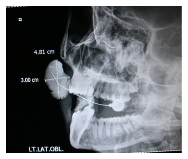
This diagnostic image is a lateral oblique sialography of the left parotid gland. The radiograph displays a radiopaque contrast medium introduced via a cannula to visualize Stensen's duct. A significant pathological finding is noted in the mid-cheek region, where the contrast medium pools into a large, well-defined, ovoid cystic lesion measuring approximately 4.81 cm x 3.00 cm. This accumulation represents focal ductal dilation or a sialocele. The contrast flows through the distal portion of the parotid duct but demonstrates no communication with the proximal glandular parenchyma, suggesting a complete obstruction or discontinuity. The anatomical landmarks of the mandible, maxilla, and dentition are visible in a lateral oblique projection, providing spatial context for the subcutaneous mass. This imaging is clinically significant for evaluating parotid duct abnormalities, such as strictures, sialolithiasis, or rare infections like rhinosporidiosis that can lead to secondary ductal ectasia.
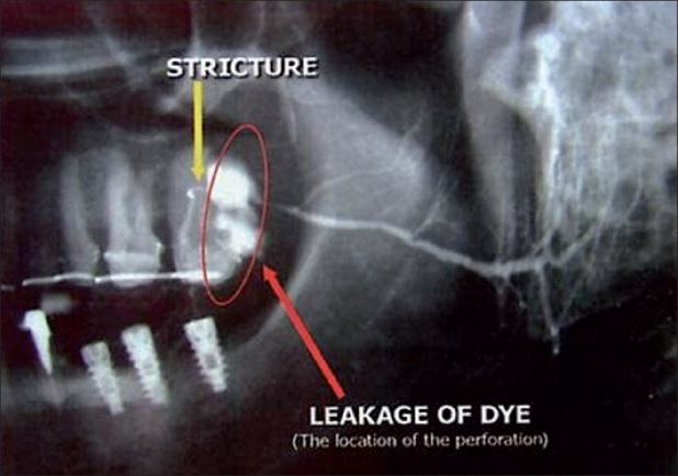
**Imaging Modality:** Sialogram (radiograph with intraductal contrast medium). **Anatomical Region:** Lateral view of the maxillofacial region, focusing on the parotid gland and Stensen’s duct (ductus parotideus). **Observed Pathology:** Perforation and stricture of the parotid duct. **Key Visual Features:** * **Contrast Extravasation:** A prominent, irregular collection of radiopaque contrast medium is visible outside the normal anatomical course of the duct (labeled "Leakage of Dye"), indicating a ductal breach or perforation. * **Ductal Stricture:** A localized narrowing of the parotid duct lumen is identified distal to the primary branching, indicated by an annotated arrow. * **Sialography Pattern:** The proximal portion of the ductal system shows normal opacification of the intraglandular branches, while the distal main duct exhibits discontinuity. * **Associated Findings:** Dental implants and restorations are visible in the mandibular and maxillary arches, providing anatomical orientation. **Diagnostic Context:** The image demonstrates a loss of ductal wall integrity (perforation) occurring in proximity to a pre-existing or concurrent stricture, leading to contrast medium leakage into the surrounding soft tissues. This is a characteristic finding in traumatic injury or iatrogenic complications of the salivary duct system.
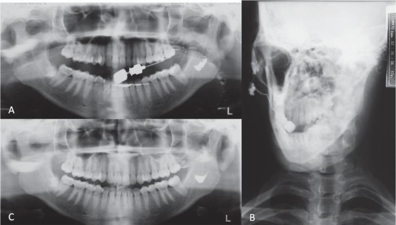
This set of diagnostic radiographs illustrates a sialography procedure of the left parotid gland. Figure A shows a panoramic view immediately following the injection of a radiopaque contrast medium (Lipiodol), with an intraoral cannula visible. Figure B provides an anteroposterior (AP) view, demonstrating the distribution of the contrast within the parotid ductal system and gland parenchyma, superimposed against the mandible and cervical spine. Figure C is a follow-up panoramic radiograph taken 24 hours later, highlighting significant contrast retention. The images reveal 'cavitary sialectasia,' characterized by irregular, globular collections of contrast medium within the gland, indicating damage to the ductal epithelium. The surrounding maxillofacial bony structures, including the mandible, maxilla, and alveolar bone, appear radiographically normal. This series is an educational example of salivary gland imaging used to diagnose chronic inflammatory conditions or obstructive sialadenitis by visualizing ductal morphology and glandular evacuation efficiency.
OPG orthopantomograph panoramic radiograph jaw teeth
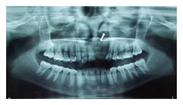
An orthopantomograph (OPG) or panoramic dental radiograph providing a comprehensive view of the maxilla, mandible, and associated dental structures. The image shows the bilateral temporomandibular joints, maxillary sinuses, nasal cavity, and alveolar processes. In the anterior maxilla (upper left quadrant), there is a notable absence of tooth 21 and tooth 22. In the region of the missing tooth 22, a diffuse, ill-defined area of radiolucency is visible, suggesting localized bone loss or a pathological lesion consistent with inflammatory fibrosis or a cystic process. The adjacent teeth show relatively normal root morphology, though there is evidence of generalized mild horizontal bone loss. A small, curved radiopaque artifact is visible in the midline area above the anterior maxillary teeth. This diagnostic image is significant for evaluating dental spacing, alveolar bone density, and the secondary effects of tooth extraction or localized soft tissue pathology in oral surgery and clinical dentistry.
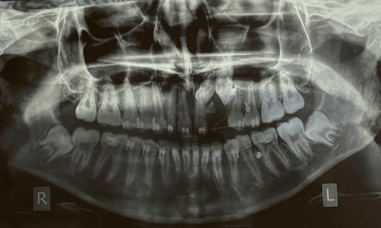
An orthopantomograph (OPG) or panoramic radiograph of a human dentition showing significant anomalies in the upper left anterior quadrant. The image demonstrates a cluster of multiple, small, radio-opaque tooth-like structures (denticles) in the region of the maxillary left lateral incisor and canine, characteristic of a compound odontoma. This calcified mass is associated with the failure of eruption and subsequent impaction of the permanent maxillary left lateral incisor (22) and canine (23), which are visible superior to the odontoma. Overretained deciduous teeth (62 and 63) are present in the corresponding arch segment. Fixed orthodontic appliances, including brackets and archwires, are visible on the maxillary and mandibular arches, indicating active orthodontic management. Other notable features include horizontally impacted mandibular third molars and evidence of orthodontic traction. This diagnostic image is significant for demonstrating the role of odontogenic tumors in tooth impaction and the utility of panoramic imaging in orthodontic treatment planning.
CBCT cone beam CT maxillofacial 3D reconstruction dental
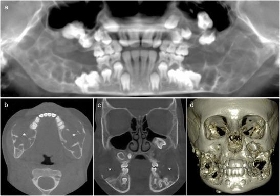
This composite of diagnostic images features Cone Beam CT (CBCT) scans and a 3D reconstruction of the skull from an 8-year-old pediatric patient, demonstrating classic radiographic features of cherubism. Panel (a) shows a thick-slab translucent CBCT reconstruction highlighting extensive, well-defined, bilateral multilocular radiolucencies causing symmetric expansion of the mandible and maxilla. Significant dental abnormalities are present, including unerupted first molars and displaced permanent teeth floating within the lesions. Axial (b) and coronal (c) cross-sections reveal pseudocystic osteolytic lesions containing fine, irregular bony septa (asterisks), with notable absence of periosteal reaction. These views further demonstrate the invasion of the inferior alveolar nerve canal and profound cortical thinning. Panel (d) provides a 3D volume-rendered reconstruction, visualizing the hypertrophic nature of the mandibular and maxillary lesions, which contribute to the characteristic 'cherubic' facial deformity and massive bony distortion. The content serves as an educational reference for diagnosing genetic fibro-osseous disorders of the jaw in pediatric oral and maxillofacial radiology.
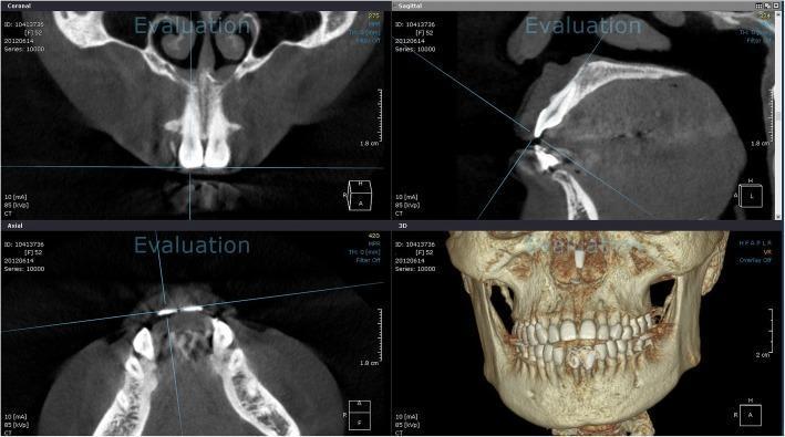
This diagnostic image displays a multi-planar reconstruction (MPR) of a maxillofacial cone beam computed tomography (CBCT) scan, categorized as Clinical Imaging for orthodontic or dental evaluation. The interface is divided into four panels: Coronal, Sagittal, Axial, and a 3D Volume Rendering. The Coronal view illustrates the maxilla, nasal septum, and inferior turbinates, with a horizontal reference line at the incisal edge. The Sagittal view shows a cross-section of a maxillary anterior tooth, with a blue software-generated line connecting the incisal edge to the root apex, facilitating tooth axis measurement. The Axial view provides a horizontal cross-section of the mandible and maxillary arch, showing the transverse relationship of the anterior dentition. The 3D reconstruction provides an external surface rendering of the skull, maxilla, and mandible, demonstrating full permanent dentition and occlusion. This setup is primarily used in dentistry for precise anatomical assessment, tooth inclination analysis, and pre-surgical planning by visualizing bone density and root morphology in relation to surrounding structures.
ORAL RADIOLOGY: SPECIAL TECHNIQUES & IMAGING - Complete Study Notes
LONG QUESTIONS
1. Sialography - Technique, Significance & Diseases of Salivary Glands (2017, 2019, 2012, 2009, 22)
Definition
Contrast Media Used
| Type | Examples | Properties |
|---|---|---|
| Oil-based (iodized oils) | Lipiodol, Ethiodol | High viscosity; better ductal coating; slower excretion; risk of granuloma; rarely used now |
| Water-soluble (iodinated) | Urografin, Conray, Omnipaque (iohexol) | Low viscosity; rapid excretion; patient-friendly; currently preferred |
- Water-soluble
- Non-irritant to ductal epithelium
- High radiopacity
- Rapid excretion
- No risk of granuloma formation
Anatomy Relevant to Sialography
- Duct: Stensen's duct (Parotid duct), ~5-6 cm long
- Opens: Opposite upper second molar (buccal mucosa) - the parotid papilla
- Orifice: Small - requires fine cannula (0.5-0.7 mm)
- Duct: Wharton's duct, ~5 cm long
- Opens: Sublingual caruncle at the floor of mouth (beside lingual frenum)
- Orifice: Larger - easier cannulation
Technique of Sialography
Pre-procedure
- Patient preparation: No oral intake for 2 hours (reduces salivary flow)
- Stimulate secretion first: Give lemon juice → stimulates flow → helps identify orifice
- Informed consent: Explain procedure, risks
- Preliminary (survey) radiographs: Taken before contrast injection to detect radiopaque calculi (they may be obscured by contrast after injection)
- Dilate orifice: Using graded lacrimal dilators if orifice is narrow
Instruments Required
- Lacrimal dilators (graded)
- Blunt-ended cannula (Rabinov or Stensen's cannula - 0-2 gauge)
- 2 mL syringe
- Contrast medium (water-soluble iodinated)
- Local anaesthetic (topical - xylocaine gel for comfort)
Parotid Sialography
- Patient seated, head turned to opposite side
- Dry the buccal mucosa near upper 2nd molar
- Identify Stensen's duct orifice (papilla opposite upper 2nd molar)
- Gently dilate with lacrimal dilators
- Cannulate with blunt-tipped cannula, advance ~1-2 cm into duct
- Inject contrast slowly (0.5-1.5 mL for parotid; 0.5-1 mL for submandibular)
- Inject until patient feels slight fullness/discomfort (overfilling causes "acinar rupture")
- Immediately take radiographs
Submandibular Sialography
- Patient seated with mouth open and tongue raised
- Identify Wharton's duct orifice at the sublingual caruncle
- Cannulate (easier due to larger orifice)
- Inject 0.5-1 mL contrast slowly
- Take radiographs
Radiographic Views Taken
| Gland | Views |
|---|---|
| Parotid | Lateral oblique, AP view, OPG |
| Submandibular | Lateral oblique jaw, Occlusal (floor of mouth - mandatory to show entire duct), OPG |
Phases
- Filling phase: Immediately post-injection (duct + gland filled)
- Secretory phase: 5 minutes post-injection (normal gland empties ~90% in 5 min)
- Emptying phase (evacuation): Patient given lemon juice → stimulates secretion → contrast expelled; repeat film at 5 min and 10 min
Normal Sialographic Appearances
- "Leafless winter tree" or "herring-bone" pattern
- Stensen's duct: smooth walls, uniform calibre ~2-3 mm
- Secondary and tertiary branches fill uniformly
- Complete evacuation within 5-10 minutes after stimulation
- "Compact tree" appearance (denser branching than parotid)
- Wharton's duct: uniform, smooth, ~2 mm calibre
- Branches shorter, more numerous than parotid
Significance in Diseases of Salivary Glands
1. Sialolithiasis (Salivary Calculi)
- Plain film first - detect radiopaque stones (85% submandibular stones are radiopaque; only 60% of parotid)
- Sialographic appearance: Complete or partial filling defect in duct; duct dilated proximal to stone; "cupping" around stone; pooling of contrast proximal to obstruction
- Significance: Confirms location, number, size of calculi; guides surgical approach
- Note: If complete obstruction, contrast may not pass beyond stone
2. Chronic Sialadenitis (Obstructive/Inflammatory)
- Appearance: Irregular duct walls; "beading" (alternating dilatation and strictures); delayed emptying; retained contrast at 10 min; punctate sialectasis in late stages
- Significance: Grades severity of inflammatory damage; guides conservative vs. surgical management
3. Sjogren's Syndrome (Autoimmune Sialadenitis)
- Classic appearance: "Fruit-laden branchless tree" / "snowstorm pattern" / "punctate sialectasia"
- Early: Salt and pepper pattern (punctate collections 1 mm - tiny pools of contrast)
- Moderate: Globular sialectasia (3-5 mm pools)
- Advanced: Cavitary sialectasia (>5 mm, large pooling)
- Ducts may be normal or irregular
- Significance: Pathognomonic appearance; helps in diagnosis of primary vs secondary Sjogren's; monitors disease progression
- Rubin and Holt Classification: Stage 0 (normal) → Stage I (punctate) → Stage II (globular) → Stage III (cavitary) → Stage IV (destructive)
4. Salivary Gland Tumors
- Benign (Pleomorphic Adenoma):
- Smooth, round, well-defined compression of duct branches ("ball in hand" appearance)
- Ducts displaced around mass; no infiltration
- Normal evacuation
- Malignant (Mucoepidermoid carcinoma, Adenoid cystic carcinoma):
- Irregular, moth-eaten duct destruction
- "Pruned tree" (abrupt cutoff of branches)
- Poor contrast filling; irregular margins
- No normal gland architecture
- Contrast leakage through destroyed duct walls
- Significance: Helps differentiate benign from malignant; delineates tumor extent
5. Duct Stricture
- Appearance: Localized narrowing of duct with proximal dilatation; smooth stricture in post-traumatic; irregular in inflammatory
- Significance: Confirms diagnosis; guides dilatation or surgery
6. Sialocele / Fistula
- Appearance: Pooling of contrast outside normal duct course; irregular collection communicating with duct
- Post-traumatic or post-surgical complication
- Significance: Confirms communication between duct and sialocele
7. Recurrent Parotitis of Childhood
- Appearance: Punctate or globular sialectasia; duct dilatations; normal-appearing major duct
- Significance: Confirms diagnosis; helps in monitoring; disease usually resolves at puberty
Contraindications to Sialography
- Acute suppurative (infective) sialadenitis - risk of spreading infection and pain; CT/US preferred instead
- Known sensitivity to iodinated contrast media
- Thyroid disease (iodine uptake)
- Complete duct obstruction (contrast cannot pass; risk of rupture)
- Pregnancy (radiation exposure)
Advantages of Sialography
- Defines ductal architecture with high detail
- Shows strictures, dilatations, calculi position
- Dynamic information (evacuation efficiency)
- Therapeutic function - hydrostatic pressure of injection can dislodge small calculi
Disadvantages
- Invasive (cannulation required)
- Radiation exposure
- Discomfort/pain for patient
- Risk of duct injury or perforation
- Cannot be done in acute infection
- Provides less information about gland parenchyma than ultrasound or MRI
- Cannot visualize sublingual gland well
2. Occlusal Radiograph for Sialolith in Submandibular Duct
Why Occlusal View?
Technique - Lower Standard (True) Occlusal
- Patient position: Seated upright, head tilted back to 45° (chin raised)
- Film placement: Size 4 (57×76 mm) occlusal film placed horizontally on the floor of the mouth with the longest dimension crosswise; patient gently occludes on film to stabilize
- Tube position: X-ray tube directed through the chin downward at 90° to the film (perpendicular), from below the chin in the midline
- Central ray: Directed through the midline of the floor of mouth
- Film-focal distance: Approximately 20 cm
What it Shows
- The entire course of Wharton's duct from sublingual caruncle to the hilum of the gland
- Calculi appear as radiopaque (white) ovoid/round shadows within or along the duct course
- ~85% of submandibular calculi are radiopaque (calcium phosphate and calcium oxalate)
- A stone in the anterior part of Wharton's duct is well shown
Supplementary Views for Sialolith
- Lateral oblique of mandible: Shows posterior/hilar calculi
- Periapical film (floor of mouth): For smaller anterior duct calculi
- OPG: Shows calculi in submandibular region
- Ultrasound: Most sensitive (detects radiolucent calculi too); non-invasive; first-line investigation today
3. OPG / Orthopantomography (2016, 13, 2020s, 21, 15, 22)
Definition
Principle of OPG
- The X-ray tube, patient, and film/receptor rotate simultaneously but at different speeds
- A narrow (slit) collimated X-ray beam rotates around the patient
- The film/detector moves in the opposite direction to the tube
- The focal trough (a curved, horseshoe-shaped layer) conforms to the approximate curvature of the dental arches
- Only structures within the focal trough are recorded sharply; structures outside are blurred (become "ghost images")
- A curved, three-dimensional zone in the shape of an arch
- Width: 25-30 mm anteriorly, 30-35 mm posteriorly
- Structures must lie within this trough to be in focus
- The patient's dental arch must be positioned within this trough

Patient Positioning for OPG
- Patient stands or sits in the machine
- Frankfort horizontal plane parallel to floor (head level, not tilted)
- Mid-sagittal plane aligned with machine midline (no rotation left/right)
- Patient bites on the chin rest/bite guide (aligns upper and lower incisors in the slot; anterior teeth slightly apart)
- Tongue pressed to palate (eliminates air shadow above tongue)
- Lips closed
- Patient holds handles to remain still during 15-20 second rotation
- Lead apron applied (no thyroid collar - would be in the image field)
Structures Seen on Normal OPG
- All teeth (crown and root) in both arches
- Alveolar bone, lamina dura, periodontal space
- Mandibular canal, mental foramen, mandibular symphysis
- Mandibular condyles, coronoid processes, TMJ area
- Maxillary sinuses (floor and walls)
- Nasal fossae, nasal septum, inferior turbinates
- Soft tissue shadows (ear lobe, nose, lips, tongue)
- Hyoid bone (sometimes)
- Styloid processes (sometimes)
Uses / Indications of OPG (16, 13, 15, 22)
- Full-mouth survey (all teeth - caries, bone levels, root morphology)
- Impacted teeth assessment (third molars especially)
- Orthodontic examination (dental development, eruption assessment, growth)
- Detection of unerupted supernumerary teeth
Advantages of OPG
- Single image showing entire dentition and surrounding structures
- Low radiation dose (~4-10 µSv) - lower than full-mouth periapical series
- Quick and easy to perform (15-20 seconds)
- Comfortable for patient (film outside mouth)
- Useful for patients with trismus, gag reflex, or limited mouth opening
- Shows structures not visible on intraoral films (condyles, lower border of mandible, maxillary sinus)
- Useful for pediatric and medically compromised patients
- Readily available in most dental practices
Disadvantages / Limitations
- Low resolution - cannot substitute periapical films for caries diagnosis
- Geometric distortion - magnification varies (1.2-1.5×); horizontal and vertical dimensions are not equal
- Superimposition - cervical spine overlaps anterior teeth; ghost images from bilateral structures
- Ghost (phantom) images - see below
- Focal trough limitations - teeth outside trough are blurred
- Positioning errors are common and reduce diagnostic quality
- Does not show periapical detail as clearly as periapical films
- No interproximal caries detection - cannot replace bitewing films
- Cannot detect small lesions (<1 cm)
Contraindications
- Patients who cannot cooperate or remain still (children <5 years, confused patients)
- Where high-resolution detail is required (must supplement with periapical/bitewing)
- First trimester pregnancy (if avoidable; though dose is low)
Ghost Image in OPG (2018, 2023)
| Feature | Real Image | Ghost Image |
|---|---|---|
| Side | Ipsilateral (same side) | Contralateral (opposite side) |
| Position | Normal anatomical level | Higher up (superior) |
| Sharpness | Sharp | Blurred, indistinct |
| Density | Denser | Less dense |
| Magnification | Normal | More magnified (wider, taller) |
- Contralateral mandibular ramus → ghost appears as blurred opacity on opposite side
- Cervical spine → midline blur over anterior teeth
- Lead apron → white band across lower image
- Earrings, neck jewelry → ghost images
- Hyoid bone ghost
- Condyle ghost
- Can mimic pathology (simulate calcifications, masses)
- Must be recognized to avoid misdiagnosis
- Identified by their characteristic location (contralateral, higher) and blurred margins
Faults in OPG Images (2020s)
| Fault | Cause | Appearance |
|---|---|---|
| Chin tilted down (Frankfort plane tilted) | Head tilted too far forward | Smiling curve; anterior teeth blurred and elongated; exaggerated curve of Spee |
| Chin tilted up | Head tilted too far back | Frowning curve; anterior teeth foreshortened; hard palate/floor of nose superimposed |
| Patient positioned too far forward | Dental arch in front of focal trough | Anterior teeth blurred, narrow, and superimposed |
| Patient positioned too far back | Dental arch behind focal trough | Anterior teeth blurred and widened |
| Patient's head rotated | Mid-sagittal plane not aligned | One side magnified, other side reduced; unequal ramus width |
| Tongue not on palate | Air shadow below hard palate | Dark shadow over maxillary teeth roots |
| Patient movement | Moving during exposure | Blurred bands across image |
| Lead apron too high | Thyroid shield left on | White band obscuring lower teeth |
| Ghost images | See above | Blurred contralateral shadows |
4. CBCT - Cone Beam Computed Tomography (2015, 2019, 20, 20s)
Definition
Principle
- Fan-shaped beam
- Multiple rotations (helical)
- Large detector arrays
- High dose
- Large gantry
- Cone-shaped beam captures the entire volume in a single rotation
- Flat-panel detector (amorphous silicon or CCD/CMOS) or image intensifier
- Single 360° (or 180°) rotation in 10-40 seconds
- Reconstruction using filtered back-projection algorithm or Feldkamp algorithm
- Produces isotropic voxels (equal in all three dimensions) - 0.076 to 0.4 mm
- Can be reformatted in any plane (axial, coronal, sagittal, oblique, cross-sectional)
- 3D volume rendering possible
- Small FOV (4×4 cm to 5×5 cm): Single tooth/implant site; endodontic assessment
- Medium FOV (8×8 cm to 10×10 cm): Single jaw; TMJ; limited craniofacial
- Large FOV (15×15 cm to 23×17 cm): Full craniofacial complex; airway
Advantages of CBCT
- True 3D imaging - eliminates superimposition completely
- Isotropic voxels - measurements accurate in all planes
- Multi-planar reformatting (MPR) - view in any plane
- Lower radiation dose than medical CT (but higher than OPG): 5-100× lower than medical CT
- High spatial resolution (0.076-0.4 mm voxels) - excellent bone detail
- Small size / chair-side units available
- Short acquisition time (10-40 seconds)
- No need for general anaesthesia (unlike MRI for children)
- Accurate 3D measurements for surgical planning
- Shows root canal morphology, cortical bone, nerve canal clearly
Disadvantages of CBCT
- Higher radiation dose than conventional radiographs and OPG (though much less than medical CT)
- Scatter artifacts from metal restorations, implants (beam hardening streaks)
- Motion artifacts if patient moves during acquisition
- Limited soft tissue contrast (not suitable for soft tissue pathology - MRI better)
- Higher cost than conventional radiography
- Requires specialized training for interpretation
- Not available in all dental practices
Indications of CBCT in Maxillofacial Region (2019)
- Implant planning - bone volume, height, density; nerve canal location
- Impacted teeth - exact 3D position, relation to adjacent structures (especially lower 3rd molars and IAN canal)
- Endodontics - complex root canal morphology, missed canals, calcifications, root fractures, periapical lesions not visible on periapical
- Orthodontics - skeletal analysis, root resorption, impacted canines, airway assessment
- Orthognathic surgery planning - skeletal measurements, cephalometric analysis, surgical simulation
- Temporomandibular joint (TMJ) - condylar erosion, subcortical cysts, flattening (bony changes only; MRI for disc)
- Cysts and tumors - extent, bone involvement, cortical perforation, multilocularity
- Facial trauma - fracture location, displacement, orbital floor blowout
- Cleft palate - bone grafting assessment, nasal septum
- Airway analysis - obstructive sleep apnea (narrowest airway cross-section)
- Periodontal - furcation defects, intrabony craters, bone topology
- Forensic odontology - 3D reconstruction, age estimation
Contraindications
- Pregnancy (relative - if avoidable)
- Patients who cannot remain still (young children, uncooperative)
- When soft tissue detail is required (MRI preferred)
- No absolute contraindications beyond these
5. Techniques of IOPA (Intraoral Periapical) Radiograph (20s, 22)
A. Bisecting Angle Technique (BAngle / BAT) (2014)
- Film placed as close to tooth as possible (touches the crown/gingiva)
- Film forms an angle with the long axis of the tooth
- Bisect this angle mentally
- Direct central X-ray beam perpendicular (90°) to the bisecting line
- No film holder needed - patient or finger can hold film
- Simple, no special equipment
- Useful when paralleling is difficult (shallow palate, tori)
- Can be used with standard film holders or finger
- Geometric distortion inevitable (elongation or foreshortening if bisector incorrectly estimated)
- Variable accuracy based on operator skill
- Not as reproducible as paralleling technique
B. Paralleling Technique (Long-cone / Right-angle technique) (2022)
- Film placed in film holder (Rinn XCP, Snap-A-Ray, Dentsply Rinn holders)
- Film positioned away from teeth (toward midline of mouth) to achieve parallelism
- Film must be parallel to long axis of tooth
- Central ray directed perpendicular to both tooth and film
- Long cone (16-inch/40 cm PID) used to reduce beam divergence (increase focus-film distance)
- Minimal geometric distortion (most accurate technique)
- Reproducible (film holders allow standardized views)
- Better detail of crestal bone levels (important for periodontics)
- Standardized vertical angulation (no elongation/foreshortening)
- Accepted as the standard technique
- Requires film holders (Rinn XCP or equivalent)
- Greater patient discomfort (film further from teeth)
- Difficult in patients with:
- Shallow palate (maxillary posterior)
- Tori (torus palatinus, torus mandibularis)
- High floor of mouth
6. TMJ Radiography (2007, 2023)
Conventional Techniques
- Most common conventional TMJ view
- Principle: Beam directed from contralateral side downward through the temporal bone to reach the condyle
- Patient position: Head tilted laterally; Frankfurt plane at 20° to film; beam angled 20-25° caudally
- Shows: Lateral aspect of condylar head, articular tubercle, glenoid fossa, and joint space (lateral pole)
- Limitation: Only shows lateral 1/3 of condyle; medial pole not seen; superimposition issues
- Beam directed from opposite side at +5° upward
- Shows medial aspect of condyle
- Less commonly used
- Patient position: Patient faces the tube; head tilted 30° forward (chin down); or in true AP (PA) position with central ray angled 35° caudally
- Purpose: Shows condylar heads from posterior - condylar neck and head clearly; excellent for condylar fractures (especially subcondylar fractures)
- Shows: Both condyles simultaneously; condylar neck and head; useful for bilateral comparison
- Towne's: beam directed anterior → posterior; Reverse-Towne's: patient faces tube, beam directed posterior → anterior (AP direction, caudal angulation)
- Simple projection; low diagnostic yield
- Replaced by transcranial in modern practice
- Shows both condyles simultaneously
- Useful for gross asymmetry, fractures, gross pathology
- Does not show joint space adequately
Advanced Techniques for TMJ
- Best for: Articular disc position and morphology, disc displacement (with/without reduction), synovial fluid, soft tissue tumors
- T1: anatomy; T2: joint effusion (bright)
- Views in both open and closed mouth positions
- No radiation
- Best for: Condylar erosion, subcortical cysts, osteophytes, cortical destruction, surface irregularity, degenerative changes
- 3D condylar morphology
- Does NOT show soft tissue disc
- Contrast injected into inferior (and/or superior) joint space
- Shows disc position and perforation
- Now replaced by MRI
7. Lateral Oblique Radiograph (Short Note - 2016)
Definition
Technique
- Film: Standard extraoral or cephalometric film
- Patient: Head rotated to place area of interest closest to film; face tilted 10° toward film
- Tube: On opposite side, aimed at 15-25° downward and horizontal angulation toward the region
Types
- Lateral oblique of mandible (body): Shows molar and premolar region
- Lateral oblique of ramus: Shows ascending ramus and condyle region
- Lateral oblique of submandibular region: Floor of mouth (for sialoliths)
Uses
- Impacted lower third molars (before OPG was widely available)
- Mandibular fractures
- Submandibular sialoliths
- Large cysts/tumors of posterior mandible
- Now largely replaced by OPG in most situations
8. Grids and Grid Ratio (2015, 23, 2018, 13, 14, 20, 28)
Definition
Purpose
- Absorbs Compton scattered X-rays (which would fog the film and reduce contrast)
- Allows primary (useful) beam to pass through
- Dramatically improves image contrast
Grid Ratio
Grid ratio = Height of lead strips (h) / Distance between lead strips (d)
- Typical grid ratios: 4:1, 5:1, 8:1, 10:1, 12:1, 16:1
- Higher grid ratio = more scatter absorbed = better contrast, BUT higher patient dose and more critical alignment required
Bucky Factor (Grid Factor)
Bucky factor = Exposure needed with grid / Exposure without grid
- This is the factor by which exposure must be increased when a grid is used to maintain the same film density
- Grid ratio 5:1: Bucky factor ~4
- Grid ratio 8:1: Bucky factor ~5-6
- Grid ratio 12:1: Bucky factor ~6-8
Types of Grids
| Type | Description |
|---|---|
| Parallel grid | Lead strips parallel to each other; simple but cuts off peripheral beam (grid cutoff) |
| Focused (converging) grid | Lead strips angled toward focus; must be used at specific focal-film distance; minimal cutoff |
| Cross-hatch grid | Two parallel grids perpendicular to each other; highest scatter removal; requires no grid movement |
| Moving grid (Potter-Bucky) | Grid moves during exposure to prevent lead strip lines appearing on film |
Grid Lines (Moiré Pattern)
- Stationary grids produce visible lead line artifacts on film
- Moving grids (Bucky grids, oscillating) eliminate this by blurring the grid lines
Selectivity and Contrast Improvement Factor
- Selectivity (Σ): Ratio of primary radiation transmitted to scattered radiation transmitted
- Contrast improvement factor (K): = Contrast with grid / Contrast without grid; typically 1.5-3.0
Not Used in Dental Radiography
- Scatter is less of a problem in intraoral dental radiography (small field size, lower kVp)
- Grids are routinely used in extraoral (skull, cephalometric, chest) views
9. SLOB Technique / Object Localization (2017, 19, 22)
Definition
Principle (Clark's Rule / Tube-shift technique)
- If the object moves in the SAME direction as the tube shift → it lies on the LINGUAL side
- If the object moves in the OPPOSITE direction to the tube shift → it lies on the BUCCAL side
Types of Object Localization Techniques
- Tube-shift (SLOB) / Clark's rule: Horizontal shift
- Right-angle technique: Use two views at 90° to each other (periapical + occlusal)
- Miller's technique: Vertical shift
- Stereoscopic radiography: Two exposures, slight horizontal tube shift, viewed stereoscopically
Uses of Object Localization
- Locating impacted/unerupted canines (buccal vs. palatal)
- Root localization for multi-rooted teeth
- Foreign body localization (needles, root fragments)
- Locating roots of maxillary molars relative to maxillary sinus
- Supernumerary teeth localization
10. PNS Radiography (Paranasal Sinuses) (2018, 22)
Standard Views for PNS
- Position: Patient faces film; neck extended; chin on film; Frankfurt plane at 45° to film; central ray perpendicular to film through occiput
- Shows: Maxillary sinuses (best view), orbital floors, frontal sinuses, zygomatic arches, nasal cavity
- Uses: Maxillary sinusitis (fluid levels, opacification), orbital floor fractures (blowout), maxillary fractures
- Position: Patient faces film; forehead and nose touching film; central ray at 23° caudal
- Shows: Frontal sinuses (best view), ethmoid air cells, orbits
- All four sinuses seen in lateral projection
- Cannot side-differentiate
- Shows fluid levels well
- Shows sphenoid sinuses, zygomatic arches (axial view)
- Patient extends neck maximally; beam directed along Frankfurt plane
- Now gold standard for PNS
- Shows all sinuses in 3D without superimposition
- Reveals polyps, mucosal thickening, bony erosions, osteomeatal complex
11. RVG - Radiovisiography (2006)
Definition
Principle
- X-rays strike the CCD/CMOS sensor
- The sensor converts X-ray photons to electrical charge
- Charge is digitized by an analog-to-digital converter (ADC)
- Image is displayed on monitor within seconds (0.1-0.5 seconds)
- No chemical processing required
Types of Digital Sensors
| Type | Description |
|---|---|
| CCD/CMOS (direct digital) | Wired sensor; immediate image; higher resolution; bulkier, less comfortable |
| PSP (Photostimulable Phosphor) plate / CR | Flexible plate similar in size to film; scanned by laser; more comfortable; requires plate reader |
| Flat panel detectors | For extraoral (OPG, CBCT, cephalometric) |
Advantages of RVG/Digital Radiography
- Radiation dose reduction: 50-80% less than conventional film
- Immediate image (no processing time, no chemicals)
- Image manipulation: Contrast, brightness, density, magnification adjustable
- No darkroom required
- No chemical waste (environmentally friendly)
- Storage and retrieval: Easy digital records, can be emailed/sent electronically
- Image enhancement: Color mapping, edge enhancement, reverse image (positive to negative)
- Measurement tools: Digital rulers for endodontic length measurement
Disadvantages
- Higher initial cost
- Sensor is rigid and thick (bulkier than film) - less comfortable for patient
- Sensor cannot be bent - limits placement in some areas
- Requires computer and software
- Infection control: sensor must be covered with barrier
12. Tomography (Short Note - 2012)
Definition
Principle
Types
- Linear tomography: Tube and film move in straight line (simple); most common
- Circular tomography: Circular motion - better blur
- Hypocycloidal: Figure-8 motion - best blur pattern; thinnest sections
- OPG is curved-layer tomography (pantomography)
Applications in Dentistry
- TMJ assessment (conventional tomography now replaced by MRI/CBCT)
- Evaluation of dental implant sites (replaced by CBCT)
- Salivary gland assessment
- OPG is the most widely used tomographic technique
13. Ultrasound in Orofacial Disorders (2003)
Principle
Advantages Over Radiography
- No radiation
- Real-time imaging
- Soft tissue differentiation (excellent)
- Dynamic imaging (tissue movement, blood flow with Doppler)
- Cheap and portable
Applications in Orofacial Region
- Salivary gland disease - first-line investigation for parotid and submandibular pathology:
- Sialolithiasis (detects radiolucent calculi missed on plain X-ray; calculus = hyperechoic with acoustic shadow)
- Sialadenitis - enlarged, hypoechoic gland
- Parotid tumors - differentiates solid from cystic; benign (smooth margins) vs. malignant (irregular)
- Sjogren's syndrome - inhomogeneous gland echotexture
- Lymph node assessment - reactive vs. malignant (size, echogenicity, hilar vascularity on Doppler)
- Soft tissue infections and abscesses - identifies collection, guides drainage
- TMJ - disc displacement (limited compared to MRI); synovial fluid
- Ranula and sublingual cysts - anechoic cystic lesion
- Hemangiomas - flow on color Doppler
- Parotid gland cysts, lipomas
14. Reverse-Towne's Projection (2005, 07, 21, 23)
Definition
Technique
- Position: Patient faces the X-ray tube (true AP); chin tilted slightly down
- Central ray: Directed caudally at 30° to the orbitomeatal line through the condylar region
- Film: Placed behind the patient's occiput
- Shows: Both condylar heads and necks simultaneously; mandibular condyles, rami
Clinical Use
- Subcondylar fractures (most important indication) - shows displacement, angulation, fragmentation
- Condylar asymmetry
- Condylar hyperplasia vs. hypoplasia (bilateral comparison)
- Large condylar tumors
- Still used in emergencies for trauma assessment; CBCT now preferred for detailed evaluation
Summary Table: Short Notes
| Topic | Key Points |
|---|---|
| Sialography | Retrograde contrast injection; oil-based or water-soluble; Stensen's/Wharton's duct; shows calculi, strictures, Sjogren's (snowstorm), tumors |
| OPG principle | Curved-plane tomography; focal trough; simultaneous rotation of tube-patient-film; single image of entire dentition |
| Ghost image | Contralateral, higher, blurred, more magnified than real image; caused by bilateral structures |
| CBCT principle | Cone beam + flat panel detector; single rotation; isotropic voxels; 3D multiplanar reconstruction |
| Bisecting angle | Cieszynski's rule; ray perpendicular to bisector of film-tooth angle; simple but distorts |
| Paralleling technique | Film parallel to tooth; beam perpendicular to both; long cone; Rinn XCP holder; most accurate |
| SLOB rule | Same Lingual Opposite Buccal; tube-shift technique for object localization |
| Reverse-Towne's | AP view; 30° caudal; shows condylar heads; best for condylar neck fractures |
| Transcranial view | Shows lateral condyle, glenoid fossa; 20-25° caudal angulation |
| Waters' view | Occipitomental; best for maxillary sinuses; neck extended 45° |
| Grids | Lead strips absorb scatter; grid ratio = h/d; higher ratio = better contrast, more dose |
| RVG | CCD/CMOS sensor; digital; 50-80% dose reduction; immediate image; image enhancement |
| Tomography | Tube + film move opposite directions; fulcrum plane in focus; blur above/below |
| Ultrasound | No radiation; real-time; excellent for salivary glands, lymph nodes, abscesses |
Write in detail about normal anatomical landmarks of maxilla and mandible with diagram. (2018) • SHORT NOTE: 2. 3. 4. 1. Lamina Dura. (13) View for maxillary sinus. (12) Normal anatomical landmarks in mandible as seen in radiograph. (09) Common radiopaque lesions of the mandible. 08) RADIOGRAPHIC INTERPRETATION BASICS: 1. Radiological features of Dentigerous Cyst. (2018) 2. Radiographic features of Radicular Cyst. (2017) 3. Pericoronal radiolucencies. (13) 4. Radiographic features of Osteomyelitis. (15) 5. Define Hamartoma. Enumerate hamartomas affecting the oral and dental tissues. Describe in brief about the clinical features, radiological features and treatment of hemangioma affecting the oral cavity. (21) 6. Principles of formulation of radiographic diagnosis 21) 7. Radiographic features of adenomatoid odontogenic tumor(2023) MISC: 1. Radioactive isotope. (12) 2. Brachytherapy. (12) 3. Advantage and disadvantage of radiovisiography. (06) 4. Stafne's Bone cyst. (20) 5. Germination, fusion and concrescence (21) 6. Osteosarcoma (21) 7. Fibrous dysplasia (21)
normal anatomical landmarks maxilla mandible periapical radiograph lamina dura
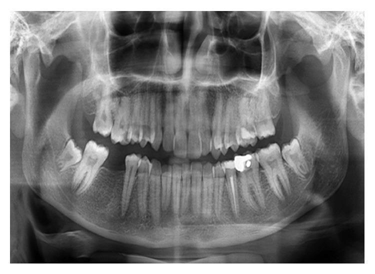
A panoramic radiograph (orthopantomogram) of the maxilla and mandible, illustrating a full dentition with various dental restorations. The image displays the anatomical landmarks including the temporomandibular joints, maxillary sinuses, and the mandibular canal. A primary focus is on the left mandibular second premolar, which features a highly radiopaque coronal restoration and a distinct radiopaque linear filling within the root canal, consistent with endodontic treatment. The periapical region of this premolar shows normal bone density and a well-defined lamina dura, indicating the complete resolution of a previous periapical lesion. Other visible features include endodontic fillings in several molars and the presence of third molars in varying stages of eruption. The cortical bone of the mandible appears continuous, and the trabecular pattern is consistent throughout, demonstrating successful post-treatment healing in a clinical follow-up context.
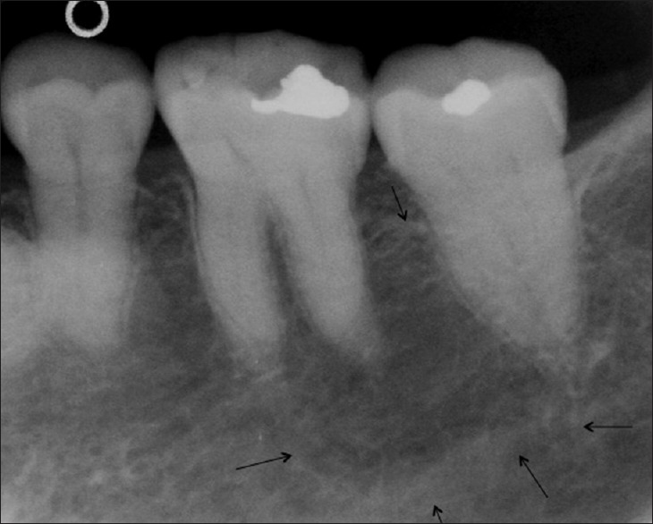
**Imaging Modality:** Periapical intraoral radiograph. **Anatomical Region:** Posterior mandible or maxilla (molar region), specifically focusing on the first and second molars. **Observed Pathology:** The radiograph demonstrates normal hard tissue structures with no overt evidence of periapical radiolucency, widening of the periodontal ligament space, or significant alveolar bone loss. The trabecular bone pattern appears within normal physiological limits. **Characteristic Visual Features:** * **Restorations:** Radio-opaque (metallic) occlusal restorations are visible on the first and second molars. * **Anatomy:** Clear visualization of the crown, pulp chamber, root canals, and lamina dura. * **Landmarks:** Black arrows at the inferior portion of the image indicate the borders of an anatomical canal, likely the inferior alveolar nerve canal (if mandibular) or a similar neurovascular landmark. * **Bone Density:** Homogeneous distribution of trabecular bone without sequestra or osteolytic lesions. **Diagnostic Context:** This image serves as a baseline radiographic study for a clinically asymptomatic tooth. The absence of radiographic inflammation or periodontal pathology supports a diagnosis of a healthy periradicular status despite the presence of dental restorations.
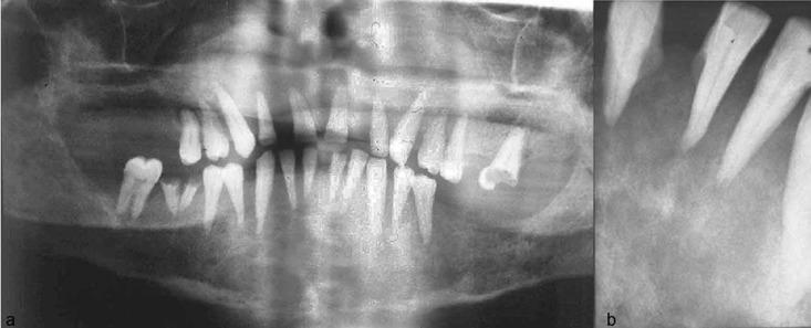
This composite diagnostic image features a panoramic radiograph (a) and a periapical radiograph (b) of the human maxilla and mandible, illustrating systemic bone pathology. Image (a) demonstrates generalized demineralization and rarefaction of the jaw bones. Notable findings include an ill-defined inferior alveolar canal, particularly on the right side, and thinning of the mandibular inferior cortex. There is evidence of cortical destruction in the posterior right region and localized resorption on the left. Anatomical landmarks such as the maxillary sinus borders and hard palate appear indistinct. Image (b) shows a detailed periapical view of the anterior incisors, highlighting a complete loss or severe lack of clarity of the lamina dura. Significant vertical bone loss is visible in the interdental regions. These radiographic features are characteristic of metabolic bone diseases or secondary hyperparathyroidism, demonstrating a loss of normal trabecular pattern and cortical definition within the craniofacial skeleton.
**Imaging Modality:** Intraoral periapical (retroalveolar) radiograph. **Anatomical Region:** Posterior mandible/maxilla, featuring a molar and adjacent edentulous space. **Observed Findings:** The radiograph displays a post-extraction tooth socket distal to a fully erupted molar. The extraction site (likely the third molar or "wisdom tooth" region) appears as a well-defined radiolucent area within the alveolar bone, corresponding to the anatomical space previously occupied by the tooth roots. The lamina dura of the socket is partially visible, indicating a recent postoperative state. **Characteristic Visual Features:** - **Primary Feature:** A distinct radiolucent void (extraction socket) with a shape mimicking the root morphology. - **Adjacent Structures:** The neighboring molar remains in situ, showing clear enamel-dentin boundaries and pulp chamber morphology. - **Bone Quality:** Surrounding trabecular bone density appears within normal limits for a postoperative site, with no immediate evidence of retained root fragments or gross pathological lesions. - **Landmarks:** The alveolar crest is visible, showing the surgical site’s relationship to the gingival margin. **Clinical Context:** Standard postoperative imaging to verify complete tooth removal and assess the integrity of the surrounding alveolar bone and adjacent dentition.
dentigerous cyst pericoronal radiolucency impacted tooth radiograph
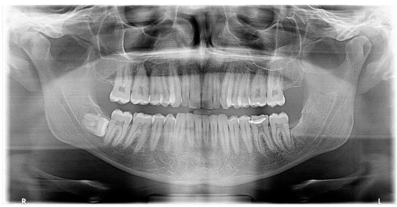
This diagnostic image is a panoramic radiograph (orthopantomogram) of the human dentomaxillofacial region. The primary finding is an impacted mandibular left third molar (tooth #38) in a horizontal and mesioangular orientation. A well-defined, unilocular pericoronal radiolucency with a thin sclerotic border is observed surrounding the crown of tooth #38, consistent with a dentigerous cyst. The lesion extends anteriorly to the distal root of the second molar (tooth #37), where localized bone resorption is present without evidence of root resorption. In the caudal region, the radiolucency appears to interfere with the inferior alveolar canal (IAC), which shows signs of inferior (caudal) displacement. The surrounding mandibular bone remains largely intact except for the area of the lesion. This radiographic presentation is typical for odontogenic cysts associated with impacted teeth and is a key educational example for dental students and maxillofacial surgeons in identifying follicular pathology and its relationship to neighboring anatomical structures like the IAC.
This diagnostic image is a periapical radiograph of the mandibular molar region, specifically focusing on an impacted third molar. The central tooth exhibits a mesioangular impaction, with its crown positioned below the occlusal plane of the adjacent second molar, which features a radiopaque dental restoration. A prominent, well-defined radiolucency is visible surrounding the crown of the impacted third molar. This pericoronal radiolucency is particularly widened on the distal aspect, as indicated by a black arrow. The radiolucent area suggests a pathological lesion such as a dentigerous cyst or enlarged dental follicle, especially as it appears to exceed 3 mm in width. The surrounding trabecular bone density and the roots of the impacted tooth are visible, though the apical morphology is less distinct due to the impaction depth. This image is used in oral and maxillofacial radiology to teach the identification of pericoronal pathology and the classification of impacted teeth using Winters or Pell and Gregory systems.
This intraoral periapical (IOPA) radiograph depicts the maxillary anterior and premolar region, specifically involving the left lateral incisor and first premolar. The primary finding is a large, ill-defined radiolucent lesion located between the roots of these teeth. The pressure from this lesion has caused significant distal and mesial deflection of the adjacent roots, increasing the inter-radicular space. Superior to this primary radiolucency, a dense, partial radiopacity is visible, suggesting the presence of an unerupted impacted tooth, possibly the maxillary canine. Additionally, a smaller, well-defined circular radiolucency is noted at the alveolar crest between the lateral incisor and first premolar. The overall radiographic presentation is highly suggestive of a pericoronal or cystic lesion, such as a dentigerous cyst, associated with an impacted tooth. The image illustrates key diagnostic features in oral and maxillofacial pathology, including root divergence and the characteristic radiolucency associated with odontogenic cysts in a pediatric or adolescent patient.
osteomyelitis jaw radiograph moth eaten bone sequestrum involucrum
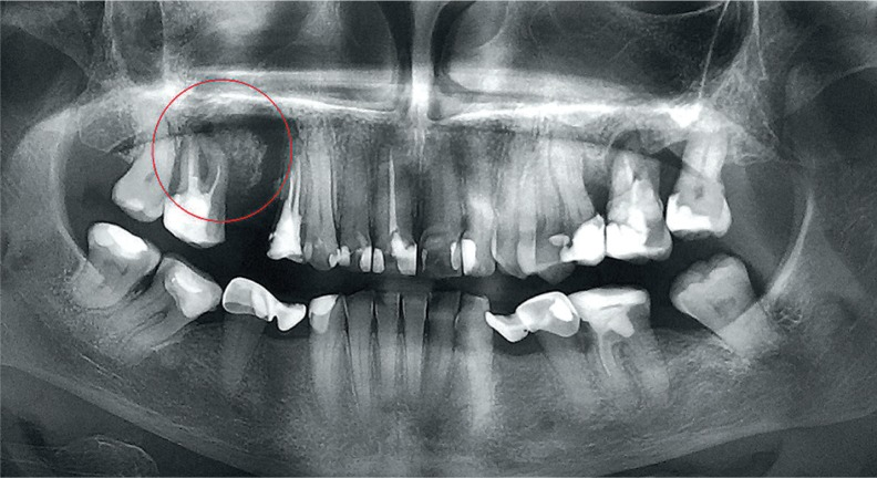
**Imaging Modality:** Panoramic Radiograph (Orthopantomogram). **Anatomical Region:** Maxillofacial region, specifically the right posterior maxilla. **Observed Pathology:** Bone sequestrum associated with chronic osteomyelitis or osteonecrosis. **Visual Features:** - **Primary Finding:** A well-defined, radiopaque island of non-vital bone (sequestrum) is visible in the right maxillary alveolar process, highlighted by a red circle. - **Surrounding Architecture:** The sequestrum is partially surrounded by a radiolucent zone (involucrum), indicating separation from the healthy bone. - **Dental Findings:** Generalized severe horizontal and vertical bone loss consistent with advanced periodontal disease. Multiple teeth exhibit extensive dental caries, restorations, and periapical radiolucencies. Several teeth are displaced or show signs of significant attachment loss. - **Maxillary Sinuses:** The right maxillary sinus floor appears closely approximated or involved by the inflammatory process. **Key Diagnostic Features:** - Presence of an isolated, dense radiopaque fragment within an area of radiolucency. - Mottled or "moth-aten" appearance of the surrounding trabecular bone pattern. - Clinical correlation typically involves Medication-Related Osteonecrosis of the Jaw (MRONJ) or chronic suppurative osteomyelitis.
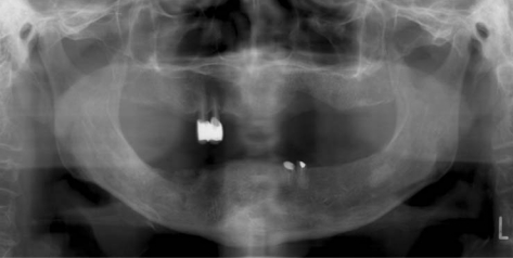
**Imaging Modality:** Panoramic radiograph (Orthopantomogram). **Anatomical Region:** Edentulous mandible and maxilla, including the temporomandibular joints and maxillary sinuses. **Observed Pathology:** Evidence of chronic osteomyelitis or osteonecrosis of the jaw. The right mandibular body and angle exhibit significant heterogeneous radiopacity and radiolucency. **Characteristic Visual Features:** * **Sequestrum:** An island of dense, non-vital bone is visible within the right mandibular body, surrounded by a radiolucent zone of granulation tissue or involucrum formation. * **Bone Density:** There is a generalized decrease in bone density (osteopenia) with moth-eaten radiolucent patterns in the affected right mandibular region, indicating active bone destruction. * **Dental Status:** Predominantly edentulous arches with a few remaining dental remnants and radiopaque restorative materials in the upper right and lower left quadrants. * **Cortical Changes:** Thinning and irregularity of the inferior border of the mandible on the right side. **Key Diagnostic Features:** The presence of a localized sequestrum associated with ill-defined osteolytic changes is highly characteristic of inflammatory or necrotic bone disease, such as Medication-Related Osteonecrosis of the Jaw (MRONJ) or actinomycotic osteomyelitis.
fibrous dysplasia jaw ground glass orange peel radiograph
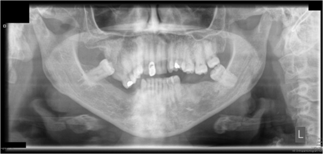
**Imaging Modality:** Orthopantomogram (OPG/Panoramic Radiograph) **Anatomical Region:** Maxillofacial region, focusing on the maxilla, mandible, and dentition. **Observed Pathology:** There is a significant expansile lesion involving the right maxillary tuberosity and posterior maxilla. The lesion demonstrates a "ground-glass" or "orange-peel" internal radiopacity, characteristic of fibro-osseous dysplasia. **Characteristic Visual Features:** * **Expansion:** Marked buccolingual and superior expansion of the right maxillary alveolar process. * **Bony Pattern:** A diffuse, poorly demarcated area of increased radiodensity with a granular texture replacing normal trabecular patterns. * **Effect on Surrounding Structures:** The lesion causes superior displacement of the floor of the right maxillary sinus. * **Dentition:** Generalized dental crowding, multiple missing teeth, and evidence of previous restorative work (radiopaque fillings). An impacted or displaced molar is visible in the right posterior maxilla, potentially associated with the expansile mass. **Diagnostic Cues:** The poorly defined borders and characteristic ground-glass opacity in a craniofacial bone are highly suggestive of fibrous dysplasia or a related fibro-osseous entity. No aggressive cortical destruction or sunburst periosteal reaction is visualized.
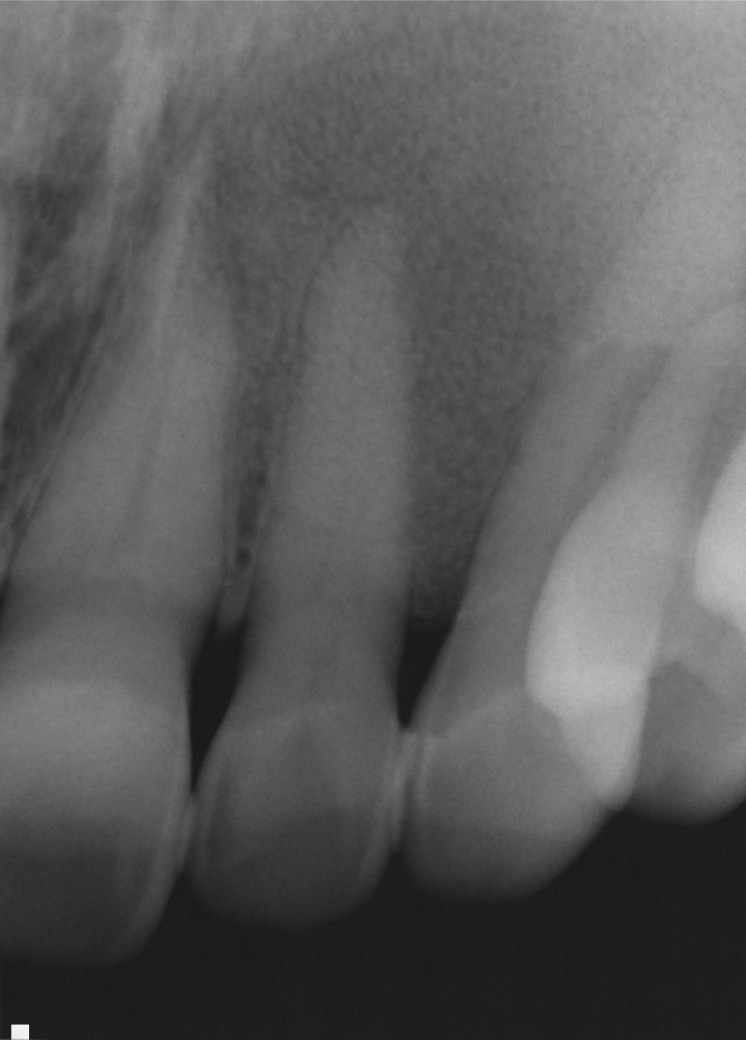
**Imaging Modality:** Intraoral periapical radiograph. **Anatomical Region:** Anterior maxillary dentition and associated alveolar bone. **Observed Pathology:** A radiopaque osseous lesion exhibiting a characteristic "ground-glass" or "orange peel" appearance. The lesion shows an ill-defined, blending anterior border that merges imperceptibly with the surrounding normal bone. **Characteristic Visual Features:** * **Trabecular Alteration:** Normal trabecular patterns are replaced by a dense network of shorter, irregularly shaped, and more numerous trabeculae, resulting in increased radiopacity. * **Periodontal Changes:** There is a generalized loss of the lamina dura surrounding the roots of the involved teeth. * **Expansion:** The image suggests a subtle expansion of the alveolar bone, though the cortical outlines remain largely intact. **Key Diagnostic Features:** The combination of a diffuse, poorly demarcated border, ground-glass internal radiopacity, and loss of lamina dura is highly suggestive of fibro-osseous dysplasia, such as fibrous dysplasia. The absence of a discrete radiolucent rim distinguishes this from typical odontogenic tumors.
osteosarcoma jaw sunray sunburst periosteal reaction radiograph
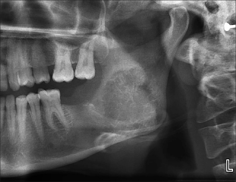
**Modality:** Orthopantomogram (OPG) / Panoramic Radiograph. **Anatomical Region:** Left posterior mandible, specifically the region of the angle and ascending ramus. **Observed Pathology:** Large, ill-defined, mixed radiolucent-radiopaque lesion involving the left mandibular ramus and angle. The lesion demonstrates aggressive features with infiltrative borders. **Characteristic Visual Features:** * **Bone Pattern:** Classic "sunburst" or "sunray" appearance characterized by divergent spicules of periosteal new bone formation radiating perpendicular to the cortical surface. * **Cortical Integrity:** Significant expansion and destruction of the buccal and lingual cortical plates. * **Dental Impact:** Displacement and potential resorption of adjacent molar roots are visible. * **Soft Tissue:** Evidence of a soft tissue mass extending beyond the bony confines of the mandible. **Clinical Context:** These radiographic findings are highly suggestive of an osteosarcoma of the mandible or a similar primary malignant bone tumor. **Key Diagnostic Features:** The presence of a poorly demarcated lesion with a radiating spiculated periosteal reaction ("sunburst" pattern) in the jaw is a hallmark for osteosarcoma, differentiating it from benign odontogenic cysts or tumors.
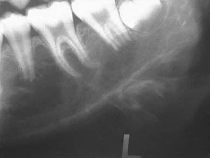
**Imaging Modality:** Intraoral periapical radiograph. **Anatomical Region:** Posterior region of the mandible, including molars and the inferior mandibular border. **Observed Pathology:** Large, ill-defined radiolucent lesion involving the body of the mandible with aggressive periosteal reaction. **Characteristic Visual Features:** * **Periosteal Reaction:** Prominent "sunray" or "sunburst" appearance characterized by divergent, thin radiopaque spicules of bone extending perpendicular to the cortical surface. * **Bone Destruction:** Irregular destruction of the medullary bone and thinning/perforation of the inferior cortical plate. * **Dental Involvement:** Displacement of the permanent molars and loss of the lamina dura surrounding the roots of the involved teeth. * **Soft Tissue Extension:** The radiating bony spicules extend into the adjacent soft tissue space. **Diagnostic Differentiating Features:** The aggressive "sunburst" periosteal pattern is a classic radiologic hallmark of osteosarcoma of the jaw, distinguishing it from benign reactive processes or chronic osteomyelitis. The combination of osteolysis and osteogenesis suggests a high-grade malignant bone tumor.
radicular cyst periapical radiolucency root apex well defined corticated
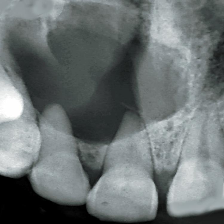
This diagnostic image is an intraoral periapical radiograph of the maxillary anterior region, specifically focusing on teeth 11, 12, and 13. The radiograph reveals a large, well-defined, and corticated radiolucency extending from the periapical region of the right maxillary central incisor (tooth 11) laterally toward the canine (tooth 13). Key pathological features include significant root resorption and a wide, blunted apex of tooth 11, which is the likely tooth of origin. Additionally, there is a distal displacement of the root of tooth 12, indicating the pressure effects of the expanding lesion. The lesion's well-circumscribed, radio-opaque border suggests a slow-growing, benign cystic process, characteristic of a radicular (periapical) cyst. This radiographic presentation is essential for distinguishing between periapical granulomas, cysts, and other odontogenic lesions such as odontogenic keratocysts or ameloblastomas. The image serves as a clinical example of the secondary effects of chronic periapical inflammation on adjacent dental and bony structures.
**Imaging Modality:** Intraoral periapical (IOPA) radiograph. **Anatomical Region:** Maxillary anterior region, specifically focusing on the central and lateral incisors. **Observed Pathology:** Large, well-defined, unilocular radiolucency with a thin, corticated border. The lesion is located periapically and appears to involve the roots of the maxillary incisors. **Characteristic Visual Features:** - **Lesion:** A prominent, circular radiolucent area in the alveolar bone, suggestive of an odontogenic cyst or granuloma (e.g., radicular cyst). - **Dental Structures:** The adjacent teeth show signs of widened periodontal ligament spaces. There is evidence of potential root resorption or displacement of the involved teeth due to the expansive nature of the lesion. - **Surrounding Bone:** The lesion exhibits a clear transition zone with a sclerotic/corticated rim, indicating a slow-growing, chronic process. The maxillary sinus floor or nasal fossa floor may be superiorly displaced. **Key Diagnostic Features:** The well-circumscribed, corticated radiolucent appearance centered at the apex of a tooth is a hallmark of a periapical cyst. This image is critical for differentiating inflammatory periapical lesions from other odontogenic tumors.
Stafne bone cyst defect mandible angle lingual salivary gland depression
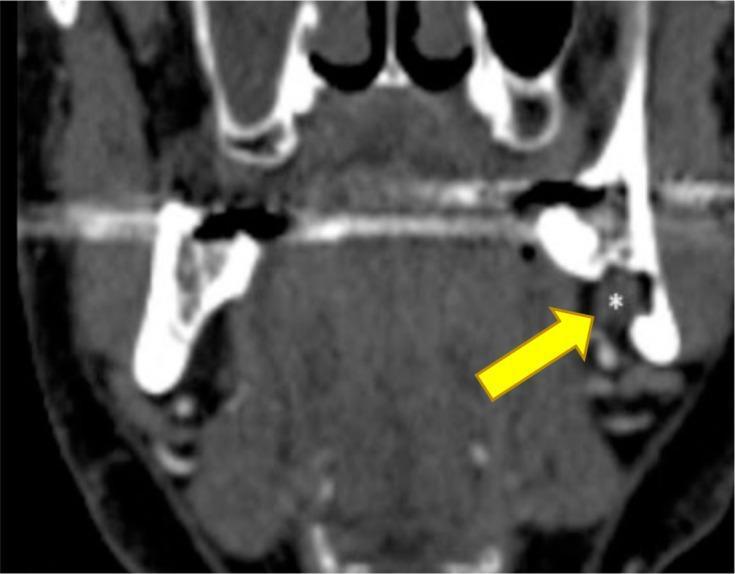
This diagnostic image is a coronal reconstruction from a CT angiogram of the head and neck, focusing on the mandible and submandibular region. The image demonstrates a well-defined Stafne bone cavity (SBC), also known as a static bone cyst or lingual mandibular bone depression. A yellow arrow points to a distinct focal concavity or discontinuity in the lingual cortical plate of the posterior mandible. Within this osseous defect, a white asterisk marks an area of soft tissue density that is continuous with the submandibular gland, confirming the herniation of glandular tissue into the mandibular depression. This finding is a classic anatomical variant rather than a true cyst or pathological lesion. The image is highly relevant for oral and maxillofacial radiology, illustrating how cross-sectional imaging can differentiate incidental radiolucent findings in the mandible from more aggressive odontogenic pathologies by identifying the presence of ectopic salivary gland tissue.
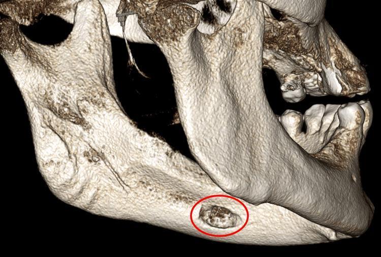
This diagnostic image is a 3D volume-rendering reconstruction of the human mandible, viewed from a posterolateral perspective focusing on the left lingual cortical surface. The primary clinical finding is a well-defined, oval-shaped concavity (highlighted by a red circle) located on the medial aspect of the mandibular body, inferior to the expected pathway of the inferior alveolar canal. While the surrounding lingual cortical bone appears dense and smooth, the bone within this indentation demonstrates an irregular, trabecular-like texture. This anatomical variation is characteristic of a Stafne defect (static bone cyst), which represents a developmental depression created by the submandibular salivary gland. The image serves as an educational tool for distinguishing benign developmental anomalies from pathological intraosseous lesions in oral and maxillofacial radiology. The anatomical landmarks shown include the mandibular ramus, angle, and body, providing context for the atypical anterior location of this specific defect.
ORAL RADIOLOGY: ANATOMY, INTERPRETATION & MISC - Complete Study Notes
SECTION 1: NORMAL ANATOMICAL LANDMARKS
1. Normal Anatomical Landmarks of Maxilla on Radiograph (2018, 2009)
MAXILLARY LANDMARKS
A. Radiopaque (White) Landmarks of Maxilla
- Location: Periapical view of maxillary incisors
- Appearance: V-shaped or arrowhead-shaped radiopaque projection at the base of the nasal septum, midline, at the floor of the nasal fossa
- Significance: Midline landmark; can mimic periapical pathology of central incisors
- Appearance: Vertical radiopaque line dividing the nasal fossae in the midline
- Seen on: Anterior maxillary periapical views, OPG
- Appearance: Thin, vertical radiolucent line flanked by radiopaque cortical bone edges, running in the midline of the palate
- Seen on: Maxillary anterior periapical, occlusal views
- Significance: Must not be confused with a root fracture
- Appearance: U-shaped or inverted "J" radiopaque band overlying the maxillary molar region; extends from the maxillary tuberosity superiorly
- Seen on: OPG, maxillary molar periapical
- Significance: Common landmark; may simulate pathology if confused with carious lesion
- Appears above the maxillary molar region on OPG as a long curving radiopaque band
- Radiopaque line running horizontally above the maxillary anterior teeth
- Below this lies the floor of the nasal fossa
- Faint radiopaque curvilinear line above the maxillary sinus on OPG and PA ceph
- Enamel: Most radiopaque (brightest white) - outermost crown layer
- Dentin: Less radiopaque than enamel - forms bulk of tooth
- Cementum: Radiopacity similar to dentin - covers root
- Pulp: Radiolucent space within the tooth
- Radiopaque line connecting the necks of adjacent teeth
- Normally 1.5-2 mm below the CEJ (cemento-enamel junction)
- Sharp, pointed (anterior) or flat (posterior)
- Radiopaque dense cortical bone lining the tooth socket (alveolus)
- Continuous white line surrounding the root
- Small hooklike bony projection at the distal end of the medial pterygoid plate, visible on maxillary molar/tuberosity periapical
- Appears as small radiopaque curved projection posterior to the maxillary tuberosity
B. Radiolucent (Dark) Landmarks of Maxilla
- Location: Molar/premolar periapical, OPG, Waters' view
- Appearance: Large, air-filled, radiolucent space with thin radiopaque cortical walls (Schneiderian membrane lining appears as thin white line when inflamed)
- The floor of the maxillary sinus lies close to the roots of upper premolars and molars
- Significance: Must be distinguished from periapical pathology; may contain mucus retention cysts (dome-shaped radiopaque shadow within sinus)
- A "halo" or teardrop shape at the floor of the sinus = mucosal retention cyst
- Bilateral symmetrical radiolucencies above the anterior teeth separated by the nasal septum
- Appear as paired oval radiolucencies above the central and lateral incisor roots
- Location: Anterior maxillary periapical, between the roots of central incisors
- Appearance: Small, round to oval, well-defined radiolucency at the midline between the roots of maxillary central incisors
- Normal size: Up to 6 mm diameter (larger may indicate nasopalatine cyst)
- The incisive canal runs from the nasal floor to this foramen
- Appears as bilateral radiolucent lines (two thin dark lines) running superiorly from the incisive foramen toward the nasal floor
- Sometimes only one line visible (when canal is single)
- Posterior hard palate - behind the last maxillary molar
- Small circular or oval radiolucency
- Rarely visible on periapical
- Faint radiopaque structures projecting into the nasal fossae - may be seen on OPG
- Fine radiolucent lines (vascular channels) within bone
- Seen in anterior maxilla
MANDIBULAR LANDMARKS
A. Radiopaque (White) Landmarks of Mandible
- Location: Mandibular molar periapical, OPG
- Appearance: Dense white oblique line running downward and forward from the anterior border of the ramus across the body of the mandible, crossing the molar region
- The external oblique ridge is the bony ridge on the outer (buccal) surface of the mandible
- Significance: Dense shadow may superimpose on the molar apices; can mimic bone sclerosis
- Location: Mandibular molar periapical, OPG
- Appears: Below and often parallel to the external oblique ridge on radiographs
- Represents the bony ridge on the inner (lingual) surface for attachment of the mylohyoid muscle
- May appear as a faint radiopaque line crossing the molar region at or below the level of the apices
- Location: Mandibular anterior periapical, occlusal
- Appearance: Small, circular or ring-shaped radiopaque structure in the midline of the mandible at the lingual surface; gives a "ring within a ring" pattern
- On standard periapical: Appears as small radiopaque "donut" at the midline
- Significance: Can mimic periapical pathology of mandibular incisors
- Midline fusion line of mandible - seen in children as faint radiolucent line; fuses by age 2 years
- Sometimes superimposed over the maxillary tuberosity on OPG/panoramic radiographs of edentulous patients
- Can mimic a large calcification
- Dense cortical radiopaque line forming the lower border on lateral oblique and OPG
- Can occasionally be seen on OPG as a curvilinear radiopaque structure below the mandible
B. Radiolucent (Dark) Landmarks of Mandible
- Location: Between roots of mandibular premolars (most commonly at or distal to apex of 2nd premolar)
- Appearance: Round or ovoid, well-defined radiolucency, 3-5 mm diameter, usually with slightly corticated borders
- Clinical significance: Can mimic a periapical pathology (granuloma or cyst) at the lower premolar apices. Test tooth vitality to differentiate
- The mental foramen has a slightly corticated border (radiopaque rim) which helps distinguish it from a periapical lesion
- Location: Within the mandibular body running from the mandibular foramen (ramus) to the mental foramen
- Appearance: Radiolucent band (dark tube) approximately 2-4 mm wide, bounded by two parallel radiopaque (white) lines (cortical walls of the canal)
- Contains the inferior alveolar nerve and artery
- Seen on: Mandibular molar periapical views, OPG - important for surgical planning (implants, extractions, osteotomies)
- On the medial aspect of the ramus
- Appears as a radiolucency on the inner surface of the ramus; overlying the antilingula anteriorly
- Seen on: OPG, lateral oblique
- Small radiolucent dot at midline of mandible anteriorly, at the center of the genial tubercle ring
- Transmits a small blood vessel and nerve
- Shallow concavity on the lingual surface of the mandible below the mylohyoid ridge for the submandibular gland
- On periapical views: Appears as a diffuse radiolucency below the mylohyoid ridge in the molar region
- Can mimic a large cystic lesion
- This is the region where Stafne's bone defect (lingual salivary gland depression) forms
- Small, fine radiolucent channels for blood vessels
- Most commonly seen in the mandibular anterior region - vertical fine dark lines between roots
Summary Table of Anatomical Landmarks
| Region | Radiopaque | Radiolucent |
|---|---|---|
| Maxilla - anterior | ANS, nasal septum, median palatine suture | Nasal fossae, incisive foramen, incisive canal |
| Maxilla - posterior | Zygoma U-shape, zygomatic arch | Maxillary sinus, greater palatine foramen |
| Mandible - anterior | Genial tubercles (ring), symphysis | Mental foramen, lingual foramen, nutrient canals |
| Mandible - posterior | External + internal oblique ridges | Mandibular canal (with corticated walls), submandibular fossa |
| Both | Enamel, dentin, lamina dura, alveolar crest | Pulp, periodontal ligament space |
2. Lamina Dura (Short Note - 2013)
Definition
Radiographic Appearance
- Thin, unbroken, white line encircling the entire root from the alveolar crest to the apex
- It is the radiographic projection of the alveolar bone proper (the cribriform plate)
- Pierced by numerous small channels (Volkmann's canals) which are too small to see radiographically
Clinical Significance
| Condition | Lamina Dura Appearance |
|---|---|
| Normal | Continuous thin white line |
| Periapical abscess/granuloma/cyst | Loss/break at apex |
| Acute apical periodontitis | Widened PDL space, early lamina dura loss |
| Hyperparathyroidism | Generalized loss of lamina dura (pathognomonic) |
| Paget's disease | Loss of lamina dura |
| Hypophosphatasia | Loss of lamina dura |
| Scleroderma | Generalized widening of PDL space with thickened lamina dura |
| Chronic osteomyelitis | Loss in affected area |
| Renal osteodystrophy | Generalized loss |
SECTION 2: RADIOGRAPHIC INTERPRETATION
3. Principles of Formulating Radiographic Diagnosis (2021)
- Age, sex, symptoms (pain, swelling, duration)
- Location in jaw (anterior/posterior, maxilla/mandible)
- Whether tooth is vital or non-vital
- Is the radiograph diagnostic quality?
- Correct exposure, processing, positioning?
- Which jaw, which region, relationship to teeth, bone
- Well-defined (corticated) = slow-growing, benign
- Well-defined (non-corticated) = benign, less certain
- Ill-defined = aggressive, malignant, inflammatory
- Radiolucent (unilocular / multilocular), radiopaque, or mixed
- Septation pattern (soap bubble, honeycomb)
- Completely radiolucent, mixed, completely radiopaque
- Tooth displacement, root resorption
- Cortical expansion, perforation
- Effect on inferior alveolar canal, maxillary sinus
- Effect on adjacent teeth (tooth at center = odontogenic)
- List most likely to least likely diagnoses
- Narrow down with clinical, laboratory, histopathology
4. Radiographic Features of Radicular (Periapical) Cyst (2017)
Definition
Radiographic Features
- Associated tooth: Non-vital tooth (carious, restored, or traumatized) - essential feature
- Location: Periapical region - centered at root apex
- Shape: Round or ovoid radiolucency
- Size: Can range from small (5 mm) to very large (several cm); large cysts may displace adjacent teeth
- Border/margin: Well-defined, corticated (white sclerotic line) - smooth, continuous radiopaque rim
- Internal structure: Uniformly radiolucent (no internal structure)
- Effect on adjacent structures:
- Displacement of adjacent teeth (not root resorption usually)
- Occasional root resorption of associated tooth
- Loss of lamina dura at apex of associated tooth
- May displace the inferior alveolar canal downward
- May push into maxillary sinus (dome-shaped into sinus floor)
- Periodontal ligament (PDL) space: Continuous with and widens at the apex
- The corticated rim is the key feature distinguishing a large cyst from a granuloma (granuloma usually <1 cm, less well-defined)
Note: Radiographic Differentiation
- Periapical granuloma: Smaller (<1 cm), ill-defined, no corticated margin; most periapical radiolucencies are granulomas
- Periapical abscess: Ill-defined, diffuse margins; acute presentation; often no clear demarcation
- Radicular cyst: Large (>1 cm), well-corticated, round; definitive diagnosis only by histopathology
5. Radiographic Features of Dentigerous (Follicular) Cyst (2018)
Definition
Radiographic Features
- Location: Always surrounds the crown of an unerupted tooth; attached at the CEJ
- Shape: Unilocular, round-to-oval pericoronal radiolucency
- Border: Well-defined with thin, corticated sclerotic border (smooth white rim)
- Internal structure: Uniformly radiolucent (fluid-filled); no internal calcifications
- Crown relationship: Crown projects into the cystic cavity - the critical diagnostic feature
- Most common sites: Lower third molar (most frequent), upper canine, lower premolar, mesiodens
- Effects:
- Displacement of associated tooth (to unusual positions - orbit, lower border of mandible)
- Displacement of adjacent teeth
- Root resorption of adjacent teeth
- Displacement/erosion of inferior alveolar canal
- Expansion and thinning of cortical plates
- Size: Can become very large - the larger the cyst, the greater risk of ameloblastomatous transformation
Pericoronal Radiolucencies (Short Note - 2013)
- Enlarged dental follicle (normal variant if <4 mm)
- Dentigerous cyst (most common pathological cause)
- Odontogenic keratocyst (OKC/KCOT) - scalloped, may have daughter cysts
- Adenomatoid odontogenic tumor (AOT) - often contains calcifications
- Ameloblastoma (unicystic type) - posterior mandible
- Calcifying epithelial odontogenic tumor (CEOT/Pindborg) - contains calcifications
6. Adenomatoid Odontogenic Tumor (AOT) - Radiographic Features (2023)
Definition
Radiographic Features
- Type A (follicular): Pericoronal radiolucency - mimics dentigerous cyst but extends below the CEJ onto the root (key distinguishing feature from dentigerous cyst which is only above CEJ)
- Type B (extrafollicular): Interradicular or periapical radiolucency NOT associated with a tooth crown
- Shape: Round/oval, well-defined
- Border: Corticated, smooth margins
- Internal calcifications: Flecks of calcification (snowflake-like radiopaque flecks) within the radiolucency - pathognomonic when present (seen in ~75%)
- Location: Anterior maxilla > anterior mandible; upper canine region most common
- Size: Usually small to moderate (<3 cm)
- Effect: Displacement of teeth; rarely root resorption
7. Radiographic Features of Osteomyelitis (2015)
Definition
Radiographic Features
Acute Osteomyelitis (early)
- No visible radiographic changes in first 1-2 weeks (bone must lose 30-50% mineral density before visible)
- Early: Subtle loss of trabecular pattern
- Then: Ill-defined radiolucency - "moth-eaten" bone destruction (irregular, poorly demarcated radiolucent areas with loss of normal trabecular architecture)
Chronic Osteomyelitis
- Moth-eaten appearance: Irregular, ill-defined radiolucencies mixed with areas of sclerosis
- Sequestrum: Dense, radiopaque island of non-vital (dead) bone separated from living bone by radiolucent zone; the sequestrum appears brighter than surrounding bone as it retains its mineral while the living bone demineralizes
- Involucrum: Layer of new bone formation around the sequestrum/abscess (appears as radiopaque envelope around the sequestrum)
- Cloaca: Radiolucent channel through the involucrum allowing pus to drain (may be invisible on plain film)
- Periosteal reaction: New bone deposition on outer surface of cortex (may appear as parallel "onion skin" layers or radiating spicules)
- Loss of lamina dura around involved teeth
- Widening of PDL space
- Sclerosis: Areas of increased density (reactive bone formation) around the lesion (Garre's osteomyelitis = predominantly sclerotic type with periosteal "onion skin")
Garre's (Chronic Sclerosing) Osteomyelitis
- In children/young adults; mandible; near infected mandibular first molar
- Periosteal new bone formation producing layered "onion skin" or "garlic-shaped" cortical duplication
- Also called: Periostitis ossificans
MRONJ (Medication-Related Osteonecrosis of the Jaw)
- Bisphosphonate or anti-RANKL therapy
- OPG shows: Sclerotic bone, sequestra, moth-eaten pattern, cortical disruption
- No periosteal reaction (unlike infectious osteomyelitis)
8. Fibrous Dysplasia (2021)
Definition
Types
- Monostotic (one bone - 70-80%)
- Polyostotic (multiple bones - 20-30%)
- McCune-Albright syndrome (polyostotic + café-au-lait spots + endocrine abnormalities)
Radiographic Features
- Internal pattern (pathognomonic): "Ground glass" or "orange peel" or "cotton wool" appearance - diffuse, granular, hazy increase in density with loss of normal trabecular pattern
- Borders: Characteristically ILL-DEFINED (key feature) - lesion blends imperceptibly into surrounding normal bone ("fades into bone"); no corticated border
- Cortical changes: Expansion and thinning of cortical plates but not destruction
- Teeth: Displacement of teeth; displacement of inferior alveolar canal; loss of lamina dura around involved teeth
- Maxillary sinus: Floor elevated/obliterated; sinus may be filled with the lesion
- Bone trabeculae: Short, irregular, numerous trabeculae replacing normal bone (histologically: C-shaped or "Chinese alphabet" pattern)
- Lesion is monostotic - limited to one bone; does not cross sutures
9. Osteosarcoma (2021)
Definition
Radiographic Features
- Location: Posterior mandible, symphysis, condyle; maxillary antrum
- Pattern: Can be:
- Predominantly radiolucent (osteolytic)
- Mixed radiolucent and radiopaque
- Predominantly radiopaque (osteoblastic / sclerotic)
- Borders: ILL-DEFINED, poorly demarcated - irregular, moth-eaten margins; bone destruction without clear boundary
- Sunburst / Sunray appearance (PATHOGNOMONIC): Radiating spicules of new bone formation perpendicular to the cortex; formed as periosteal new bone is repeatedly broken and reformed by the tumor
- Codman's triangle: Periosteal new bone at the periphery of the lesion forms triangular radiodense areas at the margins where periosteum is lifted
- Widened PDL space: Symmetrical, generalized widening of the periodontal ligament space around teeth in the affected region - early, CHARACTERISTIC sign in jaw osteosarcoma (due to tumor spreading along PDL space)
- Cortical perforation: Destruction of cortical plates with soft tissue extension
- Root resorption: Irregular resorption of adjacent tooth roots
- Displacement of inferior alveolar canal
10. Stafne's Bone Cyst (2020)
Definition
Key Features
- Truly a pseudocyst (no cystic epithelial lining) - an anatomical variant
- First described by Stafne (1942)
- Asymptomatic, found incidentally on radiograph
Radiographic Features
- Location: Below the inferior alveolar canal, at the angle of mandible, lingual surface - CLASSIC location (below mandibular canal distinguishes it from most pathological lesions which are above it)
- Shape: Well-defined, round or oval radiolucency
- Borders: Well-corticated - smooth, sclerotic white rim around the radiolucency
- Size: Usually 1-3 cm
- Content: Radiolucent (occupied by submandibular gland tissue and fat)
- Inferior alveolar canal: Displaced superiorly or passes above the defect
- Teeth: Not associated with teeth; teeth are vital and have normal periapical status
- Unilateral in most cases
Confirmation
- CT / CBCT: Shows cortical defect on lingual surface filled with soft tissue density (glandular tissue)
- Sialography or MRI: May demonstrate salivary gland tissue in the defect
- Vital teeth + characteristic location confirms it is not a pathological lesion
Clinical Significance
- Benign - no treatment required
- Must be distinguished from: Ameloblastoma, OKC, metastatic disease, giant cell lesions
11. Germination, Fusion, and Concrescence (2021)
Germination
- Large bifid crown or deep groove on crown
- Single root with single pulp canal (or partially divided canal)
- Tooth count normal (the "doubled" tooth counts as one)
- Most common in primary incisors; maxillary anterior most frequently
Fusion
- Single large tooth with two pulp chambers and usually two root canals (fused or separate)
- Groove or line visible on crown radiographically at the fusion plane
- Tooth count: One tooth missing (two have become one)
- Can occur between a normal tooth and supernumerary tooth
- Most common in primary mandibular incisors
- Fusion: Tooth count = reduced by one (normal tooth + another = 1)
- Germination: Tooth count = normal (1 tooth germ → appears as 2)
Concrescence
- Two separate crowns and pulps
- Roots joined by dense bridge of cementum at their proximal surfaces
- Most common in maxillary molar region (second and third molars)
- Diagnosis confirmed by periapical radiograph showing joined root surfaces
12. Hemangioma of Oral Cavity (2021)
Definition
Hamartoma Definition
Clinical Features of Hemangioma
- Soft tissue: Soft, compressible, bluish-purple mass; blanches on pressure (diascopy positive); may enlarge with straining/crying/bending forward
- Location: Lips, tongue, buccal mucosa, gingiva; parotid region
- Involutes spontaneously in most infantile cases (60-70% by age 7)
Radiographic Features
- Most soft-tissue hemangiomas show no bone changes
- Phleboliths (calcified thrombi): Multiple small, round, radiopaque calcifications within the soft tissue - highly suggestive of cavernous hemangioma
- Intraosseous hemangioma: Multilocular radiolucency with "honeycomb" or "soap-bubble" pattern; or "sunray" pattern (from dilated marrow spaces with bony septae perpendicular to the surface); enlarged vascular foramina
- OPG / Periapical: May show widened interdental spaces, root resorption if bone involved
- Angiography (DSA): Shows vascular blush and feeding vessels
- MRI: Best for soft tissue hemangioma - characteristically bright on T2, shows flow voids; defines extent
- Ultrasound with Doppler: High-flow lesion with multiple intralesional vessels
Treatment
- Infantile/capillary: Observation (most involute); oral propranolol (first-line for proliferating infantile hemangiomas)
- Corticosteroids: Intralesional or systemic for rapidly growing lesions
- Sclerotherapy: Injection of sclerosing agents (sodium tetradecyl sulfate, bleomycin) for cavernous type
- Nd:YAG / Pulsed dye laser
- Surgery: For symptomatic, large, or non-involuting lesions; CAUTION: risk of massive hemorrhage with intraosseous hemangioma
SECTION 3: MISCELLANEOUS TOPICS
13. Radioactive Isotope / Radionuclide (Short Note - 2012)
Definition
Key Terms
- Atomic number (Z): Number of protons; defines the element
- Mass number (A): Protons + neutrons
- Isotopes: Atoms of the same element (same Z) but different mass numbers (different number of neutrons)
- Radioisotopes (Radionuclides): Unstable isotopes that undergo radioactive decay
Types of Radioactive Decay
| Type | Particle/Ray Emitted | Penetrating Power | Use |
|---|---|---|---|
| Alpha | ²He nucleus (2p + 2n) | Least - stopped by paper | Internal therapy (Radium-226) |
| Beta | Electron (β⁻) or positron (β⁺) | Moderate - stopped by few mm Al | Thyroid therapy (I-131); PET (F-18) |
| Gamma | High-energy photon | Highest - needs lead/concrete | Imaging (Tc-99m), therapy |
Half-Life (T½)
- Tc-99m: 6 hours (most used in nuclear medicine scans)
- I-131: 8 days (thyroid therapy)
- Cs-137: 30 years
- Ra-226: 1600 years
Medical Uses
- Tc-99m bone scan: Detects osteomyelitis, metastases, Paget's disease (as "hot spots")
- I-131: Thyroid cancer treatment, hyperthyroidism
- I-125 seeds: Brachytherapy for prostate cancer
- F-18 FDG PET: Tumor staging, recurrence detection
14. Brachytherapy (Short Note - 2012)
Definition
Principle
- Short distance between source and target → inverse square law means dose falls off rapidly
- High dose to tumor, low dose to adjacent normal tissue
- Continuous or pulsed low-dose-rate irradiation
Types
| Type | Description | Examples |
|---|---|---|
| Intracavitary | Sources placed in body cavities | Cervical cancer (Manchester system) |
| Interstitial | Seeds/wires implanted directly into tissue | Oral cancer (tongue, floor of mouth), prostate |
| Surface mold | Sources on applicator placed on tissue surface | Skin, superficial lesions |
Isotopes Used
- Iridium-192 (Ir-192): Most common; gamma emitter; T½ = 74 days; used for oral cavity, pharynx cancers
- Iodine-125 (I-125): Low energy; used for prostate seeds; T½ = 60 days
- Caesium-137 (Cs-137): Intracavitary; T½ = 30 years
- Radium-226 (Ra-226): Historical; now replaced due to safety concerns
Dose Rate Classification
- LDR (Low Dose Rate): 0.4-2 Gy/hour; continuous; patient hospitalized
- HDR (High Dose Rate): >12 Gy/hour; remote afterloading; outpatient
- PDR (Pulsed Dose Rate): Intermediate
Applications in Oral and Maxillofacial Region
- Tongue carcinoma (interstitial implant with Ir-192 wires)
- Floor of mouth carcinoma
- Lip cancer
- Buccal mucosa carcinoma
- As boost to external beam RT
- Recurrent carcinomas in previously irradiated fields
Advantages
- Very high local dose to tumor
- Spares surrounding normal tissue
- Short treatment time (HDR)
- Conformal (source tracks tumor shape)
Disadvantages
- Invasive (surgical implantation)
- Risk of osteoradionecrosis, soft tissue necrosis
- Radiation hazard to staff (LDR)
- Limited to accessible, small-moderate tumors
Summary Table: All Short Notes
| Topic | Key Points |
|---|---|
| Lamina dura | Thin cortical white line around tooth socket; loss = periapical pathology, hyperparathyroidism, Paget's |
| Mental foramen | Round radiolucency between lower premolars; can mimic periapical lesion; test vitality |
| Radicular cyst | Non-vital tooth; periapical; well-corticated radiolucency; uniformly radiolucent; large (>1 cm) |
| Dentigerous cyst | Impacted tooth; pericoronal above CEJ; well-defined; unilocular; can become very large |
| AOT | Pericoronal extending below CEJ; snowflake calcifications; anterior maxilla; impacted canine |
| Osteomyelitis | Moth-eaten; sequestrum + involucrum; periosteal reaction; ill-defined |
| Fibrous dysplasia | Ground glass; ill-defined blending borders; expands; no destruction; monostotic |
| Osteosarcoma | Sunburst/sunray periosteal reaction; ill-defined; mixed density; widened PDL space (early) |
| Stafne's bone cyst | Below inferior alveolar canal; angle of mandible; lingual; well-corticated; NOT a true cyst; incidental finding |
| Radioactive isotope | Unstable nucleus; emits alpha/beta/gamma; half-life; Tc-99m most used diagnostically |
| Brachytherapy | Radioactive source inside/adjacent to tumor; Ir-192; interstitial for oral cancer |
LONG QUESTION: Enumerate developmental anomalies affecting oral cavity? Describe Amelogenesis Imperfecta. (11) SHORT NOTE: 1. Plummer-vinson syndrome. (16) Dens-in-dente. (07) 3. Cleft lip. (06) 4. Taurodontism. (06) 5. Developmental abnormalities of the condyle. (12) ULCERATIVE VESICULAR AND BULLOUS LESION: LONG QUESTION: 1. Classify vesiculobullous lesion. Describe etiology, pathogenesis, clinical features and management of erythema multiform. (2018) 2. Classify vesiculobullous lesion. Describe etiopathogenesis, clinical features, investigations and treatment of Herpes Zoster. (15) 3. 4. Describe etiology, pathogenesis, clinical features and management of erythema multiform. (14) Define and difference between a vesicle and a bulla. Classify vesiculobullous lesion having oral manifestation. Describe in detail about etiology, clinical features, investigation and management of pemphigus vulgaris. (12) 5. 6. Classify ulcerative lesions of the oral cavity. Write down in detail about herpes simplex. (20s) Classify Vesiculo bullous lesion and write in detail about etiology, clinical features and management of erythema multiforme. (20) SHORT NOTE: 1. Apthous stomatitis. (11) 2. Clinicopathological diagnostic features of pemphigus vulgaris. (07) 3. Pemphigus vulgaris. (13,19) 4. Define vesicle and bulla. (13) 5. Behcet's syndrome. (12) 6. Recurrent aphthous stomatitis. (19,22) 7. Erythema Multiforme. (20s) 8. Geographic tongue. (20s) RED AND WHITE LESIONS OF ORAL MUCOSA AND SOME PIGMENTED LESIONS: LONG QUESTION: 3. Classify Pre-cancerous lesions & condition. Describe the etiopathogenesis, C/F & management of OSMF. (2017) Classify oral ulcers. Describe the etiopathogenesis, clinical features, Investigations and treatment of oral candidiasis. (16,23) Classify white lesions of oral cavity and discuss in detail about oral leukoplakia. (2018,'14)
pemphigus vulgaris oral blisters acantholysis mucosal erosions
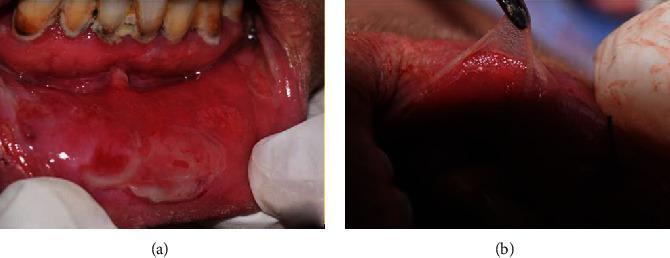
Two-panel clinical photograph illustrating severe oral manifestations of Pemphigus Vulgaris. Panel (a) shows the lower labial and gingival mucosa with extensive erythematous erosions, postbullous ulcerations, and sloughing tissue. The lesions exhibit a raw, inflamed appearance with irregular borders and whitish fibrin deposits. Visible mandibular teeth demonstrate significant plaque accumulation and poor oral hygiene. Panel (b) demonstrates a positive Nikolsky sign, a hallmark of autoimmune bullous diseases. Under tangential pressure, the superficial epithelium is being peeled away from the underlying basement membrane, creating a thin, translucent flap. This finding visually confirms acantholysis and the extreme fragility of epithelial attachment mechanisms. The images highlight the diagnostic physical exam findings in a 55-year-old patient with systemic vesiculobullous disease, emphasizing the transition from intact blisters to painful mucosal erosions.
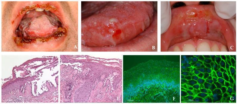
This composite image illustrates clinical and diagnostic features of Mucosal Pemphigus Vulgaris (MPV). Panels A-C are clinical photographs of the oral cavity: (A) shows extensive hemorrhagic crusting and erosions on the vermilion border of both lips; (B) demonstrates multiple small, intact blisters and shallow erosions on the lateral and dorsal tongue; (C) highlights an erosive lesion on the upper labial mucosa with a characteristic collapsed bulla roof appearing as a white slough. Panels D-E are histopathological images (H&E stain, 40x magnification) showing classic suprabasal clefting and intraepidermal bulla formation with acantholysis, where keratinocytes lose adhesion. Panels F-G present direct immunofluorescence (DIF) microscopy at 10x and 40x magnification. These panels demonstrate a diagnostic 'fishnet' or reticular staining pattern, indicating intercellular IgG/C3 deposits or Dsg3 autoantibodies along the keratinocyte cell surfaces. This collection serves as a comprehensive educational tool for the diagnosis of autoimmune blistering diseases through clinical presentation, histopathology, and immunofluorescent confirmation.
erythema multiforme target lesion iris lesion skin oral ulcers Stevens Johnson
Clinical photograph of a patient's mid-back displaying a classic targetoid (iris) lesion. The primary lesion is well-circumscribed and located on the mid-right posterior trunk. It features three distinct zones: a central dusky, possibly eroded or blistered core; an intermediate pale edematous ring; and a peripheral erythematous rim. This 'target-like' morphology is a hallmark of erythema multiforme. The surrounding skin is of medium tone with visible hair follicles and minor scattered nevi but is otherwise free of widespread exanthema in this view. The clinical context suggests an immunologic reaction, possibly associated with medication-related hypersensitivity or Stevens-Johnson Syndrome (SJS) spectrum given the reported mucosal involvement. This image serves as an educational reference for identifying characteristic skin signs in inflammatory and hypersensitivity dermatoses.
Clinical photograph of a dermatological finding on the upper right chest. The image displays a well-circumscribed, erythematous skin lesion characterized by a classic targetoid (target-like) morphology. The lesion consists of a dark red or purpuric central zone, which appears slightly raised, surrounded by a paler intermediate ring and a peripheral erythematous halo. This morphology is highly characteristic of erythema multiforme. The surrounding skin is light brown with minor scattered macules (dark spots) and appears otherwise non-inflamed. A portion of a striped hospital gown is visible in the lower right corner. The clinical context suggests an acute mucocutaneous reaction, possibly secondary to medication or infection, emphasizing the diagnostic importance of targetoid lesions in identifying hypersensitivity syndromes like Erythema Multiforme, Stevens-Johnson Syndrome (SJS), or Toxic Epidermal Necrolysis (TEN).
herpes zoster shingles dermatomal vesicular rash oral trigeminal
Clinical photograph of a male patient presenting with a localized vesicular rash on the face, characteristic of herpes zoster (shingles). The lesions consist of multiple grouped, erythematous papules and small vesicles, some showing signs of early crusting and ulceration. The distribution is strictly unilateral and dermatomal, primarily involving the mandibular (V3) and maxillary (V2) divisions of the trigeminal nerve. Clusters are most prominent around the right side of the vermillion border of the lower lip, extending inferiorly onto the chin and laterally toward the cheek. Additional smaller clusters are visible on the mid-cheek region. This image demonstrates a classic reactivation of the varicella-zoster virus (VZV), which can occur postoperatively following surgical manipulation of the trigeminal nerve, such as microvascular decompression for trigeminal neuralgia. The clinical focus is on the dermatomal pattern and the morphology of the herpetic vesicles.
Clinical photograph demonstrating herpes zoster (shingles) involving the ophthalmic division (V1) of the trigeminal nerve in an HIV-positive patient. The frontal skin of the forehead shows clustered vesicular lesions on an erythematous base with crusted erosions, most prominent near the distribution axis along the V1 dermatome, including the glabellar region and the superior eyelid. The eruption is unilateral and dermatomal, with vesicles grouped in a vesicular-rash pattern and surrounding edema. Superficial crusts indicate healing lesions; some vesicles appear partially collapsed. In immunocompromised hosts, including persons living with HIV, zoster may present with more extensive, disseminated, or atypical cutaneous involvement; caution for potential ocular involvement (herpes zoster ophthalmicus) and corneal risk, uveitis, or lid swelling. The clinical appearance is characteristic of varicella-zoster virus reactivation within the trigeminal nerve territory; differential diagnosis includes herpes simplex infection, impetigo, contact dermatitis, dermatitis herpetiformis, and cellulitis. Timely antiviral therapy (acyclovir, valacyclovir, or famciclovir) and HIV management optimize outcomes and reduce postherpetic neuralgia risk. This image is intended for educational illustration of zoster dermatomes patterns, HIV-associated immunosuppression effects on cutaneous viral infections, dermatology teaching, and clinical decision support in infectious disease and ophthalmology contexts. Recognize signs early to prevent complications and optimize antiviral management in HIV.
oral submucous fibrosis OSMF interincisal opening fibrous band trismus
A frontal clinical photograph of an adult male patient demonstrating maximum mouth opening during a one-year postoperative follow-up for oral submucous fibrosis (OSMF). The patient is positioned in a dental chair with eyes closed. The oral cavity is visible, showing a functional interincisal distance of approximately 30 mm, consistent with successful surgical management of trismus. The upper and lower dentition are visible, alongside the tongue and oral mucosa. This image illustrates the long-term efficacy of surgical fibrous band excision and skin grafting in restoring masticatory function and mandibular mobility. The mucosal tissues appear pink and healthy without evidence of recurrent restrictive scarring or acute inflammation. This procedural outcome image is relevant for oral and maxillofacial surgery education, specifically regarding the rehabilitation of patients with progressive trismus secondary to betel nut or pan chewing habits.
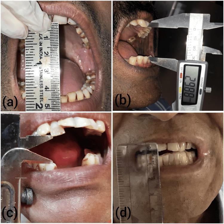
A composite of four clinical photographs (a-d) demonstrating the progressive stages of trismus (restricted mouth opening) in patients with Oral Submucous Fibrosis (OSMF). The images illustrate the clinical measurement of maximum interincisal distance using different tools: (a) a vertical stainless steel ruler, (b) a digital caliper showing a measurement of 29.88 mm, (c) a manual vernier-style caliper, and (d) a transparent plastic ruler. The series depicts a clear reduction in the oral aperture from stage I to advanced stage IV. Key visible features include blanched, pale, and opaque oral mucosa in the later stages, which replaces the healthy pink vascular appearance seen in earlier stages. The alignment of the teeth and the health of the gingiva are increasingly difficult to visualize as the disease progresses and the interincisal gap narrows. This progression highlights the fibrous band formation characteristic of OSMF, leading to significant functional impairment in jaw mobility.
oral leukoplakia white patch tongue floor of mouth precancerous
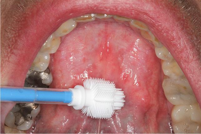
This intraoral clinical photograph displays the floor of the mouth in an adult patient, oriented with the tongue likely elevated. The mucosa of the anterior floor of the mouth exhibits areas of homogeneous leukoplakia—distinct white, well-demarcated patches—on both the left and right sides. The surrounding tissue shows generalized erythema and an irregular texture, suggesting chronic irritation or a precancerous state. An Orcellex cell collector (a specialized white-bristled brush with a blue handle) is positioned over the lesion on the left side, demonstrating a liquid-based cytology collection procedure for diagnostic screening. The visible mandibular dentition shows signs of dental wear, attrition, and significant restorative work, including a large silver-colored amalgam filling on a posterior molar. This image is used for educational purposes to illustrate the clinical appearance of potentially malignant oral mucosal lesions and the methodology for non-invasive brush biopsy sampling.
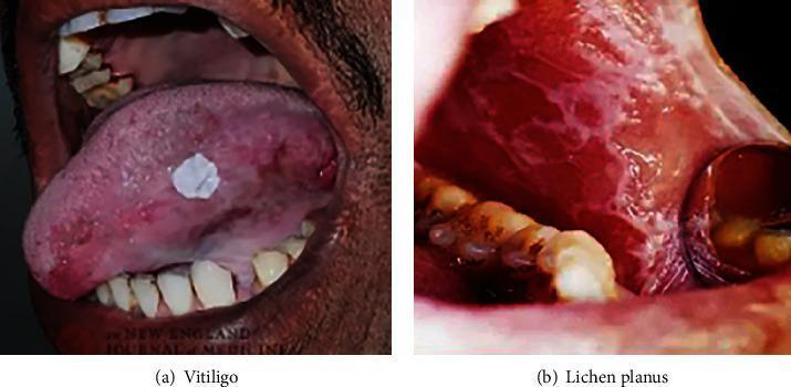
This clinical photograph displays two distinct oral precancerous and mucosal conditions within the oral cavity. Figure (a) illustrates a case of oral leukoplakia (captioned as Vitiligo in some contexts but clinically described here as a precancerous lesion). The lesion is characterized by a solitary, well-demarcated, irregularly shaped white plaque on the dorsal surface of the tongue. The patch is slightly raised with a roughened texture, contrasting with the surrounding erythematous and pinkish mucosa. Figure (b) demonstrates oral lichen planus affecting the buccal mucosa. The visual features include diffuse erythema and inflammation of the soft tissue, overlaid with characteristic Wickham striae—white, lace-like, or web-like reticular patterns. The adjacent gingiva also appears swollen and erythematous. These images serve as educational tools for identifying common oral pathological states that require clinical monitoring for potential malignant transformation into oral cancer.
recurrent aphthous stomatitis minor aphthous ulcer oral mucosa
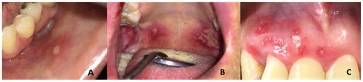
This composite clinical photograph illustrates the three primary classifications of Recurrent Aphthous Stomatitis (RAS) within the oral cavity. (A) Minor RAS: A single, small, well-defined oval ulcer on the buccal mucosa characterized by a central yellowish-white fibrinous pseudomembrane and a prominent peripheral erythematous halo. (B) Major RAS: Large, irregular, and deep ulcerations located on the soft palate and oropharynx. These lesions exceed 1 cm in diameter, exhibiting extensive tissue destruction, yellowish necrotic centers, and significant surrounding inflammation. (C) Herpetiform RAS: Multiple, pinpoint-sized (1-3 mm) ulcers clustered in a dense distribution along the gingival and labial mucosa. These lesions present as numerous discrete, intensely erythematous points that can coalesce into larger irregular areas. This comparative series serves as a diagnostic educational tool for distinguishing between aphthous variants based on ulcer size, morphology, quantity, and anatomical distribution in dental and dermatological clinical practice.
This clinical photograph provides an intraoral view of a minor Recurrent Aphthous Ulcer (RAU) located on the labial mucosa of the upper right side lip. The lesion is a solitary, shallow, ovoid ulcer approximately 5mm in diameter, characterized by a necrotic pseudomembranous center with a whitish-yellow hue. It is demarcated by a prominent, well-defined erythematous halo, indicating active inflammation. The surrounding oral mucosa appears mildly hyperemic. Visible anatomical structures include the maxillary gingiva and teeth, with fixed orthodontic appliances (braces) present. In the clinical context of an immunocompromised patient (HIV+), such lesions are significant as they may represent a manifestation of decreased immune surveillance and can severely impact quality of life due to associated pain and dysphagia. The image demonstrates classic morphological features used in the diagnosis of minor aphthous stomatitis, distinguishing it from major or herpetiform variants.
ORAL PATHOLOGY: DEVELOPMENTAL ANOMALIES, VESICULOBULLOUS & MUCOSAL LESIONS - Complete Study Notes
SECTION 1: DEVELOPMENTAL ANOMALIES
1. Developmental Anomalies Affecting Oral Cavity - Classification
A. Anomalies of Teeth
- Hypodontia (oligodontia) - fewer teeth
- Anodontia - no teeth
- Hyperdontia - extra (supernumerary) teeth (mesiodens, paramolar, distomolar)
- Macrodontia (megadontia)
- Microdontia (peg laterals)
- Gemination - single bud → bifid crown, single root
- Fusion - two buds → one tooth
- Concrescence - union by cementum
- Dilaceration - angulation/bend in root
- Taurodontism - elongated pulp chamber, short roots
- Dens invaginatus (Dens-in-dente)
- Dens evaginatus (Leong's premolar)
- Talon cusp
- Peg-shaped lateral incisor
- Amelogenesis imperfecta (enamel)
- Dentinogenesis imperfecta (dentin)
- Dentin dysplasia
- Regional odontodysplasia ("ghost teeth")
- Fluorosis
- Tetracycline staining
- Congenital syphilis (Hutchinson's incisors, mulberry molars)
- Transposition
- Impaction
- Ankylosis
- Eruption cysts
B. Anomalies of Soft Tissues and Jaws
- Cleft lip and/or palate
- Bifid tongue, fissured tongue, geographic tongue, macroglossia, microglossia
- Ankyloglossia (tongue tie)
- Fordyce's spots (ectopic sebaceous glands)
- Tori (torus palatinus, torus mandibularis)
- Eagle's syndrome (elongated styloid)
- Hemifacial microsomia (developmental anomaly of condyle)
- Condylar hyperplasia / aplasia
- Pierre Robin sequence, Treacher Collins syndrome
2. Amelogenesis Imperfecta (AI) (2011)
Definition
Genetics
- Incidence: 1:700 (Sweden) to 1:1200 (USA)
- Multiple inheritance patterns: Autosomal dominant (most common), autosomal recessive, X-linked
- Gene mutations: AMELX (amelogenin - X-linked), ENAM (enamelin - AD), MMP20 (enamelysin), KLK4 (kallikrein-4), FAM20A, WDR72
- Depends on which stage of enamel formation is affected: matrix secretion, maturation, or calcification
Classification (Witkop's Classification - 4 major types)
Type I: Hypoplastic AI
- Defect: Insufficient enamel matrix secretion (reduced quantity of enamel)
- Enamel: Thin but normally hard and translucent; pits, grooves, or absent enamel
- Clinical: Small teeth, diastemas (spaces between teeth); enamel may be pitted, locally absent, or smooth; normal hardness
- Radiographic: Thin enamel layer, contrast with dentin maintained
- Multiple subtypes: IA (pitted, autosomal dominant), IB (local, AD), IC (local, AR), ID (smooth, AD), IE (smooth, X-linked), IF (rough, AD), IG (enamel agenesis, AR)
Type II: Hypocalcified AI
- Defect: Defective mineralization/calcification of enamel matrix (enamel quantity normal, quality poor)
- Enamel: Normal thickness but soft, poorly calcified, chalky; wears off rapidly
- Clinical: Teeth appear opaque white to yellow-brown at eruption; enamel is soft, friable, easily scraped off with instrument; rapid attrition; hypersensitivity; open bite may develop
- Radiographic: Enamel radiodensity equal to or less than dentin (normally enamel is MORE radiopaque) - pathognomonic finding
- This is the most severe type clinically
Type III: Hypomaturation AI
- Defect: Defective maturation of enamel crystallites (crystals formed but don't grow properly)
- Enamel: Normal thickness, but mottled, opaque, yellowish-white; softer than normal; "snowcapped" enamel in some subtypes
- Clinical: Teeth appear mottled; enamel can be pierced by an explorer point
- Radiographic: Enamel similar in density to dentin
- Special subtype: Snow-capped teeth (IIIA) - white opaque enamel on cuspal and incisal portions
Type IV: Hypomaturation-Hypoplasia with Taurodontism
- Combined features of Types I and III
- Associated with taurodontism (enlarged pulp chambers)
Clinical Features of Amelogenesis Imperfecta (General)
- Both dentitions (primary and permanent) affected
- Sensitivity: All types sensitive to temperature change (exposed dentin)
- Aesthetics: Discoloured teeth (white, yellow, brown); significant psychological impact
- Anterior open bite: Common in hypocalcified type
- Enamel shows absent, thin, pitted, or rough surface depending on type
- No systemic associations (unlike fluorosis, which is systemic)
Radiographic Features
- Hypoplastic: Thin enamel coat; preserved enamel-dentin contrast
- Hypocalcified: Enamel radiodensity = or < dentin (normally enamel is brighter); enamel layer of normal thickness but poorly radiopaque
- Hypomaturation: Enamel slightly less radiopaque than normal; similar density to dentin
Treatment
| Age | Approach |
|---|---|
| Primary dentition | Stainless steel crowns; composite resin; relief of sensitivity |
| Mixed dentition | Monitor; temporary composite coverage |
| Permanent dentition | Full-coverage cast metal or ceramic crowns; bleaching for mild cases; complete porcelain or PFM crowns for moderate-severe; all-on-implants for complete rehabilitation in severe cases |
- Psychological counselling - aesthetic concerns
- Genetic counselling for family members
3. Dens-in-Dente (Dens Invaginatus) (Short Note - 2007)
Definition
Classification (Oehlers, 1957)
| Type | Description |
|---|---|
| Type I (Coronal) | Enamel-lined pit confined within the crown; does not extend beyond CEJ |
| Type II (Radicular mild) | Invagination extends apically beyond CEJ; remains within root; ends blindly |
| Type III (Radicular severe) | Invagination extends through the root to form a separate foramen at apex or lateral surface; no connection to main canal |
Clinical Features
- Most common in maxillary lateral incisors (permanent); bilateral in some cases
- Deep foramen cecum; peg-shaped or barrel-shaped tooth possible
- Usually asymptomatic unless pulp involved
- Invagination acts as a plaque trap → rapid caries → early pulp necrosis even in young patients
Radiographic Features
- "Tooth within tooth" appearance - radiopaque enamel-lined invagination visible within crown/root
- Type I: Teardrop-shaped radiopacity within crown
- Types II/III: Linear radiopaque density extending into root
Treatment
- Type I: Prophylactic sealing of invagination; regular monitoring
- Type II with pulp involvement: Root canal treatment
- Type III: Complex endodontic management; surgical treatment; extraction if untreatable
4. Taurodontism (Short Note - 2006)
Definition
Classification
- Hypotaurodont: Mild - pulp floor slightly below normal
- Mesotaurodont: Moderate - pulp floor at mid-root level
- Hypertaurodont: Severe - pulp floor near the apex
Associations
- May occur as isolated finding or as part of syndromes: Klinefelter syndrome (XXY), Tricho-dento-osseous (TDO) syndrome, Mohr syndrome, Down syndrome, associated with Amelogenesis imperfecta (Type IV)
Radiographic Features
- Elongated rectangular pulp chamber ("rectangular" rather than trapezoidal shape on periapical)
- Furcation displaced apically
- Short, stubby roots with widely open apex
- The tooth body appears taller and longer
5. Cleft Lip (Short Note - 2006)
Definition
Classification
- Unilateral (left > right) or bilateral
- Complete (extending into nasal floor) or incomplete
- May be isolated or associated with cleft palate
Incidence
- 1:1000 live births; Asian and Native American highest; African lowest
- Males > females for cleft lip; Females > males for isolated cleft palate
Etiology
- Multifactorial: genetic + environmental
- Genes: IRF6, MSX1, FOXE1
- Environmental: Folic acid deficiency, anti-epileptic drugs (phenytoin, valproate), maternal smoking, alcohol, corticosteroids
Clinical Features
- Unilateral cleft: Groove from lip to nostril on one side
- Bilateral cleft: Bilateral grooves with prolabium (central lip segment) protruding
- Associated nasal deformity (alar cartilage displacement)
- Feeding difficulties, speech problems
Management (Timing - "Rule of 10s")
- Surgery at 10 weeks, 10 lbs weight, hemoglobin 10 g/dL
- Lip repair: Millard's rotation-advancement technique (most common) or Tennison-Randall technique
- Alveolar cleft: Bone grafting at age 9-11 (before canine eruption)
- Multidisciplinary team: Surgeon, orthodontist, speech therapist, ENT
6. Developmental Abnormalities of the Condyle (Short Note - 2012)
A. Condylar Hyperplasia
- Unilateral excessive growth of condyle continuing beyond normal growth cessation
- Results in progressive facial asymmetry (chin deviates to opposite side), unilateral cross-bite, open bite
- Most common in females, 2nd-3rd decade
- Radiographic: Enlarged condyle, elongated condylar neck, mandibular asymmetry
- Treatment: Condylectomy + orthognathic surgery after growth cessation
B. Condylar Hypoplasia/Aplasia
- Underdevelopment or absence of condyle
- Causes: Congenital, trauma in childhood (fracture), radiation in childhood, infection (ankylosis)
- Results in mandibular deficiency, facial asymmetry (chin deviation to affected side)
- Associated with: Hemifacial microsomia (HFM / Goldenhar syndrome), Treacher Collins syndrome, Pierre Robin sequence
- Radiographic: Small or absent condyle, retrognathic mandible on affected side
C. Bifid Condyle
- Condylar head divided into two portions by a sagittal groove or notch
- Usually an incidental radiographic finding
- May cause clicking, pain, or restriction
- Diagnosis on OPG, CT
D. Condylar Ankylosis
- Bony or fibrous fusion of condyle to glenoid fossa
- Causes: Trauma, infection (otitis media, mastoiditis), RA, ankylosing spondylitis
- Results in severe trismus, mandibular hypoplasia (in children), facial asymmetry
- Treatment: Gap arthroplasty + reconstruction (condylar prosthesis or costochondral rib graft)
E. Condylar Fractures
- Most common mandibular fracture site
- Subcondylar, condylar neck, intracapsular
- Seen on: OPG, Reverse-Towne's, CBCT
SECTION 2: VESICULOBULLOUS AND ULCERATIVE LESIONS
7. Definition: Vesicle and Bulla (Short Note - 2013, 2012)
| Feature | Vesicle | Bulla |
|---|---|---|
| Size | < 5 mm diameter | > 5 mm diameter |
| Content | Clear serous fluid | Clear serous or serosanguinous fluid |
| Levels | Intraepithelial or subepithelial | Same, but larger |
| Example | Herpes simplex, herpes zoster, varicella | Pemphigus vulgaris, bullous pemphigoid, TEN |
8. Classification of Vesiculobullous Lesions (2018, 2015, 2012, 2020)
A. Viral (Intraepithelial)
- Primary herpetic gingivostomatitis (HSV-1)
- Recurrent herpes labialis / intraoral herpes (HSV-1)
- Herpes zoster / shingles (VZV reactivation)
- Varicella (chickenpox - primary VZV)
- Herpangina (Coxsackievirus A)
- Hand, Foot, and Mouth disease (Coxsackievirus A16, Enterovirus 71)
B. Autoimmune / Immune-Mediated (Intraepithelial)
- Pemphigus vulgaris
- Pemphigus vegetans
- Paraneoplastic pemphigus
C. Autoimmune / Immune-Mediated (Subepithelial)
- Mucous membrane pemphigoid (cicatricial pemphigoid)
- Bullous pemphigoid
- Dermatitis herpetiformis
- Linear IgA disease
- Epidermolysis bullosa acquisita
D. Hypersensitivity / Immune-mediated
- Erythema multiforme (EM minor, EM major)
- Stevens-Johnson syndrome (SJS)
- Toxic Epidermal Necrolysis (TEN)
- Behcet's syndrome
E. Genetic (Hereditary)
- Epidermolysis bullosa (various types)
F. Traumatic
- Traumatic ulcers (bullae from trauma)
9. Pemphigus Vulgaris (PV) (2018, 2012, 13, 19)
Definition
Etiopathogenesis
- Autoantibody: IgG antibodies against Desmoglein-3 (Dsg-3) (mucous membrane) and Desmoglein-1 (Dsg-1) (skin)
- Desmoglein is a cadherin-type adhesion molecule within desmosomes
- IgG + Dsg-3/1 → disruption of desmosomal adhesion → keratinocytes lose cohesion (acantholysis) → intraepithelial cleft formation → blister
- Complement may also be activated
- The basal cell layer remains attached to the basement membrane ("row of tombstones")
Clinical Features
- Frequently precede skin lesions by weeks to months
- Thin-walled, flaccid bullae that rupture readily → painful, irregular erosions
- Any site: buccal mucosa (most common), soft palate, gingiva, lip, tongue
- Erosions persist - do not heal spontaneously
- Covered by grayish pseudomembrane

- Flaccid bullae on any skin surface (scalp, trunk, flexures)
- Bullae rupture leaving raw, painful erosions
- Nikolsky sign positive: Application of lateral pressure to normal-appearing skin/mucosa causes shearing of epithelium - confirms active acantholysis
Investigations
- Histopathology (H&E):
- Suprabasal intraepithelial cleft/bulla
- Acantholytic cells (Tzanck cells) - rounded, detached keratinocytes with large nuclei in the cleft
- Basal cells remain attached ("tombstone" appearance)
- Inflammatory cells: eosinophils in dermis/lamina propria
- Direct Immunofluorescence (DIF) - Gold Standard:
- IgG and C3 deposition in the intercellular spaces of the epithelium
- "Fishnet/chicken wire" pattern (intercellular staining around each keratinocyte)
- Indirect Immunofluorescence (IDIF):
- Circulating IgG antibodies in serum detected on monkey esophagus substrate
- ELISA: Serum anti-Dsg-3 and anti-Dsg-1 antibody titers
- Tzanck smear: Cytologic smear from blister floor - shows rounded acantholytic Tzanck cells (Tzanck test) - rapid but not specific
Management
- Systemic corticosteroids (first-line): Prednisolone 1-2 mg/kg/day initially, then taper; or pulsed IV methylprednisolone
- Steroid-sparing immunosuppressants:
- Azathioprine (most common)
- Mycophenolate mofetil
- Cyclophosphamide
- Rituximab (anti-CD20): Now considered first-line with corticosteroids in moderate-severe; targets B cells producing autoantibodies
- IVIG (Intravenous immunoglobulin): For refractory cases
- Dapsone: For mild disease
- Local care: Topical steroids for oral lesions (triamcinolone acetonide); antiseptic mouthwashes; adequate nutrition
10. Erythema Multiforme (EM) (2018, 2014, 2020, 20s)
Definition
Spectrum (Bastuji-Garin Classification)
| Type | Mucosal sites | BSA detached | Trigger |
|---|---|---|---|
| EM minor | 0-1 site | <10% | Usually HSV (90%) |
| EM major | ≥2 sites | <10% | HSV, drugs |
| SJS (Stevens-Johnson Syndrome) | ≥2 sites | <10% but widespread | Drugs (mainly) |
| SJS-TEN overlap | ≥2 sites | 10-30% | Drugs |
| TEN (Toxic Epidermal Necrolysis) | ≥2 sites | >30% | Drugs |
Etiopathogenesis
- Infections (most common): Herpes Simplex Virus (HSV) types 1 and 2 (responsible for 70-90% of recurrent EM minor); Mycoplasma pneumoniae (especially EM major in children); other infections
- Drugs (for SJS/TEN): Sulfonamides, NSAIDs (oxicams), antiepileptics (carbamazepine, phenytoin, lamotrigine), allopurinol, antibiotics (penicillin)
- Other: Radiotherapy, pregnancy, idiopathic
- In HSV-triggered EM: HSV DNA fragments transported by CD34+ dendritic cells to skin → HSV-specific T lymphocytes accumulate → Interface dermatitis → Apoptosis of keratinocytes
- In drug-triggered SJS/TEN: Cytotoxic T-cell (CD8+) attack on drug-hapten-modified keratinocytes → massive keratinocyte apoptosis → full-thickness epidermal necrosis and detachment
Clinical Features
- Target (iris) lesion: Pathognomonic - three concentric zones: (1) central dark/blistered zone, (2) pale edematous middle ring, (3) peripheral erythematous rim
- Distribution: Acral (hands and feet - dorsum), extensor forearms, face
- Macules → papules → vesicles/bullae → crusts
- Symmetric, often bilateral
- Severe, painful ulcerations and erosions; often covered by hemorrhagic crusts on lips
- Multiple sites: Lips (hemorrhagic crusting - CLASSIC), buccal mucosa, tongue, palate
- Pseudomembranes
- Difficulty swallowing, eating; drooling
- Labial hemorrhagic crusting is the hallmark oral sign
Histopathology
- Interface dermatitis / lichenoid reaction
- Vacuolar degeneration of basal cells
- Subepidermal blister / intraepidermal blister
- Necrotic keratinocytes (apoptotic bodies)
- Superficial perivascular T-lymphocytic infiltrate
- Direct immunofluorescence: Negative (distinguishes from pemphigus/pemphigoid - these are positive)
Management
- Symptomatic: Topical analgesics (viscous lidocaine), chlorhexidine mouthwash
- Acyclovir / Valacyclovir: Prophylactic continuous antiviral if ≥6 recurrences/year
- Short course systemic prednisolone for severe episodes (controversial)
- Analgesics, fluids, soft diet
- Immediate hospitalization (burns unit for TEN)
- Stop offending drug immediately (drug-triggered)
- IV fluids, electrolyte management, nutritional support
- Systemic corticosteroids (prednisolone 1-2 mg/kg/day) - controversial for SJS; beneficial if given early
- Cyclosporin - may be beneficial in SJS
- IVIG - for SJS/TEN
- Ophthalmology review; eye lubricants; prevent adhesions
- Wound care (non-adherent dressings for TEN)
- Antibiotics for secondary infection
11. Herpes Zoster (Shingles) (2015)
Etiopathogenesis
- Caused by Varicella-Zoster Virus (VZV) - reactivation of latent VZV in dorsal root ganglia or cranial nerve ganglia (especially trigeminal ganglion)
- Primary infection → varicella (chickenpox) → virus becomes latent in sensory ganglia
- Reactivation triggers: immunosuppression (HIV, chemotherapy, corticosteroids), old age, stress, malignancy
Clinical Features
- Burning, pain, paresthesia along dermatome
- In oral region: Severe unilateral toothache/jaw pain (can mimic odontogenic pain)
- Strictly unilateral and dermatomal distribution
- Vesicles on erythematous base → pustules → crusts
- Follows one or two dermatomes
- V1 (ophthalmic): Forehead, scalp, tip of nose, eye (herpes zoster ophthalmicus - risk of blindness)
- V2 (maxillary): Upper lip, cheek, nose; oral vesicles/ulcers on hard palate, maxillary gingiva
- V3 (mandibular): Lower lip, chin, tongue; oral ulcers on anterior tongue, floor of mouth, mandibular gingiva
- Ramsay Hunt Syndrome: VZV in geniculate ganglion (facial nerve) → facial nerve palsy + vesicles in ear canal + loss of taste anterior 2/3 tongue
- Unilateral oral ulcers that do not cross the midline = key clinical sign of intraoral zoster
- Persistent pain along the dermatome for months-years after rash resolves
- Most important complication; more common in elderly
- Severe, burning, neuropathic pain
Investigations
- Clinical diagnosis usually sufficient
- Tzanck smear: Multinucleated giant cells (also in HSV - not specific)
- PCR of vesicular fluid: Most sensitive and specific
- DFA (Direct fluorescent antibody) staining
- ELISA serology (IgM for acute)
Management
- Acyclovir 800 mg 5× daily for 7 days (oral); or IV for severe/ophthalmic
- Valacyclovir 1000 mg TDS for 7 days (better bioavailability)
- Famciclovir 500 mg TDS for 7 days
12. Recurrent Aphthous Stomatitis (RAS) (Short Note - 2011, 19, 22)
Definition
Types
| Feature | Minor (MiRAS) | Major (MaRAS) | Herpetiform (HuRAS) |
|---|---|---|---|
| Frequency | 80% | 10% | 10% |
| Size | <1 cm (<10 mm) | >1 cm (can be >3 cm) | 1-3 mm (multiple) |
| Number | 1-5 | 1-3 | 10-100 |
| Site | Labial/buccal mucosa, floor of mouth, tongue | Lips, soft palate, fauces, pharynx | Any non-keratinized site |
| Healing | 7-14 days (no scar) | Weeks to months (with scarring) | 10-14 days |
| Recurrence | 3-4 weeks | Months | Frequent |
Etiology (Multifactorial)
- Local factors: Trauma (biting), SLS (sodium lauryl sulfate in toothpaste)
- Systemic: Nutritional deficiencies (B12, folate, iron - 20% of patients); celiac disease; Crohn's disease; menstrual cycle (luteal phase)
- Immune: T-cell mediated cytotoxicity; TNF-alpha; IL-1, IL-6 elevated
- Genetic: Family history in ~40%
- Stress: Triggers recurrences
- Not infectious: Not contagious; no viral cause identified
Management
- Topical corticosteroids: Triamcinolone acetonide in Orabase; clobetasol gel (most effective for minor RAS)
- Topical analgesics: Viscous lidocaine 2%; benzydamine mouthwash
- Chlorhexidine mouthwash: 0.2% twice daily; reduces secondary infection, may reduce frequency
- Nutritional supplementation: B12, folate, iron if deficient
- Colchicine / Dapsone: For severe recurrent cases
- Systemic corticosteroids: Prednisolone - short course for major aphthae
- Thalidomide: For HIV-associated major aphthae (teratogenic - restricted use)
- Avoid triggers: SLS-free toothpaste, dietary triggers (chocolate, nuts, tomatoes)
SECTION 3: RED, WHITE & PIGMENTED LESIONS
13. Classification of White Lesions of Oral Cavity (2018, 14)
A. Developmental/Hereditary
- Leukoedema
- White sponge nevus
- Hereditary benign intraepithelial dyskeratosis
B. Traumatic/Reactive
- Frictional keratosis
- Chemical burn (aspirin burn, phenol burn)
- Nicotine stomatitis
C. Infective
- Oral candidiasis (pseudomembranous, hyperplastic)
- Hairy leukoplakia (EBV)
- Syphilitic mucous patch
D. Potentially Malignant Disorders
- Oral leukoplakia
- Erythroplakia
- Speckled leukoplakia (erythroleukoplakia)
E. Immune-mediated
- Oral lichen planus (reticular/plaque type)
- Discoid lupus erythematosus
- Mucous membrane pemphigoid (white areas post-blister)
F. Premalignant / Malignant
- Oral squamous cell carcinoma (verrucous carcinoma can appear white)
14. Oral Leukoplakia (2018, 14)
Definition (WHO 2017)
Epidemiology
- Most common potentially malignant oral disorder (PMD)
- Prevalence: 0.5-3.5% worldwide; higher in South-East Asia
- Males > females (4:1); usually >40 years
Etiology
- Tobacco (most important): Smoking and smokeless tobacco; strong dose-response relationship
- Alcohol: Synergistic with tobacco
- HPV (especially HPV-16): Associated with non-tobacco leukoplakia; higher malignant transformation rate
- Candida albicans: Candidal leukoplakia (non-scrapable)
- Chronic friction (denture trauma) - causes frictional keratosis
- Idiopathic (no identifiable cause) - higher malignant potential
Classification (Axell, WHO)
| Type | Description | Malignant Potential |
|---|---|---|
| Homogeneous | Uniform, flat, white; thin, smooth or slightly wrinkled surface | Low (1-3%) |
| Non-homogeneous | ||
| - Verrucous | Exophytic, warty, corrugated surface | Moderate |
| - Nodular | Small rounded red/white protuberances | High |
| - Erythroleukoplakia (speckled) | White patches on red base | Highest (>40%) |
| Proliferative verrucous leukoplakia (PVL) | Multifocal, progressive; high malignant transformation | Very high (>70%) |
Histopathology
- Hyperkeratosis (hyperorthokeratosis or hyperparakeratosis)
- Acanthosis (thickened epithelium)
- Epithelial dysplasia (mild, moderate, severe - most important prognostic factor)
- Dysplasia features: Nuclear pleomorphism, increased mitoses, loss of polarity, drop-shaped rete ridges, keratin pearls within rete ridges
Malignant Transformation Rate
- Overall: ~0.13-17.5% over 5 years
- Homogeneous: 1-3%
- Non-homogeneous/speckled: 15-40%
- PVL: >70%
- Idiopathic leukoplakia in non-tobacco user: Higher risk than tobacco-associated
Management
- Remove causative factors: Cessation of tobacco, alcohol
- Biopsy - mandatory for definitive diagnosis; determine dysplasia grade
- Monitor without treatment if no dysplasia - 3-6 monthly follow-up
- Treat dysplasia:
- Surgical excision (gold standard for localized lesions)
- CO₂ laser excision (preferred for multiple/large lesions)
- Cryotherapy
- Chemopreventive agents (limited evidence):
- Retinoids (vitamin A analogues - 13-cis retinoic acid): Response rate 50-67% but relapse on stopping
- Lycopene
- Bleomycin (topical)
- Antifungal if Candida associated (fluconazole)
- Long-term surveillance mandatory - risk of recurrence and transformation
15. Oral Candidiasis (2016, 23)
Etiopathogenesis
- Causative organism: Candida albicans (most common - 90%); also C. tropicalis, C. glabrata, C. krusei
- C. albicans is a normal commensal in 50% of population; becomes pathogenic when host defenses are compromised
- Robbins Pathology: "Factors that influence likelihood of infection include the strain of C. albicans, oral microbiome composition, and the individual's immune status."
Predisposing Factors
Classification and Clinical Features
| Type | Clinical Features |
|---|---|
| Pseudomembranous (Thrush) | Most common; removable creamy-white curd-like plaques; underlying erythematous base; any mucosa |
| Erythematous (Atrophic) | Red, flat, painful; loss of tongue papillae (median rhomboid glossitis); denture stomatitis (under upper denture) |
| Hyperplastic (Candidal Leukoplakia) | Thick white plaques; CANNOT be scraped off; buccal mucosa; highest malignant potential |
| Angular Cheilitis | Erythema, fissuring, crusting at oral commissures; often mixed candida + staph infection |
Investigations
- KOH (potassium hydroxide) smear: Shows pseudohyphae (filamentous) and budding yeast cells - rapid, simple
- PAS stain (Periodic Acid-Schiff): Stains fungal elements magenta; used on histological sections
- Culture: Sabouraud's dextrose agar - cream-colored colonies
- Histopathology: Candidal hyphae invading surface epithelium with inflammatory response
- HIV testing if recurrent/unusual candidiasis
Management
- Nystatin: Mouthwash (100,000 U/mL), pastilles, or gel; rinse and swallow; 4-6 weeks
- Clotrimazole: Troches (lozenges); 10 mg 5× daily
- Fluconazole (azole): 100-200 mg/day for 7-14 days; drug of choice
- Itraconazole: Fluconazole-resistant cases
- Amphotericin B: Severe/systemic candidiasis; IV
16. OSMF - Oral Submucous Fibrosis (2017)
Definition
Etiopathogenesis
- Alkaloids (arecoline, arecaidine) from areca nut → stimulate fibroblasts → increased collagen synthesis; inhibit collagen breakdown (MMP inhibition)
- Arecoline → upregulates TGF-β → fibroblast proliferation and collagen deposition
- Juicy concentration of copper in areca nut → copper upregulates lysyl oxidase → cross-links and stabilizes collagen
- Immune-mediated: T-cell mediated reaction to betel nut components
- Nutritional deficiencies (iron, B12) impair epithelial repair
- Resulting in: Progressive fibrosis of lamina propria + submucosa → stiffness, paleness, bands
Classification (Pindborg and Sirsat)
- Grade I (Faucial fibrosis): Only fauces; no trismus
- Grade II (Faucial + Oral): Buccal mucosa involved; opening 25-35 mm
- Grade III (Oral + Oropharyngeal): Severe; opening <20 mm; tongue mobility reduced
| Stage | Mouth Opening | Features |
|---|---|---|
| I | >35 mm | Burning, early mucosal changes |
| IIA | 26-35 mm | Single fibrous band |
| IIB | 15-25 mm | Multiple fibrous bands |
| III | <15 mm | Severe trismus; wooden hard bands |
| IV | <15 mm | Grade III + leukoplakia/erythroplakia |
Clinical Features
- Burning sensation (earliest symptom) - especially with spicy food
- Blanching of oral mucosa - pale, marbled appearance
- Fibrous bands - palpable vertical bands (buccal, lingual, labial, faucial)
- Trismus (progressive reduced mouth opening) - hallmark feature
- Stiffness of tongue, difficulty protruding tongue
- Pigmentation loss → pale mucosa
- Intolerance to spicy/hot food
- Dysphagia if pharynx involved
- Vesicle formation, petechiae in early stages
- Malignant transformation rate: 7.6% overall (up to 30% in some series)
Investigations
- Biopsy and histopathology: Juxtaepithelial hyalinization; dense collagen bands; atrophic/hyperplastic epithelium; epithelial dysplasia grading
- Mouth opening measurement (clinical staging)
- Serum iron, B12, folate
- Blood picture
Management
- Cessation of areca nut habit (mandatory)
- Intralesional injections (into fibrous bands):
- Hyaluronidase (1500 IU in 1.5 mL lidocaine) - breaks hyaluronic acid in collagen
- Triamcinolone acetonide (40 mg/mL, 1 mL) - anti-inflammatory, reduces fibroblast activity
- Often used in combination (hyaluronidase + steroid alternating)
- Intralesional interferon-γ
- Placental extract
- Systemic: Pentoxifylline (inhibits TGF-β), lycopene, turmeric (anti-fibrotic), iron/vitamin supplements
- Physiotherapy: Mouth opening exercises; Therabite jaw exerciser
17. Classify Pre-cancerous Lesions & Conditions (2017)
Pre-cancerous (Potentially Malignant) Disorders (WHO 2007 → "PMD" terminology)
- Oral leukoplakia
- Erythroplakia (highest malignant potential - 50-90%)
- Speckled leukoplakia (erythroleukoplakia)
- Palatal keratosis associated with reverse smoking (snuff dipper's lesion)
- Sublingual keratosis (floor of mouth leukoplakia)
- Oral submucous fibrosis (OSMF)
- Oral lichen planus (erosive/atrophic > 1%)
- Oral discoid lupus erythematosus
- Sideropenic dysphagia (Plummer-Vinson syndrome)
- Syphilitic glossitis
- Xeroderma pigmentosum
- Actinic cheilitis (lower lip - sun exposure)
- Dyskeratosis congenita
- Epidermolysis bullosa
18. Plummer-Vinson Syndrome (Short Note - 2016)
Definition
Clinical Features
- Middle-aged women predominantly
- Iron deficiency anemia: Pallor, fatigue, brittle nails (koilonychia), hair loss
- Dysphagia: Due to postcricoid esophageal web or spasm; intermittent, initially for solids
- Oral manifestations:
- Glossitis (atrophic, smooth, depapillated tongue - Hunter's glossitis)
- Angular cheilitis
- Oral mucosal atrophy
- Achlorhydria
- Splenomegaly (rare)
Pre-malignant Status
- Associated with increased risk of squamous cell carcinoma of the pharynx and upper esophagus (postcricoid carcinoma)
- Malignant transformation rate: ~10%
Management
- Iron supplementation (oral ferrous sulfate) → anemia resolves; dysphagia may improve
- Esophageal dilation for persistent dysphagia
- Monitor for malignant transformation
19. Behçet's Syndrome (Short Note - 2012)
Definition
Diagnostic Criteria (International Study Group, 1990)
- Recurrent genital ulceration
- Eye lesions (anterior/posterior uveitis, retinal vasculitis)
- Skin lesions (erythema nodosum, pseudofolliculitis, acneiform nodules)
- Positive pathergy test (pustule at skin prick site after 48 hours)
Etiology
- Unknown; HLA-B51 strongly associated (especially in Middle East/Japan)
- Abnormal T-cell activation; neutrophil hyperfunction
- "Silk Road disease" - prevalent in Turkey, Iran, Japan, Korea
Oral Ulcers
- Identical to major or minor aphthae
- Round, painful, well-defined
- Heal within 1-2 weeks; recur frequently
Management
- Colchicine (prevents neutrophil function) - first-line for mild
- Thalidomide, dapsone
- Azathioprine, cyclosporin - for severe systemic disease
- Anti-TNF agents (infliximab, adalimumab) - refractory/severe eye disease
20. Geographic Tongue (Erythema Migrans / Benign Migratory Glossitis) (Short Note - 2020s)
Definition
Features
- Asymptomatic in most cases; some patients report mild burning/sensitivity (especially to spicy foods)
- Erythematous patches = atrophied filiform papillae; fungiform papillae preserved (red dots within lesion)
- White/yellow raised margins = regenerating filiform papillae
- Lesions come and go, change shape daily-weekly
- Association with fissured tongue (50%); psoriasis (geographic tongue may be oral psoriasis)
- No malignant potential
Management
- Reassurance (benign)
- Avoid trigger foods (spicy, acidic)
- Topical steroids or antihistamines for symptomatic cases
Summary Quick Reference Table
| Topic | Key Points |
|---|---|
| Amelogenesis imperfecta | 14 types; Hypoplastic/Hypocalcified/Hypomaturation; enamel only; AMELX, ENAM genes |
| Dens-in-dente | Invagination of enamel organ; lateral incisor; Types I-III; early caries → endodontics |
| Taurodontism | Enlarged pulp chamber, short roots; Klinefelter, TDO syndrome |
| Pemphigus vulgaris | IgG anti-Dsg-3/1; suprabasal acantholysis; Tzanck cells; fishnet DIF; Nikolsky +ve; prednisolone + rituximab |
| Erythema multiforme | Target lesion; EM minor (HSV) vs EM major/SJS (drugs); hemorrhagic lip crusting; acyclovir prophylaxis |
| Herpes zoster | VZV reactivation; unilateral dermatomal; trigeminal; PHN; acyclovir within 72 hrs |
| RAS | Most common oral ulcer; minor/major/herpetiform; multifactorial; topical steroids |
| Leukoplakia | White patch; most common PMD; biopsy mandatory; homogeneous vs non-homogeneous; dysplasia grade determines management |
| OSMF | Areca nut; arecoline → TGF-β; trismus; fibrous bands; 7.6% malignant transformation; intralesional steroids + hyaluronidase |
| Oral candidiasis | C. albicans; pseudomembranous/erythematous/hyperplastic/angular cheilitis; KOH smear; nystatin topical; fluconazole systemic |
| Plummer-Vinson | Iron deficiency anemia + dysphagia + esophageal web; glossitis; pharyngeal carcinoma risk |
. Enumerate autoimmune disease. Discuss about etlopathogenesis, clinical features, differential diagnosis and management of lichen Planus. (14,23) 5. What is pre-cancerous lesion? Describe a pre-cancerous lesion with its differential diagnosis and management. (12) 6. Classify white lesion of the oral cavity. Why the lesion appears white? Describe in detail about Oral lichen planus. (19,22) 7. Classify various pigmented lesions of the oral cavity. Describe in detail about malignant melanoma.19 & Classify white lesion of the oral cavity. Why the lesion appears white? Describe etiology, clinical features and management of OSMF. (20s) 9. Classify pigmented lesions affecting oral mucosa. Write a brief discussion about the etiopathogenesis, clinical features, diagnosis and treatment of oral malignant melanoma. (21) SHORT NOTE: 1. Aphthous Ulcer. (2018) 2. Lichen Planus. (16,21) 3. Hairy-Leukoplakia. (15,22) 4. Diagnosis and treatment of oral lichen Planus. (08,09) 5. Oral thrush. (14) 6. Oral candidiasis. (12,21) 7. Amalgam tattoo. (14) 8. Hairy tongue. (13) 9. Leukemia. (12,16) 10. Nicotina stomatitis. (12) 11. Clinical features and management of Moniliasis. (19) 12. Peutz-Jeghers Syndrome. (20s) 13. Trismus. (20s) 14. Clinical features and management of oral thrush. (20) 15. Geographic tongue (22) 16) Medical management of OSMF(23) ORAL CANCER: 1. TNM Classification of malignancy of Oral Cavity. (2017,23) PHARMACOLOGY: 1. Antifungal Drugs. (2018) - 2. Antibiotic in dentistry. (12,13) BONY DISORDER: 1. Albright syndrome. (11) 2. Diagnosis and treatment of carcinoma of tongue. (09) 3. Corticosteroid in dentistry. (19,22) 2. Features of osteomyelitis of mandible. (09) 3. Write about Exfoliative cytology. (08) 4. Lip print. (15)
oral lichen planus Wickham striae reticular erosive buccal mucosa
This clinical intraoral photograph demonstrates reticular oral lichen planus (OLP) on the buccal mucosa, categorized as a Thongprasom score 1. The primary visual finding is a network of delicate, white, lacy thread-like lines known as Wickham striae. These striae exhibit a classic arborescent or branching morphology against a background of slightly erythematous but otherwise intact mucosa. The lesion is well-distributed across the mucosal surface without evidence of ulceration, erosion, or significant atrophy, which is characteristic of the asymptomatic reticular subtype. Anatomical landmarks visible include the retracted labial commissure (using a clear plastic retractor), the maxillary and mandibular teeth, and a portion of the tongue. The image serves as a high-quality educational example of the classic diagnostic presentation of reticular OLP in oral medicine and dermatology, highlighting the distinction between white reticulated patterns and the more severe erosive forms of the disease.
This clinical photograph displays a high-resolution intraoral view of the right buccal mucosa, demonstrating the classic presentation of Oral Lichen Planus (OLP), reticular type. The image highlights Wickham striae—a characteristic network of interlacing white, thread-like keratotic lines. These lesions appear as slightly elevated, white, reticular patterns distributed irregularly across an erythematous base. The surrounding mucosal tissue is primarily pink and smooth, though there is evidence of mild perilesional inflammation. This visual serves as a primary educational example for diagnosing mucocutaneous diseases in oral medicine and dentistry, emphasizing the morphological distinction between reticular and erosive forms of Lichen Planus. The absence of ulceration or overt atrophy reinforces the classification as a stable reticular manifestation of the condition.
A clinical photograph displaying the buccal mucosa of a patient with oral lichen planus (OLP). The image shows a classic presentation of the reticular form of the disease, characterized by Wickham striae—a network of fine, white, slightly raised interlacing lines forming a lace-like pattern. These white lines are superimposed on an erythematous (reddened) mucosal background, indicating active inflammation. Within the center of the reticular network, a small, circular, pale-colored lesion is visible, alongside areas suggestive of early erosive or atrophic changes. The distribution of the striae is non-uniform, with denser concentrations in specific mucosal regions. This visual representation serves as a key educational example of an oral potentially malignant disorder (OPMD) and highlights the characteristic autoimmune-mediated mucosal changes necessary for clinical diagnosis and dermatological/stomatological assessment.
oral malignant melanoma pigmented lesion palate black brown
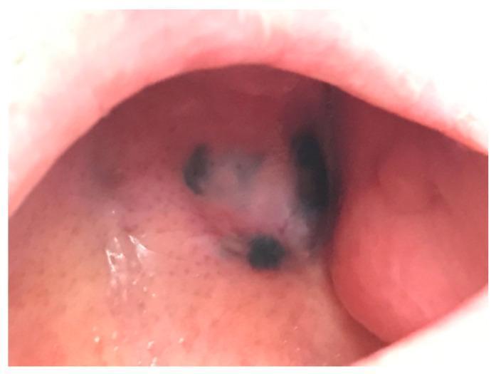
This clinical photograph shows a case of oral mucosal melanoma, an aggressive pigmented neoplasm. The image displays a large, irregular nodular lesion located on the mucosal surface of the oral cavity, likely involving the hard palate or alveolar ridge. The lesion is characterized by heterogenous pigmentation, featuring deep black to dark-brown areas interspersed with focal regions of depigmentation or amelanotic tissue. The borders of the lesion are poorly defined and asymmetric, which is a hallmark of malignancy. The surface appears slightly elevated and nodular with an irregular texture. This visual serves as an educational example of mucosal melanoma's presentation, highlighting the importance of inspecting oral pigmentation for signs of asymmetry, color variation, and nodularity. It is relevant for dental and dermatological oncology, emphasizing the distinction between benign melanotic macules and malignant mucosal lesions.
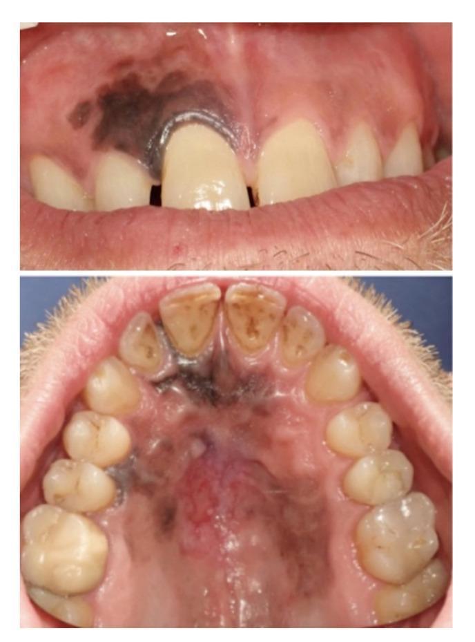
This clinical intraoral photograph set displays pigmented lesions in the maxillary gingiva and hard palate of a 45-year-old male. The upper image shows a large, irregularly shaped, dark brown-to-black macule on the anterior maxillary gingiva, primarily affecting the right central and lateral incisor regions. The pigmentation extends to the gingival margin and interdentally. The lower image provides a palatal view, revealing a more extensive, heterogeneous pigmented lesion covering a significant portion of the hard palate. This lesion exhibits variegated coloring, including deep black, brown, and bluish-grey tones, with ill-defined, asymmetric borders. A central area of erythematous change suggests focal ulceration. These clinical features are highly suspicious for mucosal melanoma. This content is intended for dental and oncology students to illustrate the clinical presentation of oral malignant melanocytic lesions, highlighting the importance of evaluating lesion color, border irregularity, and anatomical extent across the palatal and gingival mucosa.
hairy leukoplakia lateral border tongue EBV HIV white corrugated
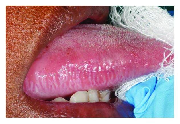
This clinical intraoral photograph displays a localized white lesion on the lateral border of the tongue, characteristic of oral hairy leukoplakia. The lesion is primarily hyperkeratotic and exhibits a distinct corrugated or 'hairy' surface texture with prominent vertical ridges. It is situated along the right lateral margin, showing a clear demarcation from the surrounding normal, pinkish-red tongue mucosa. The image provides clinical context for evaluating opportunistic infections in immunocompromised patients, specifically those with HIV/AIDS. The presence of these vertically oriented white plaques is strongly associated with Epstein-Barr virus (EBV) replication within the oral epithelium. Educational focus includes recognizing the morphology, anatomical site predilection, and clinical significance of this benign but diagnostic marker of immune suppression.
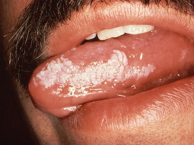
Intraoral clinical photography (high‑resolution, color-standardized) of a mucosal lesion on the tongue. The image highlights a white, shaggy, non‑scrapable plaque along the lateral border of the tongue, consistent with hairy leukoplakia caused by Epstein–Barr virus (EBV) infection in a setting of immunosuppression. Hairy leukoplakia is the most common EBV‑related lesion in HIV‑positive patients and is considered AIDS‑defining; it can be bilateral along the tongue’s lateral margins and may extend to dorsal tongue surfaces, buccal mucosa, soft palate, pharynx, or esophagus in advanced disease. Early lesions show vertical keratin streaks perpendicular to the tongue axis; later stages may appear extensive, shaggy, or corrugated, with a white or pale pink appearance and a fissured surface. The lesion does not rub off and is not a true candidiasis; its presence signals significant immune compromise and warrants HIV testing if status is unknown, or a check of CD4 count and antiretroviral therapy status. Differential diagnoses include candidiasis, true leukoplakia, lichen planus, frictional keratosis. The clinical significance lies in prognosis and management: reduce immunosuppression, treat HIV if present, monitor for dysplasia not typically associated with HL; biopsy is reserved for atypical features. This image is educational for oral medicine, infectious disease, dermatology, dentistry, and pediatrics.
oral squamous cell carcinoma tongue ulcer indurated margin
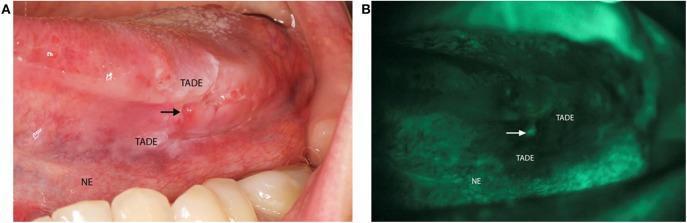
This dual-panel clinical photograph demonstrates oral squamous cell carcinoma screening using conventional white light and VELscope® tissue autofluorescence imaging. Panel A is a clinical photograph of the left lateral border of the tongue showing an indurated, erythematous ulcerated lesion (marked by an arrow) consistent with biopsy-proven squamous cell carcinoma. The surrounding area is labeled as Tumor Adjacent Dysplastic Epithelium (TADE), which appears clinically suspicious, while the more distal tissue is labeled as Normal Epithelium (NE). Panel B illustrates the same anatomical region under VELscope® examination. The malignant ulcer and the surrounding TADE exhibit significant fluorescence visualization loss (appearing as dark, non-fluorescent patches), whereas the NE maintains normal pale green autofluorescence. This comparison highlights the clinical utility of autofluorescence imaging in delineating surgical margins and identifying dysplastic changes that may not be fully apparent under standard illumination. The image serves as an educational tool for oral oncology, pathology, and diagnostic screening techniques.
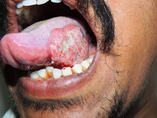
Intraoral clinical photograph of the left lateral tongue demonstrating an ulcerating, nodular mass approximately 3.0 cm in diameter. The lesion arises from the lingual mucosa along the left tongue margin and presents as a pink-erythematous, exophytic mass with focal surface ulceration. The patient is a 38-year-old male with over five years of chewing tobacco, a well-known risk factor for oral squamous cell carcinoma. The imaging modality is a standard white-light clinical photograph, acquired to document lesion morphology, localization, and size for baseline assessment and interdisciplinary discussion. The mass appears firm and indurated with surface irregularity and surrounding mucosal inflammation. Differential considerations include squamous cell carcinoma versus verrucous carcinoma or traumatic ulcer; however, the clinical history and lesion characteristics strongly support a malignant process. Definitive diagnosis requires histopathology from a biopsy, along with staging workup. This image is relevant for cancer screening, patient education, surgical and oncologic planning, and educational case discussions in otolaryngology/head-and-neck oncology. Keyword-rich descriptors: oral cancer, tongue cancer, oral SCC, left lateral tongue, intraoral lesion, ulcerating mass, tobacco use, risk factors, biopsy, histopathology, imaging documentation, staging, prognosis, therapy planning. Clinical images like this facilitate rapid recognition and timely management decisions. They also support patient counseling, multidisciplinary coordination, and education of trainees.
ORAL MEDICINE: LICHEN PLANUS, PIGMENTED LESIONS, ORAL CANCER, PHARMACOLOGY - Complete Study Notes
SECTION 1: LICHEN PLANUS & WHITE LESIONS
1. Why Does a Lesion Appear White? (2019, 22)
- Thickened keratin layer (hyperkeratosis/parakeratosis): Keratin is white and opaque; light is scattered rather than transmitted through the vascular lamina propria. This is the most common reason (leukoplakia, lichen planus, frictional keratosis, nicotine stomatitis)
- Epithelial edema/intracellular fluid accumulation: Water-logged cells scatter light (leukoedema, white sponge nevus, traumatic chemical burn)
- Fibrin coating/pseudomembrane: Dead tissue + fibrin = white coating over ulcer (candidiasis-thrush, chemical/traumatic ulcers)
- Submucosal fibrosis: Pale avascular fibrous tissue seen through thinned epithelium (OSMF)
- Reduced vascularity: Underlying ischemia makes overlying epithelium appear pale (OSMF, scar tissue)
- Necrotic tissue: Dead cells appear white/gray (necrotizing ulcerative gingivitis pseudomembrane)
- Fungal organisms: Fungal pseudohyphae + fibrin in thrush
- Calcification: Rare (calcinosis cutis)
2. Oral Lichen Planus (OLP) (2014, 23, 16, 21, 19, 22)
Definition
Enumerate Autoimmune Diseases (2014)
- Autoimmune hemolytic anemia
- Idiopathic thrombocytopenic purpura (ITP)
- Hashimoto's thyroiditis, Graves' disease
- Type 1 diabetes mellitus
- Addison's disease
- Pernicious anemia (anti-intrinsic factor)
- Myasthenia gravis
- Multiple sclerosis
- Pemphigus vulgaris, bullous pemphigoid
- Systemic lupus erythematosus (SLE)
- Rheumatoid arthritis
- Sjogren's syndrome
- Systemic sclerosis (scleroderma)
- Dermatomyositis / Polymyositis
- Mixed connective tissue disease (MCTD)
- Oral lichen planus
- Behcet's syndrome
- Psoriasis (partially autoimmune)
Etiopathogenesis of Oral Lichen Planus
- Unidentified antigen (possibly altered keratinocyte surface proteins, stress proteins - HSP-60, viral antigens) presented on MHC Class II by Langerhans cells or keratinocytes to CD4+ T helper cells
- CD4+ T cells (Th1 type) activated → release IFN-γ, TNF-α
- Chemokines (CCL5/RANTES, CXCL9, CXCL10) attract CD8+ cytotoxic T lymphocytes (CTLs) to the epithelium
- CD8+ CTLs predominate in the band-like subepithelial infiltrate
- CD8+ CTLs recognize MHC Class I-antigen complexes on basal keratinocytes
- CTLs release perforin/granzyme B → basal cell apoptosis → liquefaction degeneration of basal cell layer (called "civatte bodies" = apoptotic keratinocytes)
- Matrix metalloproteinases disrupt the basement membrane
- This produces the histological hallmarks: Band-like T-cell infiltrate + basal cell degeneration
- Stress (psychological)
- Dental materials: Amalgam (mercury - lichenoid reaction); gold, composite, cobalt
- Drugs: Beta-blockers, ACE inhibitors, NSAIDs, antimalarials, allopurinol, oral hypoglycemics → "Lichenoid drug reaction"
- Hepatitis C virus (HCV) - strong association, especially in Mediterranean populations (HCV prevalence in OLP patients: 20-40%)
- Diabetes mellitus and hypertension ("Grinspan's syndrome" - controversial triad)
- Genetic predisposition: HLA-DR1, HLA-DR9, HLA-DQ3
Clinical Features of OLP
- Interlacing white lines/striae = Wickham striae
- Lace-like, annular, or arborescent pattern
- Bilateral in almost all cases (bilaterality helps distinguish from leukoplakia)
- Usually asymptomatic
- Buccal mucosa most common site
- Small white papules (1-2 mm); usually in conjunction with reticular striae
- Homogeneous white plaques; dorsum of tongue and buccal mucosa
- Resembles leukoplakia clinically; must be distinguished
- Red, thinned mucosa (atrophied epithelium)
- Usually surrounded by reticular striae
- Symptomatic: burning, soreness, sensitivity to spicy food
- Gingiva → desquamative gingivitis (erythematous gingiva that bleeds/sloughs on gentle pressure)
- Central ulceration with pseudomembranous coating
- Surrounded by striae radiating outward
- Severely painful; eating and speaking affected
- May cause significant morbidity
- Higher malignant transformation potential
- Large bullae on buccal mucosa or tongue
- Rupture leaving erosions
- Rare; associated with more severe disease
- Buccal mucosa (most common, bilateral) > tongue (lateral border, dorsum) > gingiva (desquamative gingivitis) > palate > vermilion border of lip > floor of mouth
- Pruritic, violaceous (purple), flat-topped papules on flexor surfaces of wrists, ankles
- The 4 P's: Purple, Polygonal, Pruritic, Papules
- Wickham striae visible on skin surface with oil immersion
- Involvement of scalp (lichen planopilaris - scarring alopecia), nails (nail dystrophy)
Histopathology
- Saw-tooth rete ridges (pointed, irregular rete ridges)
- Band-like (lichenoid) lymphocytic infiltrate in the superficial connective tissue - closely hugs the epithelium
- Liquefaction (vacuolar) degeneration of basal cell layer - loss of normal columnar basal cells
- Civatte bodies (Colloid/Hyaline bodies) - apoptotic eosinophilic keratinocytes in basal layer and lamina propria
- Hyperkeratosis / Parakeratosis - thickened keratin layer
- In erosive type: Subepithelial clefting (Max-Joseph spaces)
- Non-specific: Fibrinogen deposits along basement membrane (shaggy fluorescence)
- Unlike pemphigus (fishnet IgG) and pemphigoid (linear IgG at BMZ)
Differential Diagnosis of OLP
| Condition | Differentiating Features |
|---|---|
| Leukoplakia | Unilateral usually; no reticular pattern; no bilateral striae; biopsy shows dysplasia |
| Lichenoid drug reaction | Drug history; often unilateral; resolves on drug withdrawal |
| Mucous membrane pemphigoid | Desquamative gingivitis; subepithelial blisters; DIF = linear IgG at BMZ |
| SLE | Discoid lesion with central atrophy; systemic features; ANA positive |
| White sponge nevus | Familial; generalized; from childhood; biopsy diagnostic |
| Oral candidiasis (hyperplastic) | Non-scrapable white; culture positive; antifungal responsive |
| GVHD (Graft vs Host Disease) | History of bone marrow transplant; resembles OLP |
| Hairy leukoplakia | Lateral tongue; HIV patient; EBV positive; vertical ridges; can't scrape |
Malignant Transformation
- Overall rate: ~0.5-1% per year (1-2% lifetime)
- Higher in erosive/atrophic type, plaque type
- Tongue, floor of mouth sites at higher risk
- WHO considers OLP a potentially malignant disorder
- Long-term surveillance mandatory (6-12 monthly)
Management of OLP (2008, 2009, 2014, 16, 21)
Asymptomatic Reticular OLP
- Reassurance + explanation
- Identify and remove triggers (amalgam, drugs)
- Regular review (6-monthly)
- No treatment required
Symptomatic (Erosive/Atrophic OLP) - Step-up Approach
- Triamcinolone acetonide 0.1% in Orabase (Kenalog) - applied 3-4× daily to affected area
- Clobetasol propionate 0.05% gel/ointment - stronger; for severe erosive OLP
- Betamethasone mouthwash or pellets (dissolve in water as mouthwash)
- Avoid swallowing; rinse with water after; risk of oral candidiasis with prolonged use (give antifungal prophylaxis)
- Tacrolimus 0.1% ointment - effective; steroid-sparing
- Cyclosporin 100 mg/mL oral rinse
- Black box warning: Theoretical malignant transformation risk
- Prednisolone 0.5-1 mg/kg/day for 1-2 weeks then taper
- For severe/widespread oral or mucocutaneous OLP
- Risks: Cushingoid effects, adrenal suppression, osteoporosis with long-term use
- Azathioprine, mycophenolate mofetil (steroid-sparing)
- Hydroxychloroquine (particularly for erosive)
- Methotrexate (for refractory)
- Analgesic mouthwashes (benzydamine, lidocaine viscous)
- Antifungal prophylaxis if on topical steroids (nystatin/fluconazole)
- Treatment of HCV if associated
3. Hairy Leukoplakia (Short Note - 2015, 22)
Definition
Features
- Site: Lateral border of the tongue (almost exclusively); may be bilateral
- Appearance: White, corrugated ("hairy"), non-scrapable plaques with vertical ridges/folds
- Cannot be scraped off (unlike thrush) - critical distinguishing feature
- Painless - discovered incidentally
Association
- HIV/AIDS (most common - CD4 <200/mm³ predicts faster HIV progression to AIDS)
- Other immunosuppression: Transplant recipients, chemotherapy, corticosteroids
- In pre-ART era: Marker of HIV disease progression
- Still requires HIV testing if found in patient of unknown HIV status
Histopathology
- Hyperparakeratosis with "balloon cells" (vacuolated koilocyte-like cells) in upper spinous layer
- EBV viral particles detectable by in-situ hybridization (EBER)
- No dysplasia (not premalignant)
Management
- No treatment needed if asymptomatic in HIV-positive patient
- ART (antiretroviral therapy): Suppresses HIV → immune recovery → lesion resolves
- Topical/systemic antiviral (acyclovir, ganciclovir, valacyclovir): May reduce lesion size transiently but recurs on stopping
- Monitor for superimposed candidal infection
4. Nicotina Stomatitis (Short Note - 2012)
Definition
Clinical Features
- Hard palate shows diffuse whitish/gray keratosis with characteristic small red dots (inflamed minor salivary gland duct openings)
- Palate appears "cobblestoned" or cracked
- Usually asymptomatic
- Lesion is NOT premalignant (unlike reverse smoking variety)
Significance
- Nicotine stomatitis from conventional smoking: NOT premalignant (reactive keratosis)
- Reverse smoking (lighted end inside mouth) as practiced in India/Philippines: The palatal lesions DO carry malignant potential and dysplasia may develop
- Management: Stop smoking; lesion usually resolves spontaneously
SECTION 2: PIGMENTED LESIONS OF ORAL MUCOSA
5. Classification of Pigmented Lesions of Oral Mucosa (2019, 21)
A. Exogenous (Non-Melanotic)
- Amalgam tattoo - most common
- Graphite tattoo - pencil injury to palate
- Heavy metal pigmentation: Lead line (Burton's line on gingiva), bismuth, arsenic, mercury (blue-gray gingival pigmentation)
- Drug-induced: Minocycline (blue-gray), antimalarials (chloroquine), zidovudine, ketoconazole
B. Endogenous - Melanotic
- Racial/ethnic melanosis (physiological pigmentation - symmetric, diffuse, brown)
- Post-inflammatory pigmentation
C. Vascular
- Hemangioma (red-purple, blanches on pressure)
- Varix/Varicosity
- Purpura, petechiae, ecchymosis (non-blanching)
- Kaposi's sarcoma (red-purple nodules - AIDS-related)
6. Oral Malignant Melanoma (2019, 21)
Definition
Epidemiology
- Rare (0.2-8% of all oral malignancies)
- Males > females; usually >40 years
- More common in Japan and Africa
- Most common site: Hard palate > maxillary gingiva > buccal mucosa > mandibular gingiva
- No relation to sunlight exposure (unlike cutaneous melanoma)
Etiopathogenesis
- Arises from melanocytes present in oral mucosal epithelium (most numerous in hard palate and maxillary gingiva)
- Exact cause unknown
- Precursor lesion: Oral melanotic macule or oral melanosis may rarely undergo malignant transformation; but most arise de novo
- Risk factors: Racial/ethnic melanosis, smoking (controversial), dental irritation
Clinical Features
Primary Melanoma (Most are Pigmented)
- Asymmetric brown-black-gray macule/patch that progressively enlarges
- Color heterogeneity: Multiple colors - black, brown, blue, gray, red, white (amelanotic areas)
- Irregular borders: Poorly defined, ragged edges
- Nodularity: May become elevated, nodular, or ulcerated with time
- Pain: Usually painless initially; pain develops with ulceration or bone invasion
- Bleeding: Ulceration causes bleeding; friable surface
- Tooth mobility: Bone invasion causes mobility
- Satellite lesions: Small pigmented macules surrounding the main lesion
- Asymmetry
- Border irregularity
- Color variation
- Diameter >6 mm (or growing)
- Evolution (change over time)
Types of Oral Melanoma
- In situ melanoma: Radial growth phase; flat, pigmented macule; better prognosis
- Invasive melanoma: Vertical growth phase; nodular component; poorer prognosis
Stages (Prasad's Staging)
- Stage I: Purely intramucosal (in situ)
- Stage II: Invasive with no regional lymph node metastasis
- Stage III: Regional lymph node metastasis
- Stage IV: Distant metastasis
Investigations
- Incisional biopsy - from most nodular/suspicious area; not excisional (may seed)
- Histopathology: Malignant melanocytes at various epithelial levels; marked pleomorphism; melanin pigment; abnormal mitoses; Pagetoid spread (cells within epithelium); S-100, HMB-45, Melan-A, SOX10 immunohistochemistry markers
- Immunohistochemistry: S-100, HMB-45, Melan-A (MART-1) - confirms melanocytic origin
- CT chest, abdomen, pelvis: For staging (metastases)
- MRI head and neck: For local extent, bone invasion, neck nodes
- PET scan: For distant metastases
- SLNB (Sentinel lymph node biopsy): For staging
Management
- Wide surgical excision with 2 cm margins (minimum) - primary treatment
- Partial/total maxillectomy or mandibulectomy if bone involved
- Neck dissection for nodal disease
- Radiotherapy: Adjuvant post-surgery; not curative alone
- Immunotherapy (modern - first-line for advanced):
- Checkpoint inhibitors: Pembrolizumab (anti-PD-1), nivolumab - significant survival benefit
- Ipilimumab (anti-CTLA-4)
- Targeted therapy: BRAF inhibitors (dabrafenib + trametinib) if BRAF V600E mutation present
- Chemotherapy: Dacarbazine - limited response; mainly palliative now
- Prognosis: Very poor; 5-year survival <20-25%; oral melanoma is more aggressive than cutaneous
7. Amalgam Tattoo (Short Note - 2014)
Definition
Clinical Features
- Site: Gingiva, alveolar mucosa (adjacent to amalgam restorations); buccal mucosa, floor of mouth
- Appearance: Flat, blue-gray or black macule; well-defined; typically <1 cm
- Painless, non-progressive (stable, does not grow)
Diagnosis
- Clinical diagnosis in most cases
- Radiograph: May show small radiopaque particles within soft tissue
- If large particles: Visible on periapical film
- Biopsy if uncertain (to rule out melanoma or melanotic nevus)
Histopathology
- Amalgam particles (black granules) scattered in connective tissue
- Along collagen fibers and blood vessels
- May have inflammatory infiltrate or foreign body giant cell reaction
Management
- No treatment required (benign)
- If cosmetically unacceptable: Surgical excision
- Must be distinguished from melanoma (melanoma grows, changes color, irregular borders; amalgam tattoo is stable)
8. Peutz-Jeghers Syndrome (Short Note - 2020s)
Definition
Genetics
- STK11/LKB1 gene mutation (serine/threonine kinase 11) on chromosome 19p13.3
- Autosomal dominant inheritance
Oral/Skin Features
- Multiple brown-black melanotic macules (lentigines) on:
- Lips (especially lower lip, vermilion border) - most distinctive
- Buccal mucosa
- Perioral skin (around mouth, nose)
- Fingers and toes (periungual)
- Tongue, palate
- Lesions may fade after puberty (especially skin but oral lesions persist)
- Oval or round, 1-5 mm macules
GI Features
- Hamartomatous polyps in small intestine (jejunum most common), stomach, colon
- Polyps can cause: Intussusception, obstruction, bleeding, abdominal pain
Cancer Risk
- Significantly increased risk of GI cancers (small bowel, colon, stomach)
- Extraintestinal: Breast, ovarian, cervical, uterine, testicular (Sertoli cell tumor), pancreatic cancer
- Overall lifetime cancer risk: ~90%
Management
- Genetic testing for STK11 mutation
- Regular surveillance: Upper and lower GI endoscopy (from age 8-10)
- Polypectomy for large polyps
- Surveillance for extraintestinal malignancies
- No treatment for pigmented macules (benign)
9. Hairy Tongue (Short Note - 2013)
Definition
Causes (Predisposing Factors)
- Broad-spectrum antibiotics (disrupts oral flora; allows chromogenic bacteria/fungi to proliferate)
- Bismuth-containing products (Pepto-Bismol → black hairy tongue)
- Hydrogen peroxide mouthwashes
- Smoking and tobacco
- Heavy tea/coffee consumption (staining)
- Poor oral hygiene
- Xerostomia
- Head and neck radiation
- Immunosuppression
Clinical Features
- Elongated, matted, hair-like projections on dorsal tongue
- Color varies: Black (black hairy tongue), brown, yellow, green (depending on chromogenic bacteria, food pigments, tobacco)
- May cause: Gagging sensation, nausea, altered taste (dysgeusia), halitosis
- Usually asymptomatic otherwise
Management
- Identify and remove causative factors
- Good oral hygiene - tongue scraping/brushing
- Stop antibiotics if implicated; use probiotics
- Antifungals if Candida associated
- Spontaneous resolution usually after removing cause
SECTION 3: ORAL CANCER
10. TNM Classification of Malignancy of Oral Cavity (2017, 23)
The TNM System (AJCC/UICC 8th Edition, 2017)
T - Primary Tumor
| Stage | Criteria |
|---|---|
| Tx | Primary tumor cannot be assessed |
| T0 | No evidence of primary tumor |
| Tis | Carcinoma in situ |
| T1 | Tumor ≤2 cm; DOI (depth of invasion) ≤5 mm |
| T2 | Tumor ≤2 cm with DOI >5 mm and ≤10 mm, OR tumor >2 cm and ≤4 cm with DOI ≤10 mm |
| T3 | Tumor >4 cm, OR any tumor with DOI >10 mm |
| T4a | Moderately advanced: Invades cortical bone, maxillary sinus, skin of face |
| T4b | Very advanced: Invades masticator space, pterygoid plates, skull base, or encases carotid artery |
N - Regional Lymph Nodes
| Stage | Criteria |
|---|---|
| Nx | Cannot be assessed |
| N0 | No regional lymph node metastasis |
| N1 | Metastasis in a single ipsilateral node, ≤3 cm, ENE (-) |
| N2a | Metastasis in single ipsilateral node >3 cm but ≤6 cm, ENE (-) |
| N2b | Metastasis in multiple ipsilateral nodes, all ≤6 cm, ENE (-) |
| N2c | Metastasis in bilateral or contralateral nodes, all ≤6 cm, ENE (-) |
| N3a | Metastasis in any node >6 cm, ENE (-) |
| N3b | Metastasis in any node with ENE (+) |
M - Distant Metastasis
| Stage | Criteria |
|---|---|
| M0 | No distant metastasis |
| M1 | Distant metastasis present |
Overall Stage Grouping
| Stage | TNM |
|---|---|
| Stage I | T1N0M0 |
| Stage II | T2N0M0 |
| Stage III | T3N0M0 or T1-3 N1M0 |
| Stage IVA | T4a, N0-1, M0 or T1-4a, N2, M0 |
| Stage IVB | T4b, any N, M0 or any T, N3, M0 |
| Stage IVC | Any T, any N, M1 |
11. Carcinoma of the Tongue - Diagnosis and Treatment (2009)
Clinical Features
- Most common site: Lateral border and ventral surface of tongue (anterior 2/3 - oral tongue)
- Early: Painless ulcer, white/red patch, indurated nodule
- Late: Painful ulcer with indurated, everted margins; fixation to floor of mouth; trismus; cervical lymphadenopathy
- Risk factors: Tobacco, alcohol (synergistic), betel nut, HPV-16/18, oral submucous fibrosis, syphilitic glossitis, iron deficiency
Diagnosis
- Incisional biopsy + histopathology (definitive)
- CT neck with contrast: Primary tumor extent, cortical bone invasion, neck nodes
- MRI: Soft tissue extent, perineural invasion, tongue base involvement
- PET-CT: Distant metastases staging
- FNAC of cervical nodes: Cytological confirmation
- Direct laryngoscopy + panendoscopy: Second primary tumors of upper aerodigestive tract
Treatment (Based on Stage)
| Stage | Treatment |
|---|---|
| Stage I-II | Surgery alone (hemiglossectomy) ± selective neck dissection |
| Stage III-IVA | Surgery (hemiglossectomy/total glossectomy + neck dissection) + adjuvant RT ± chemotherapy |
| Stage IVB-C | Chemoradiotherapy (concurrent cisplatin + RT); palliative |
SECTION 4: PHARMACOLOGY
12. Antifungal Drugs (2018)
Classification of Antifungal Drugs
A. Polyene Macrolides
| Drug | Route | Uses |
|---|---|---|
| Nystatin | Topical only (too toxic for systemic) | Oral candidiasis (mouthwash/pastilles/gel); skin candidiasis; NOT absorbed from GI tract |
| Amphotericin B | IV (systemic); also topical | Invasive/disseminated candidiasis, cryptococcal meningitis, aspergillosis, severe fungal infections |
| Amphotericin B lipid formulations | IV | Reduced nephrotoxicity; ABLC, ABCD, liposomal AmB (L-AmB) |
B. Azoles (Inhibit Ergosterol Synthesis)
| Drug | Use |
|---|---|
| Clotrimazole | Oral candidiasis (lozenges/cream); vaginal candidiasis |
| Miconazole | Topical oral gel; denture adhesive with antifungal |
| Ketoconazole | Oral; now mainly topical (hepatotoxic); seborrheic dermatitis shampoo |
| Drug | Use |
|---|---|
| Fluconazole | Drug of choice for oral/esophageal/vaginal candidiasis; 100-200 mg/day oral; well tolerated; single 150 mg for vaginal candidiasis |
| Itraconazole | Fluconazole-resistant candidiasis; dermatophytes; histoplasmosis; requires acid for absorption |
| Voriconazole | Invasive aspergillosis (first-line); fluconazole-resistant Candida |
| Posaconazole | Aspergillus prophylaxis; zygomycosis |
| Isavuconazole | Aspergillosis, mucormycosis |
C. Echinocandins (Inhibit Cell Wall)
- Drugs: Caspofungin, Micafungin, Anidulafungin
- Uses: Invasive candidiasis (especially for Candida krusei, C. glabrata resistant to azoles); aspergillosis
- Route: IV only (not oral bioavailability)
- Well tolerated; minimal drug interactions
D. Allylamine (Inhibit Ergosterol Synthesis - Different Step)
- Terbinafine: Oral/topical; onychomycosis (nail infection), tinea; excellent for dermatophytes
E. Flucytosine (5-FC)
- Uses: Candida, Cryptococcus (combined with AmB)
- Not used alone (rapid resistance develops)
- Toxicity: Bone marrow suppression
F. Griseofulvin
- Uses: Dermatophyte infections (ringworm, tinea capitis)
- Given with fatty food (increases absorption)
Summary Table for Dentistry
| Indication | Drug of Choice |
|---|---|
| Mild oral candidiasis (immunocompetent) | Nystatin mouthwash OR clotrimazole troches |
| Moderate-severe oral candidiasis | Fluconazole 100-200 mg/day × 7-14 days |
| Denture stomatitis | Miconazole gel; nystatin; treat denture with antifungal |
| Angular cheilitis | Miconazole cream or nystatin cream |
| HIV candidiasis (CD4 <200) | Fluconazole systemic |
| Fluconazole-resistant candidiasis | Itraconazole, voriconazole, caspofungin |
13. Antibiotics in Dentistry (2012, 2013)
Classification
| Drug | Use in Dentistry |
|---|---|
| Amoxicillin | First-line dental infections (mild-moderate); 500 mg TDS × 5 days; periodontal disease |
| Amoxicillin + Clavulanate (Co-amoxiclav) | Beta-lactamase producing organisms; severe odontogenic infection |
| Penicillin V | Odontogenic infections; infective endocarditis prophylaxis historically |
| Ampicillin | Broader spectrum; IV for severe infections |
| Drug | Use in Dentistry |
|---|---|
| Azithromycin | Penicillin allergy alternative; 500 mg OD × 3-5 days; good tissue penetration |
| Clarithromycin | Penicillin-allergic patients; twice daily |
| Erythromycin | Older option; penicillin allergy; GI side effects |
| Drug | Use in Dentistry |
|---|---|
| Metronidazole | Anaerobic bacteria (dominant in periodontal, endodontic, pericoronitis); ANUG/ANUP; often combined with amoxicillin; 400 mg TDS × 5 days |
| Tinidazole | Similar to metronidazole; once daily; better tolerated |
| Drug | Use in Dentistry |
|---|---|
| Doxycycline | Periodontal disease (host modulation - inhibits MMP at sub-antimicrobial dose); Actinomyces; NOT in children <8 yrs (tooth staining) |
| Minocycline | Periodontal; sub-antimicrobial dose capsules |
- Penicillin-allergic patients; bone infections (good bone penetration)
- Anaerobic coverage; IE prophylaxis in penicillin allergy
- Risk: Clostridioides difficile colitis (pseudomembranous colitis)
- Ciprofloxacin: Actinomycosis; Gram-negative infections
- Moxifloxacin: Broad-spectrum; dental infections when other agents fail
- Gentamicin: Combined with amoxicillin for IE prophylaxis in high-risk patients
Infective Endocarditis (IE) Prophylaxis (Current NICE/AHA Guidelines)
- AHA/NICE 2007 guideline revision: Prophylaxis NOT routinely recommended for dental procedures for most patients
- Only for high-risk cardiac conditions: Prosthetic valves, previous IE, congenital cyanotic heart disease (unrepaired)
- Amoxicillin 2 g oral 30-60 min before procedure (or ampicillin IV/IM)
- Penicillin allergy: Clindamycin 600 mg (UK/older) or azithromycin 500 mg or clarithromycin 500 mg
14. Corticosteroids in Dentistry (2019, 22)
Classification
- Triamcinolone acetonide 0.1% (Kenalog in Orabase) - OLP, RAS, pemphigus
- Clobetasol propionate 0.05% gel - severe erosive OLP
- Betamethasone mouthwash (0.5 mg/5 mL) - OLP, pemphigus
- Fluocinolide 0.05% gel
- Triamcinolone acetonide 10-40 mg/mL (Kenalog) injection
- Uses: OSMF, OLP, recurrent aphthae, hemangioma, epulis
- Prednisolone, dexamethasone, hydrocortisone
- Methylprednisolone (IV pulse)
Uses of Corticosteroids in Dentistry
| Condition | Corticosteroid Use |
|---|---|
| Oral Lichen Planus | Topical (triamcinolone, clobetasol), systemic prednisolone for severe |
| Pemphigus vulgaris | Systemic prednisolone (1-2 mg/kg/day) - cornerstone |
| MMP (mucous membrane pemphigoid) | Topical + systemic |
| Erythema multiforme | Systemic short course prednisolone |
| Aphthous stomatitis | Topical triamcinolone; systemic for major |
| OSMF | Intralesional triamcinolone + hyaluronidase |
| Dental anxiety / premedication | Dexamethasone pre-operatively |
| Post-extraction swelling | Dexamethasone 8 mg pre-op (especially wisdom teeth) |
| Dry socket pain | Steroid-impregnated dressings |
| Bell's palsy | Prednisolone 60 mg/day × 5 days then taper |
| Temporal arteritis | High-dose prednisolone (to prevent blindness) |
| Pulpitis (pulp capping - historical) | Corticosteroid-antibiotic paste |
Precautions / Side Effects in Dentistry
- Adrenal suppression in patients on long-term steroids → risk of adrenal crisis during surgery → steroid cover (doubling dose rule)
- Oral candidiasis with topical steroids → antifungal prophylaxis
- Impaired healing, increased infection risk
- Mask signs of infection
- Osteoporosis with long-term systemic use
Steroid Cover for Dental Surgery
- Minor procedure: Double their morning steroid dose
- Major surgery: IV hydrocortisone 25-50 mg pre-op then taper
- Must not stop steroids abruptly (Addisonian crisis risk)
15. Exfoliative Cytology (Short Note - 2008)
Definition
Technique
- Lesion surface cleaned with gauze (remove debris)
- Scrape with a metal spatula, tongue depressor, or Orcellex brush
- Smear immediately on labelled glass slide
- Fix immediately in 95% ethyl alcohol (or spray fixative) - to prevent drying artifact (air drying causes changes)
- Stain: Papanicolaou stain (PAP stain); also PAS, H&E
Cytological Classes (Papanicolaou System)
- Class I: Normal
- Class II: Atypical (inflammatory changes)
- Class III: Dysplastic cells (equivocal)
- Class IV: Strongly suspicious for malignancy
- Class V: Malignant
Uses in Oral Pathology
- Screening for malignancy (oral leukoplakia, erythroplakia)
- Diagnosis of candidiasis (KOH/PAS staining of scraping)
- Herpes simplex / herpes zoster (Tzanck smear - multinucleated giant cells)
- Pemphigus (Tzanck smear - acantholytic cells)
- Monitoring response to treatment
Limitations
- Not a substitute for biopsy - lower sensitivity and specificity
- Cannot assess tissue architecture (only surface cell morphology)
- False negatives possible (subsurface lesions may not exfoliate)
- Requires expertise for interpretation
16. Lip Print Analysis (Cheiloscopy) (Short Note - 2015)
Definition
Classification (Suzuki and Tsuchihashi)
- Type I: Clear-cut grooves (vertical lines) running across entire lip
- Type I': Partial vertical grooves
- Type II: Branching grooves (fork-like)
- Type III: Intersecting/crossing grooves (rhombus pattern)
- Type IV: Reticular (irregular network)
- Type V: Undetermined
Forensic Uses
- Identification of individuals in forensic investigation (crime scenes, disasters)
- Lip prints found on cups, glasses, documents, cigarettes at crime scenes
- Sex determination: Female: Types I and I' predominate; Males: Types III and IV predominate
- Age estimation (limited accuracy)
- Part of forensic odontology
Collection Methods
- Lipstick method (apply lipstick, press lips on paper)
- Direct photography
- Cyanoacrylate fuming (latent lip prints)
- Fluorescent powder dusting
Summary Quick Reference
| Topic | Key Points |
|---|---|
| Oral lichen planus | CD8+ T cell-mediated; Wickham striae; bilateral; types: reticular/papular/plaque/atrophic/erosive/bullous; DIF = fibrinogen at BMZ; topical clobetasol/triamcinolone first-line |
| Oral malignant melanoma | Rare, aggressive; hard palate/maxillary gingiva; ABCDE rule; S-100/HMB-45/Melan-A +; wide excision + immunotherapy; <20-25% 5-year survival |
| Amalgam tattoo | Most common oral pigmented lesion; stable blue-gray macule; no treatment |
| Peutz-Jeghers | STK11 mutation; perioral melanotic macules + GI polyps + cancer risk |
| TNM oral cavity | T1(≤2cm,DOI≤5mm) to T4b (skull base); N0-N3b (ENE now included); Stage I-IVC |
| Antifungals | Nystatin (topical only); Azoles (fluconazole = first choice oral); Echinocandins (cell wall); AmB (invasive disease) |
| Antibiotics in dentistry | Amoxicillin (1st line); Metronidazole (anaerobes); Clindamycin (penicillin allergy + bone infections); IE prophylaxis = amoxicillin 2 g |
| Corticosteroids in dentistry | OLP, pemphigus, OSMF (intralesional); pre-op dexamethasone (3rd molar); steroid cover for adrenal suppression patients |
| Exfoliative cytology | Papanicolaou stain; scrape and fix immediately; screening only - not substitute for biopsy |
| Hairy leukoplakia | EBV; lateral tongue; non-scrapable; HIV marker; treat with ART |
QUESTIONS: 1. Classify salivary gland diseases. Discuss in detail about etiology, clinical features, investigation and treatment of Sjogren's syndrome. (20) SHORT NOTES: 1. Sjogrens Syndrome. (2017,2019) 2. Ramsay-Hunt's Syndrome. (2017) 3. Xerostomia. (11,14) DIABETES MELLITUS AND ENDOCRINE DISEASE: LONG QUESTION: 1. Classi Diabetes. Describe the etiopathogenesis, C/F, Investigation & treatment of the same. (2017) 2. What is the manifestation of diabetes mellitus? Describe the dental management of such patients.. (12) SHORT NOTE: 1. Oral manifestation of diabetes mellitus. (12,14) 2. Corticosteroid in dentistry. (09,16) 3. Discuss Clinicopathological features of hyperparathyroidism. (06) 4.. Hyperparathyroidism (21) OROFACIAL PAIN: LONG QUESTION: I. SHORT NOTE: Describe in details the common cause of orofacial pain and its management. (13) Burning mouth syndrome. (12) Myofascial pain dysfunction syndrome (2023) INFECTIOUS DISEASE: LONG QUESTION: 1. Describe herpes simplex infection with its management. (13) SHORT NOTE: 1. Bacterial Endocarditis. (2018) 2. Cellulitis. (2017) 3. Phoenix Abscess. (15) 4. Features of congenital syphilis. (11,15)| 5. Oral manifestation of AIDS. (06) 6. Herpes simplex. (14,16) 7. Oral manifestation of secondary syphilis. (14) 8. AIDS. (12) 9. Gumma. (20s) 10. Oral manifestation of AIDS (20) NEUROMUSCULAR DISEASE: LONG QUESTION: 1. Classify facial pain. Write in details about the causes, clinical features, differential diagnosis and treatment of Trigeminal Neuralgia.
Sjogren syndrome dry eyes dry mouth salivary gland lymphocytic infiltration
A diagnostic B-mode ultrasound image of a major salivary gland, likely the parotid gland, demonstrating severe pathological changes characteristic of chronic inflammatory conditions such as Sjögren's syndrome. The image exhibits Grade 3 severe architectural distortion, characterized by a diffuse, inhomogeneous parenchymal echotexture. Multiple well-defined anechoic to hypoechoic (darker) areas are distributed throughout the entire surface of the gland, representing lymphocytic infiltration or cystic dilatation of the ducts. These dark regions are separated and surrounded by hyperechoic (bright) linear bands, which represent fibrotic tissue or thickened septa. The glandular margins are poorly defined as the abnormal echogenicity extends into the surrounding soft tissue. This visual pattern is a key diagnostic indicator in rheumatology and oral medicine for evaluating the extent of glandular destruction and assessing systemic autoimmune disease involvement.
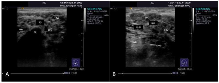
This diagnostic image displays two grayscale ultrasound frames (A and B) showing advanced-stage Sjögren's Syndrome in the major salivary glands. Panel A depicts the left parotid gland (PG), characterized by a markedly heterogeneous parenchyma. The normal glandular echotexture is replaced by numerous hypoechoic to anechoic spot-like formations and larger space-occupying lesions (SoL), representing lymphocytic infiltration or sialectatic ducts. Anatomical landmarks include the masseter muscle (MM) and the mandible (M), which presents with a sharp acoustic shadow. Panel B illustrates the left submandibular gland (SMG), showing similar diffuse heterogeneity and parenchymal rarefaction with multiple internal anechoic spots. Surrounding structures identified include the mylohyoid muscle (MHM) located superiorly, the tongue (TONG), and the tonsillar region (TONS). Both glands demonstrate pathologic enlargement. This image serves as a clinical example of chronic autoimmune sialadenitis where secretory tissue is progressively replaced by cystic and solid inflammatory changes.
trigeminal neuralgia trigger zone facial pain V2 V3 distribution
Diagnostic imaging montage featuring multi-planar MRI scans (axial, coronal, and sagittal views) for dosimetric planning of repeat Gamma Knife Radiosurgery (GKRS) in a 48-year-old male with recurrent right trigeminal neuralgia (V3 distribution). The images illustrate the spatial relationship between two separate treatment targets along the cisternal segment of the right trigeminal nerve. A blue marker indicates the initial target from October 2010 (39 Gy at the 50% isodose line, 0.1 cc volume). A yellow marker identifies the second target from March 2016 (25 Gy at the 50% isodose line, 0.1 cc volume), following pain recurrence in August 2015. The second procedure demonstrates an intentional 'Anterior Target Shift' relative to the original treatment site to avoid excessive cumulative radiation dose to the same nerve segment while addressing the recurrence. The MRI sequences provide high-resolution visualization of the brainstem, cerebellopontine angle, and nerve root entry zone, essential for precise stereotactic localization in neurosurgical oncology and pain management.
This clinical photograph illustrates a medical procedure for trigeminal neuralgia (TN) management. The image displays a close-up of a patient's lower face, specifically the cheek and mandibular region. A hand is seen holding an injection needle, which is being inserted into the skin. A black, hand-drawn grid-like pattern is visible on the surface of the cheek within an oval border, used as a topographical guide for precise intradermal administration. This grid serves to standardize the distribution of the therapeutic agent, identified as incobotulinumtoxin A (Xeomin), at a concentration of 50 units per square centimeter. The visual demonstrates the practical application of botulinum toxin as an interventional pain management technique for neurological facial pain conditions. Key anatomical landmarks include the perioral area, nasolabial fold, and mandible, indicating the target zone for local nerve-related symptom relief.
primary herpes simplex gingivostomatitis oral vesicles ulcers gingival
A clinical photograph of the oral cavity focusing on the lateral and dorsal aspect of the tongue, illustrating features of primary herpetic gingivostomatitis. The image demonstrates a cluster of multiple small, discrete, and coalescing whitish-yellow vesicles and ulcers located on the lateral border of the tongue. These lesions are slightly raised with irregular margins. A white arrow highlights a prominent ulcerated area within the cluster. The surrounding mucosa is markedly erythematous and inflamed, providing a sharp contrast to the lighter-colored lesions. This visual represents the acute phase of a viral infection (Herpes Simplex Virus Type 1), focusing on the presentation of painful intraoral vesicles that rupture to form fibrin-covered ulcers. It is a key educational example for dental and medical students in recognizing oral manifestations of primary viral stomatitis and differentiating it from other ulcerative conditions like aphthous ulcers.
This clinical photograph shows the labial mucosa of the upper and lower lips, demonstrating characteristic signs of primary herpetic gingivostomatitis. The image reveals multiple, discrete and coalescing vesicles and small, shallow ulcers. On the upper labial mucosa near the oral commissure, a cluster of small, whitish lesions is visible, marked by an arrow. On the lower lip, similar lesions are present, with some areas showing coalescence into larger, irregular ulcerations with yellowish-white fibrinopurulent bases. All lesions are surrounded by a distinct erythematous halo, indicating acute inflammation. The presentation is typical for an initial infection with Herpes Simplex Virus (HSV), illustrating the transition from vesicles to painful ulcerations on non-keratinized oral mucosa. This visual is suitable for dental and medical education regarding viral orofacial infections and differential diagnosis of vesiculobullous diseases.
oral manifestations AIDS HIV Kaposi sarcoma hairy leukoplakia candidiasis
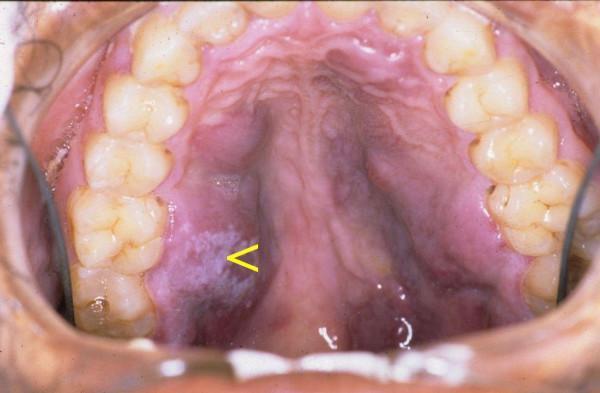
This clinical intraoral photograph displays the hard palate and maxillary dentition of a patient, demonstrating classic oral manifestations of advanced immunodeficiency. The primary findings are multifocal, irregular, purple-to-red maculopapular lesions consistent with Kaposi sarcoma (KS). These vascular lesions exhibit poorly defined margins and vary in texture, appearing as both flat discolorations and slightly elevated plaques across the palatal mucosa. Additionally, a distinct white, patchy, friable-looking area is visible on the left side of the hard palate (indicated by a yellow arrow), characteristic of pseudomembranous candidiasis. The concurrent presentation of oral Kaposi sarcoma and candidiasis is highly suggestive of HIV/AIDS-related complications. The photograph serves as an educational resource for identifying opportunistic infections and neoplastic conditions within the oral cavity, highlighting the clinical appearance of vascular tumors and fungal overgrowth in immunocompromised individuals.
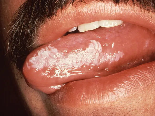
Hairy leukoplakia (HL) of the oral mucosa is a chronic EBV-driven epithelial lesion seen most commonly in severely immunosuppressed individuals, particularly people living with HIV/AIDS. It presents as a painless, pale white plaque on the tongue, usually along the lateral borders, with a shaggy, corrugated surface that cannot be scraped off. In early stages, one notes vertical keratin streaks or fissures oriented perpendicular to the tongue’s long axis. In advanced disease, the involvement can extend to the dorsal tongue, bilateral lateral surfaces, buccal mucosa, soft palate, pharynx, or even the esophagus. HL is considered an AIDS-defining opportunistic infection and serves as a clinical marker of severe immunosuppression; however, it does not confer increased risk for dysplasia or squamous cell carcinoma, unlike true leukoplakia. Clinically, HL must be differentiated from oral candidiasis, true leukoplakia, lichen planus, and other mucosal white plaques. The diagnosis is typically clinical but may be confirmed by EBV in situ hybridization or PCR if tissue is obtained; HIV management with antiretroviral therapy often leads to regression. This lesion carries educational significance for clinicians, dentists, and trainees, illustrating the spectrum of EBV-related oral disease in HIV, transplant recipients, and occasionally immunocompetent hosts. Recognition aids diagnosis and treatment planning.
congenital syphilis Hutchinson incisors mulberry molars saddle nose
This clinical photograph displays a 5-month-old infant with systemic features indicative of a congenital infectious or metabolic syndrome, specifically congenital syphilis in this clinical context. Facial dysmorphism is prominent, including frontal bossing (prominent forehead), a depressed nasal bridge (saddle nose deformity) with a rounded nasal tip, a thin upper lip, and a small chin (micrognathia). The infant exhibits signs of significant nutritional compromise and malabsorption, characterized by pale skin, sparse hair on the scalp, and sparse eyebrows. The abdomen is markedly globose and distended, suggesting hepatosplenomegaly or ascites. The limbs appear dystrophic with evidence of muscle wasting and diminished subcutaneous fat. A medical bandage is visible on the left foot, likely for intravenous access or monitoring. This image serves as an educational reference for recognizing the systemic manifestations and physical stigmata of neonatal infectious diseases and associated growth failure.
This composite clinical photograph displays the primary phenotypic manifestations of Nance-Horan syndrome (NHS) in a 15-year-old male. Panel A consists of slit-lamp photographs demonstrating bilateral congenital nuclear cataracts, characterized by central lens opacification. Panels B, C, and D show oral clinical findings. Panel B highlights screwdriver-shaped (tapered) permanent incisors with narrowed incisal edges. Panel C identifies a missing maxillary second molar and dental crowding. Panel D illustrates mulberry-like molars featuring irregular occlusal surfaces and multiple small cusps. Collectively, these images illustrate the diagnostic triad of ocular, dental, and craniofacial anomalies associated with NHS, a rare X-linked disorder. The presentation is essential for clinical genetics and pediatric ophthalmology/dentistry education, emphasizing the multisystem nature of congenital cataract syndromes.
ORAL MEDICINE: SALIVARY GLAND DISEASE, DIABETES, OROFACIAL PAIN, INFECTIOUS DISEASE, NEUROMUSCULAR - Complete Study Notes
SECTION 1: SALIVARY GLAND DISEASES
1. Classification of Salivary Gland Diseases
A. Developmental
- Aplasia / hypoplasia of salivary glands
- Aberrant salivary gland tissue (ectopic)
- Salivary gland cysts (ranula, mucous retention cyst)
B. Obstructive
- Sialolithiasis (salivary calculi)
- Mucous retention cyst (mucocele)
- Ranula
- Duct stricture
C. Inflammatory / Infective (Sialadenitis)
- Acute suppurative parotitis (bacterial - Staphylococcus aureus)
- Epidemic parotitis (Mumps - paramyxovirus)
- Recurrent parotitis of childhood
- Chronic sclerosing sialadenitis (Küttner tumor - submandibular)
- Granulomatous sialadenitis (TB, sarcoidosis, actinomycosis)
D. Autoimmune
- Sjogren's syndrome (primary and secondary)
- Sarcoidosis (Heerfordt syndrome - uveoparotid fever)
- IgG4-related sclerosing disease
E. Neoplastic
- Benign: Pleomorphic adenoma (most common, 60-70%), Warthin's tumor (papillary cystadenoma lymphomatosum), oncocytoma, myoepithelioma, basal cell adenoma
- Malignant: Mucoepidermoid carcinoma (most common malignant), adenoid cystic carcinoma (most common malignant in submandibular/minor glands), acinic cell carcinoma, carcinoma ex pleomorphic adenoma, polymorphous low-grade adenocarcinoma
F. Systemic/Metabolic
- Alcoholic cirrhosis (bilateral parotid enlargement - sialosis)
- Bulimia (bilateral parotid enlargement)
- Diabetes mellitus
- Malnutrition
- HIV-associated salivary gland disease
G. Drug-induced
- Xerostomia: Anticholinergics, antihistamines, tricyclic antidepressants, diuretics, antihypertensives
- Salivary gland swelling: Iodine compounds, heavy metals
2. Sjogren's Syndrome (2020, 2017, 2019)
Definition
Types
- Primary Sjogren's Syndrome (pSS): Salivary and lacrimal gland dysfunction alone, without another connective tissue disease
- Secondary Sjogren's Syndrome (sSS): Features of Sjogren's PLUS another autoimmune disease (rheumatoid arthritis most common; also SLE, scleroderma, polymyositis, primary biliary cholangitis)
Epidemiology
- 2nd most common systemic autoimmune disease (after RA)
- Female:Male = 9:1
- Peak age: 40-60 years (but can occur at any age)
- Prevalence: ~0.5-1% of population
Etiopathogenesis
- HLA-DR3, HLA-DQ2 associated
- IRF5, STAT4 gene variants
- Viral infections (EBV, Coxsackievirus B, HCV, HIV) proposed as trigger in genetically predisposed individuals
- Hormonal factors (predominantly female, worsens post-menopause)
- Viral/environmental trigger activates innate immunity → production of type I interferons (IFN-α, IFN-β) ("interferon signature")
- Innate immune activation → triggers adaptive immune response
- CD4+ T helper cells (Th1 and Th17) infiltrate salivary glands
- B cells activated → produce anti-SSA/Ro and anti-SSB/La autoantibodies (ribonuclear proteins)
- Lymphocytic infiltration → destruction of acinar cells → reduced secretion
- Loss of AQP5 (Aquaporin-5 water channel) expression in ductal cells of salivary glands → further impairs water transport
- Loss of Cl-/HCO₃⁻ exchanger in striated duct cells
- Result: Progressive exocrine gland atrophy and fibrosis → xerostomia, xerophthalmia
Clinical Features
Glandular (Exocrine)
- Dry mouth - inability to speak continuously without sipping water
- Difficulty chewing and swallowing dry food (dysphagia)
- Altered taste (dysgeusia)
- Burning sensation in mouth
- Smooth, dry, erythematous oral mucosa
- Filiform papillae atrophy (depapillated tongue)
- "Lipstick sign" - lipstick sticks to dried teeth
- Lobulated, mirror-dry tongue
- Rapid dental caries (especially cervical caries) - due to loss of saliva's protective functions
- Bilateral parotid gland enlargement (episodic or chronic) - soft, non-tender
- Secondary oral candidiasis (Candida thrives in dry mouth)
- Angular cheilitis
- Dry eyes - gritty, sandy sensation ("feels like sand in the eyes")
- Photophobia
- Reduced tear film → corneal erosions, risk of blindness
- Positive Schirmer's test: Filter paper strip in lower conjunctival sac - wetting <5 mm in 5 minutes = abnormal
- Rose Bengal staining: Stains devitalized corneal/conjunctival cells → positive = abnormal
Extraglandular Features
| System | Features |
|---|---|
| Musculoskeletal | Arthralgia, arthritis (non-destructive), myalgia |
| Respiratory | Dry cough, pleuritis, interstitial lung disease |
| Renal | Renal tubular acidosis type I (distal), interstitial nephritis |
| Peripheral nervous system | Sensory neuropathy, mononeuritis multiplex |
| Skin | Palpable purpura (vasculitis), annular erythema, Raynaud's phenomenon |
| GI | Dysphagia (esophageal dysmotility), primary biliary cholangitis |
| Lymphoproliferative | 40× increased risk of Non-Hodgkin lymphoma (MALT lymphoma of salivary gland) |
Investigations
- Anti-SSA/Ro antibodies: 70-90% sensitivity; marker of pSS
- Anti-SSB/La antibodies: 40-60% sensitivity; more specific than anti-SSA
- ANA (antinuclear antibodies): Positive in 80%
- Rheumatoid factor: Positive in 75% (even without RA)
- Elevated ESR, CRP
- Hypergammaglobulinemia
- Anemia, leukopenia, thrombocytopenia (10-20%)
- Serum protein electrophoresis (polyclonal gammopathy)
Diagnostic Criteria (ACR/EULAR 2016)
| Item | Score |
|---|---|
| Labial salivary gland biopsy with LFS ≥1/4 mm² | +3 |
| Anti-SSA/Ro positive | +3 |
| Abnormal ocular staining score ≥5 (eye) | +1 |
| Schirmer's test ≤5 mm/5 min (eye) | +1 |
| Unstimulated salivary flow rate ≤0.1 mL/min | +1 |
Management
- Oral hygiene: Fluoride supplements, chlorhexidine mouthwash; regular dental check-ups (every 3-4 months)
- Salivary substitutes/artificial saliva: Carboxymethylcellulose or hydroxypropyl cellulose gels (Biotene, Oasis); spray, gel, lozenges
- Salivary stimulants (sialogogues):
- Pilocarpine (muscarinic agonist M3) 5 mg TID/QID - most effective; side effects: sweating, urinary frequency
- Cevimeline (M3 agonist) 30 mg TID - fewer side effects
- Sugar-free chewing gum, lemon/sour candy, malic acid sprays
- Antifungals for oral candidiasis (nystatin/fluconazole)
- Salivary moisturizing gels at night
3. Xerostomia (Short Note - 2011, 14)
Definition
Normal Saliva
- Production: ~1.5 L/day (0.5 mL/min unstimulated; up to 7 mL/min stimulated)
- Functions: Lubrication, digestion (amylase, lipase), antimicrobial (lysozyme, IgA, lactoferrin), buffering (bicarbonate), remineralization (fluoride, calcium), taste
Causes
| Category | Examples |
|---|---|
| Medications (most common) | Anticholinergics (atropine, hyoscine), antihistamines (chlorphenamine), tricyclic antidepressants, SSRIs, antihypertensives (β-blockers, ACE inhibitors), diuretics, benzodiazepines, opioids, antipsychotics |
| Autoimmune | Sjogren's syndrome (most important pathological cause) |
| Radiation | Head and neck RT involving salivary glands (irreversible damage ≥26 Gy to parotid) |
| Dehydration | Inadequate fluid intake, fever, diarrhea |
| Systemic disease | Diabetes mellitus, renal failure, HIV/AIDS |
| Anxiety/stress | Psychogenic xerostomia |
| Nerve damage | Post-surgical, Bell's palsy, CN VII damage |
Clinical Consequences
- Dental caries (especially cervical/root caries) - loss of salivary buffering and antimicrobial action
- Oral candidiasis (Candida thrives in dry mouth)
- Difficulty eating, chewing, swallowing
- Dysgeusia (altered taste)
- Burning mouth
- Angular cheilitis
- Difficulty wearing dentures
- Halitosis
Management
- Treat underlying cause where possible
- Stop/change causative medications
- Pilocarpine 5 mg TDS (if residual gland tissue present)
- Saliva substitutes (Biotene, Oasis spray)
- Frequent sips of water; humidifiers
- Sugar-free gum, xylitol products
- Fluoride supplements (toothpaste, varnish, mouthwash) for caries prevention
- Regular dental review
4. Ramsay-Hunt Syndrome (Short Note - 2017)
Definition
- Ipsilateral lower motor neuron facial nerve palsy (Bell's palsy-type)
- Otalgia (severe ear pain)
- Vesicular eruption in the ear canal (and/or auricle, and/or oral cavity)
Clinical Features
- Facial palsy: Unilateral LMN facial nerve palsy - inability to close eye (lagophthalmos), drooping of mouth corner, loss of forehead wrinkles, Bell's phenomenon
- Ear pain: Severe, burning, unilateral otalgia
- Vesicles: In external auditory canal (pathognomonic), pinna (particularly conchal bowl), tympanic membrane; may spread to ipsilateral tongue, palate, floor of mouth
- Loss of taste anterior 2/3 of tongue (chorda tympani involvement)
- Dry eye/mouth (if greater superficial petrosal nerve involved)
- Sensorineural hearing loss + tinnitus + vertigo (if cochlea/vestibule involved - most severe form)
- Lymphadenopathy (preauricular, cervical)
Diagnosis
- Clinical diagnosis
- PCR of vesicular fluid for VZV DNA
Treatment
- Antiviral + corticosteroid combination (best outcomes)
- Acyclovir 800 mg 5× daily OR valacyclovir 1 g TDS for 7-10 days (start immediately)
- Prednisolone 60 mg/day × 5 days then taper over 10 days
- Eye care: Lubricating drops (lagophthalmos risk of corneal exposure keratopathy)
- Analgesia: Gabapentin/pregabalin for neuropathic pain; opioids if severe
Prognosis
- Worse prognosis than Bell's palsy (idiopathic facial palsy)
- Complete recovery in ~50%; partial recovery in ~30%; permanent palsy in ~20%
- Early treatment (within 72 hours) improves outcome
SECTION 2: DIABETES MELLITUS
5. Diabetes Mellitus - Classification, Etiopathogenesis, Clinical Features, Investigations & Treatment (2017)
Definition
Classification (WHO/ADA)
| Type | Description |
|---|---|
| Type 1 DM | Autoimmune destruction of β-cells → absolute insulin deficiency; usually onset <30 years |
| Type 2 DM | Insulin resistance + progressive β-cell failure; accounts for ~90%; onset usually >40 years |
| Gestational DM | Glucose intolerance first detected during pregnancy |
| Other specific types | MODY (maturity-onset diabetes of youth, genetic defects); drug-induced (corticosteroids, thiazides, antipsychotics); pancreatic disease (pancreatitis, cystic fibrosis); endocrinopathies (Cushing's, acromegaly, phaeochromocytoma) |
Type 1 DM - Etiopathogenesis
- Autoimmune: CD4+ and CD8+ T cells destroy pancreatic β-cells (insulitis)
- Autoantibodies: Anti-GAD65 (glutamic acid decarboxylase), anti-IA2, anti-insulin (IAA), anti-ZnT8
- Genetic: HLA-DR3, HLA-DR4 (strongly associated); also non-HLA genes (PTPN22, INS, CTLA4)
- Trigger: Viral infection (enteroviruses - Coxsackievirus B, mumps), environmental factors, gut microbiome
- Result: Absolute insulin deficiency → no glucose uptake → hyperglycemia + ketoacidosis
Type 2 DM - Etiopathogenesis
- Insulin Resistance: Peripheral tissues (muscle, fat, liver) insensitive to insulin → compensation by increased insulin secretion
- β-cell exhaustion: Chronic demand leads to progressive β-cell failure
- Central obesity: Adipokine dysregulation (↓ adiponectin, ↑ TNF-α, ↑ IL-6, ↑ resistin) → insulin resistance
- Glucotoxicity and lipotoxicity: Chronic hyperglycemia and excess FFA worsen β-cell function
- Genetics: Polygenic - TCF7L2, PPARG, KCNJ11 among many risk genes
- Risk factors: Obesity (BMI >30), physical inactivity, family history, age, polycystic ovary syndrome (PCOS), gestational diabetes, ethnicity (South Asian, African, Hispanic)
Clinical Features
- 3 P's: Polyuria, Polydipsia, Polyphagia (classic triad)
- Rapid weight loss despite polyphagia
- Fatigue, weakness
- Diabetic ketoacidosis (DKA): Nausea, vomiting, abdominal pain, Kussmaul respiration, ketotic breath (fruity odor), dehydration, confusion → coma
- May be asymptomatic for years (discovered on screening)
- Classic 3 P's (milder)
- Fatigue, blurred vision
- Recurrent infections (skin, UTI, candidiasis)
- Slow-healing wounds
Chronic Complications
| Type | Complications |
|---|---|
| Microvascular | Diabetic retinopathy (leading cause of blindness in adults), Nephropathy (leading cause of CKD/ESRD), Peripheral neuropathy |
| Macrovascular | Coronary artery disease (MI), Peripheral arterial disease, Stroke (2-4× risk) |
| Other | Autonomic neuropathy (gastroparesis, orthostatic hypotension, ED), diabetic foot ulcer, Charcot's arthropathy, infections |
Oral Manifestations of Diabetes Mellitus (2012, 14)
- Periodontal disease (most significant): "6th complication of diabetes"; bidirectional relationship - DM worsens periodontitis and vice versa; accelerated bone loss; increased pocket depth; more severe and rapid progression
- Xerostomia and reduced salivary flow (reduces salivary antimicrobial function)
- Increased dental caries (due to xerostomia + salivary glucose content)
- Oral candidiasis (Candida thrives in high glucose environment + reduced immunity)
- Burning mouth / burning tongue (diabetic neuropathy)
- Impaired wound healing post-extraction or surgery
- Parotid enlargement (sialosis) - bilateral, non-tender, soft
- Lichen planus-like lesions (lichenoid reaction)
- Taste disturbances (dysgeusia)
- Acetone/fruity breath in uncontrolled DM (ketoacidosis)
- Increased susceptibility to infections (periapical, deep space infections, osteomyelitis)
- Gingival abscesses more common
- Neuropathic pain (diabetic neuropathy affecting oral/facial structures)
Investigations
| Test | Normal | Pre-diabetes | Diabetes |
|---|---|---|---|
| Fasting plasma glucose | <5.6 mmol/L (<100 mg/dL) | 5.6-6.9 mmol/L | ≥7.0 mmol/L (≥126 mg/dL) |
| 2-hour OGTT (75g glucose) | <7.8 mmol/L | 7.8-11.0 mmol/L | ≥11.1 mmol/L |
| Random plasma glucose | - | - | ≥11.1 mmol/L with symptoms |
| HbA1c | <5.7% | 5.7-6.4% | ≥6.5% |
- Reflects average blood glucose over previous 8-12 weeks
- Target: <7.0% (53 mmol/mol) for most patients
- Most useful for monitoring glycaemic control
- Not reliable in haemolytic anaemia, sickle cell, recent blood transfusion
- Urine dipstick: Glycosuria, ketonuria
- C-peptide (Type 1: very low/absent; Type 2: normal/elevated)
- Anti-GAD antibodies (Type 1)
- Fasting lipids, renal function (eGFR, urine albumin:creatinine ratio)
- HbA1c monitoring every 3 months until stable, then every 6 months
Treatment
- Insulin (mandatory - absolute deficiency)
- Regimens: Basal-bolus (most physiological), twice-daily, pump therapy (CSII)
- Types: Rapid-acting (NovoRapid/aspart, Humalog/lispro), short-acting (Actrapid), intermediate (NPH/Insulatard), long-acting (Lantus/glargine, Levemir/detemir, Tresiba/degludec)
- Blood glucose monitoring (finger-prick or continuous glucose monitor - CGM)
- Diabetic education, carbohydrate counting
| Step | Treatment |
|---|---|
| 1st line | Metformin (biguanide; decreases hepatic glucose output; improves insulin sensitivity; first-line if tolerated); weight loss; lifestyle modification |
| 2nd line (add-on) | SGLT2 inhibitors (empagliflozin, dapagliflozin - reduce CV risk + weight + renal protection); GLP-1 agonists (semaglutide, liraglutide - weight loss + CV benefit); DPP-4 inhibitors (sitagliptin - neutral weight); sulfonylureas (glibenclamide, glipizide - cheap, risk hypoglycemia); pioglitazone |
| 3rd line | Triple therapy; add basal insulin |
| 4th line | Intensification of insulin regimen |
- HbA1c <7.0% (individualized)
- Blood pressure <140/90 mmHg (ideally <130/80)
- LDL <2.6 mmol/L (statin therapy for most T2DM)
Dental Management of Diabetic Patient (2012)
-
Pre-operatively:
- Know patient's HbA1c (control): HbA1c >9% = poorly controlled; elective procedures best deferred
- Morning appointments (cortisol peaks, reducing risk of hypoglycaemia)
- Patient should have eaten normally and taken usual medications
- Check blood glucose before procedure (ideal: 6-10 mmol/L)
-
During procedure:
- Have glucose (sugar) available for hypoglycaemia emergency
- Signs of hypoglycaemia: Trembling, sweating, confusion, loss of consciousness → give 15-20 g fast-acting glucose
- Minimize stress and anxiety (pain causes hyperglycaemia)
- Use vasoconstrictor in LA (adrenaline) judiciously - adrenaline raises blood glucose
-
Post-operatively:
- Wound healing impaired → close follow-up
- Prophylactic antibiotics for major surgical procedures
- NSAIDs caution (renal effects in nephropathy)
- Advise normal eating to maintain glucose levels
-
Emergency:
- Hypoglycaemia (low blood sugar): More dangerous in dental chair - give glucose immediately
- Hyperglycaemic crisis (DKA/HHS): Medical emergency - refer urgently
SECTION 3: OROFACIAL PAIN
6. Trigeminal Neuralgia (Tic Douloureux) - Classification of Facial Pain (Long Question)
Classification of Orofacial Pain
- Dental pulp: Pulpitis (reversible/irreversible), pulp necrosis
- Periapical: Apical periodontitis, apical abscess
- Periodontal pain
- Post-operative pain
- Trigeminal neuralgia (TN) - most important
- Glossopharyngeal neuralgia (CN IX)
- Post-herpetic neuralgia
- Burning mouth syndrome (BMS)
- Atypical facial pain / Persistent idiopathic facial pain
- Temporomandibular Joint disorders (TMD) - most common chronic orofacial pain
- Myofascial pain dysfunction syndrome (MPDS)
- Masticatory myalgia
- Migraine with facial referral
- Cluster headache (Horton's neuralgia) - periorbital/facial
- Temporal arteritis
- Cardiac pain referred to mandible/neck
- Cervical spine referred to face
- Sinonasal pain
Trigeminal Neuralgia
Definition
Epidemiology
- Incidence: 4-5 per 100,000/year; more common in women (3:2) and >40 years
- Right side > left side; bilateral in <3% (bilateral raises suspicion of MS)
Etiopathogenesis
Classical TN (Idiopathic/Vascular compression)
- Most common form
- Neurovascular compression: An aberrant blood vessel (usually Superior Cerebellar Artery, or less commonly Anterior Inferior Cerebellar Artery or a vein) pulsates against the trigeminal nerve root entry zone (REZ) at the pons
- Mechanical pulsatile compression → focal demyelination of trigeminal nerve → ephaptic transmission (cross-talk between pain fibers) → paroxysmal pain
- Evidence: MRI FIESTA/CISS sequences show vascular contact at REZ; microvascular decompression (MVD) surgery provides long-term relief
Secondary TN
- Multiple sclerosis (demyelinating plaques at trigeminal nucleus/root entry zone) - important in young patients
- Tumor (acoustic neuroma, meningioma, epidermoid cyst, cholesteatoma at CPA/posterior fossa)
- Brainstem lesions: Infarct, AVM, cavernoma
- Always exclude with MRI before diagnosing classical TN
Peripheral TN
- Dental infection or trauma → sensitization of peripheral branches
Clinical Features (Must Know)
- Paroxysmal - sudden onset and offset; no background pain between attacks
- Brief duration - seconds to 2 minutes (usually <1 minute); multiple attacks can cluster
- Severe intensity - described as "electric shock," "lancinating," "stabbing," "burning," "like a hot poker"; one of the most severe pains known
- Unilateral - strictly follows trigeminal dermatome (almost always unilateral)
- Distribution: V2 (maxillary - upper lip, cheek, nose, upper teeth) AND/OR V3 (mandibular - lower lip, chin, lower teeth, tongue) most common; V1 (ophthalmic) rare; V2+V3 most common combination
- Trigger zones/factors: Specific trigger areas on face, lips, gingiva, teeth; triggered by: touching face, washing face, shaving, eating, chewing, talking, smiling, wind, cold air, dental examination; the trigger zone is NOT the site of most intense pain
- Pain-free intervals between attacks (though can become more frequent over time)
- Neurological examination: NORMAL - no sensory deficit, no facial weakness (key feature distinguishing from secondary TN, which may have sensory loss)
Differential Diagnosis
| Condition | Distinguishing Features |
|---|---|
| Dental pulpitis | Persistent ache; associated tooth; responsive to hot/cold; alleviated by treatment |
| Post-herpetic neuralgia | History of zoster; constant burning background pain; allodynia; sensory loss |
| Cluster headache | Periorbital; severe autonomic (lacrimation, rhinorrhea, Horner's); 15-180 min duration |
| Atypical facial pain | Diffuse, constant, bilateral; no trigger zone; depression history |
| MS-related TN | Young patient; bilateral; may have other neurological features |
| SUNCT/SUNA | Short duration, periorbital/temporal, with autonomic features; more frequent |
| TMD/Myofascial pain | Bilateral/unilateral; constant ache; clicking jaw; muscle tenderness |
Investigations
- MRI brain (with special sequences: FIESTA, CISS, 3D-SPACE TOF): Mandatory to:
- Exclude MS, tumors, AVM
- Identify neurovascular compression (for surgical planning)
- Dental periapical radiographs: Rule out odontogenic cause
- Nerve conduction studies if atypical
Management
Medical Treatment (First-line)
- Sodium channel blocker (blocks voltage-gated Na+ channels → reduces neuronal firing)
- Start: 100 mg BD, increase by 100-200 mg every 3-5 days to response
- Effective dose: 400-1600 mg/day in divided doses
- Response rate: 70-80% - partial/complete pain relief; response to carbamazepine is also diagnostic of TN
- Side effects: Drowsiness, ataxia, diplopia (dose-dependent); rash; hyponatraemia (SIADH); bone marrow suppression (rare); Genetic testing for HLA-B*1502 before starting (Stevens-Johnson risk in Han Chinese/South-East Asian populations)
- Monitor: FBC, LFTs, sodium levels; drug levels
- "Carbamazepine is, in fact, the drug of choice in the treatment of these neuralgias" - Henry's Clinical Diagnosis
- Similar to carbamazepine; better tolerated; 300-600 mg BD
- Better side effect profile; increasing preference as first-line
- Baclofen (GABA-B agonist): 10-30 mg TDS; good as add-on
- Lamotrigine: 25-400 mg/day; add-on; sodium channel
- Gabapentin/Pregabalin: Calcium channel α2δ ligands; also useful in atypical cases
- Phenytoin: Older agent; IV for acute attacks
Interventional/Surgical Treatment (for refractory/intolerant cases)
- Peripheral nerve block (local anaesthetic/alcohol/glycerol) at V2/V3 foramina
- Cryotherapy of peripheral branch
- Peripheral neurectomy
- Side effect: All cause some degree of facial numbness (numbness>pain tradeoff)
- Posterior fossa craniotomy; Teflon sponge placed between offending vessel and trigeminal nerve
- Long-term success: 80-90% at 5 years; lowest recurrence rate
- Preserves neurological function (no numbness)
- Complication: Hearing loss (~1%), facial weakness, CSF leak, stroke (<1%)
7. Burning Mouth Syndrome (Short Note - 2012)
Definition
Features
- Burning tongue (glossodynia) most common; also lips, palate, gingiva
- Intensity: Moderate-severe; described as "scalded feeling"
- Pattern: Often absent in morning, worsens through day, peaks in evening; relieved by eating/drinking (Type I = most common pattern)
- No mucosal lesion visible
- Normal salivary flow (subjective dry mouth possible)
- Associated: Dysgeusia (altered/metallic taste), xerostomia (perceived), anxiety, depression
Etiology
- Neuropathic component (most evidence): Small fiber neuropathy of oral mucosa; central sensitization
- Nutritional deficiencies: B12, B2, B6, folate, iron, zinc
- Hormonal (post-menopausal women - most common demographic)
- Psychological: Anxiety, depression, cancer phobia
- Denture-related factors (type IV hypersensitivity to denture materials)
- Medications (ACE inhibitors - captopril, enalapril)
Management
- Treat nutritional deficiencies
- Clonazepam (topical: suck and spit; or systemic 0.5-2 mg/day) - most evidence for pain relief
- Cognitive behavioral therapy (CBT) - addresses psychological component
- Tricyclic antidepressants (amitriptyline 10-75 mg at night)
- Gabapentin/pregabalin
- Alpha lipoic acid (antioxidant)
- Capsaicin mouth rinse
- Stop causative medication if possible (ACE inhibitor switch to ARB)
8. Myofascial Pain Dysfunction Syndrome (MPDS) (2023)
Definition
Features
- Most common form of TMD; females > males (3:1)
- Cardinal signs: Dull aching facial/jaw pain; tenderness in masticatory muscles (especially masseter, temporalis); restricted mouth opening (<35 mm); clicking/popping noises (not always)
- Trigger points: Hypersensitive spots in muscle that, when pressed, produce local tenderness AND referred pain
- No organic joint changes (distinguishes from internal derangement or osteoarthritis)
Etiology (Laskin's Psychophysiologic theory)
- Chronic occlusal parafunctions (bruxism, clenching) → muscle fatigue/spasm
- Emotional stress → muscle hyperactivity → TrP formation
- Contributing: Poor posture, malocclusion, sleep disorders, anxiety
Management
- Patient education and reassurance
- Soft diet - reduce muscle loading
- Occlusal splint (bite guard): Stabilization splint worn at night - reduces bruxism forces; most effective conservative treatment
- Physiotherapy: Hot/cold packs, TENS, ultrasound, massage, exercise
- Pharmacological: NSAIDs (ibuprofen), muscle relaxants (cyclobenzaprine, diazepam short-term), tricyclic antidepressants
- Trigger point injection: Local anaesthetic (0.5% bupivacaine) + corticosteroid into trigger point
- Botulinum toxin injections: Into masseter/temporalis for refractory bruxism-related MPDS
- CBT for stress management
SECTION 4: INFECTIOUS DISEASES
9. Herpes Simplex Infection (2013, 14, 16)
Virology
- Herpes Simplex Virus Type 1 (HSV-1): Primarily oral (>90% of oral herpes); also causes genital herpes
- Herpes Simplex Virus Type 2 (HSV-2): Primarily genital; can cause oral herpes
- Double-stranded DNA virus; enveloped
- Primary infection → latency in sensory ganglia → reactivation
- Oral HSV-1: Latent in trigeminal ganglion
Primary Herpetic Gingivostomatitis
- Prodrome (1-2 days): Fever, malaise, headache, lymphadenopathy (cervical), sore throat
- Rapidly developing painful vesicles on any oral mucosal surface (both keratinized and non-keratinized - distinguishes from RAS)
- Gingiva: Bright erythematous "fiery red" generalized gingivitis - highly characteristic
- Vesicles (1-2 mm) → rupture → shallow, round, painful ulcers with yellow-gray fibrinous base and erythematous halo
- Lesions may coalesce → large irregular ulcers
- Affected sites: Lips, buccal mucosa, palate, tongue, pharynx
- Extraoral: Vesicles on perioral skin; regional tender lymphadenopathy
- Duration: 7-14 days (self-limiting)
- Complications: Dehydration (in children due to painful swallowing), herpetic whitlow (thumb-sucking child), eczema herpeticum, keratoconjunctivitis, viral encephalitis (rare)
Recurrent Herpes (Reactivation)
- Prodrome: Burning, tingling, itching at vermilion border 24-48 hours before vesicles
- Cluster of vesicles on vermilion border of lip
- Progresses: Vesicles → pustules → crusts → heals in 7-10 days
- Same site each recurrence (fixed location)
- Only on keratinized mucosa (hard palate, attached gingiva) - distinguishes from aphthous ulcers which are on non-keratinized mucosa
- Small clusters of ulcers preceded by vesicles
- Often triggered by dental procedure
Investigations
- Clinical diagnosis usually sufficient
- Tzanck smear: Multinucleated giant cells (also in VZV - not specific to HSV alone)
- Viral culture: Gold standard; swab vesicular fluid; 48-72 hours
- PCR: Most sensitive and specific; rapid (same day for encephalitis diagnosis)
- Direct fluorescent antibody (DFA) test
- Serology (IgM/IgG): Not useful for acute oral diagnosis; useful for seroprevalence
Management
- Acyclovir (main antiviral):
- Children <6 years: 200 mg 5× daily (or 15 mg/kg/dose) for 7 days
- Adults: 400 mg TDS or 200 mg 5× daily for 7 days
- Most effective if started within 72 hours of symptom onset
- Valacyclovir (better bioavailability prodrug of acyclovir): 500 mg BD or 1 g BD for 7 days
- Supportive care:
- Analgesics (paracetamol, ibuprofen) - pain management essential
- Topical analgesic mouthwash (viscous lidocaine, benzydamine)
- Chlorhexidine 0.2% mouthwash (prevent secondary infection)
- Adequate fluid intake (prevent dehydration - IV fluids if severe/child)
- Soft liquid diet
- Antivirals NOT needed for mild cases (self-limiting)
- Topical acyclovir 5% cream: Applied 5× daily × 4-5 days; reduces duration by ~1 day if applied at prodrome
- Topical penciclovir 1% cream: Better efficacy than topical acyclovir
- Oral valacyclovir/acyclovir: For severe episodes or frequent recurrences
- Suppressive therapy: Acyclovir 400 mg BD or valacyclovir 500 mg OD continuously; for >6 recurrences/year
10. Features of Congenital Syphilis (Short Note - 2011, 15)
Pathology
Divided into:
- Early congenital syphilis (<2 years of age)
- Late congenital syphilis (>2 years - after treponeme-rich phase)
Signs of Early Congenital Syphilis
- Rhinitis ("snuffles") - early, persistent, purulent/bloody nasal discharge
- Maculopapular rash on palms and soles
- Condylomata lata at mucocutaneous junctions
- Periostitis (painful limb pseudoparalysis = Parrot's sign)
- Hepatosplenomegaly, jaundice
- Anaemia
Stigmata of Late Congenital Syphilis (Hutchinson's Triad)
- Hutchinson's teeth (incisors) - pathognomonic: Permanent maxillary central incisors are barrel-shaped, smaller than normal, notched at central portion of incisal edge (screwdriver/peg shape), widely spaced; notch due to inhibition of enamel formation by T. pallidum
- Interstitial keratitis - corneal inflammation, photophobia, epiphora, may lead to blindness
- Sensorineural hearing loss (8th nerve deafness) - high-tone loss, bilateral
- Mulberry molars (Moon's molars): First permanent molars with multiple small cusps on the occlusal surface resembling a mulberry; due to damage to the enamel organ
- Hutchinson's molars = same as mulberry molars
- Dome-shaped (Moon's) incisors
- Saddle nose deformity: Collapsed nasal bridge (destruction of nasal septum)
- Rhagades: Linear scars radiating from corners of mouth
- Frontal bossing and high-arched palate
- Saber shins (anterior tibial bowing)
- Clutton's joints (painless synovitis of knees)
Treatment
- Benzathine penicillin G (preferred); or crystalline penicillin IV × 10-14 days
- Aqueous penicillin G IV in congenital syphilis with neurological involvement
- Penicillin allergy (non-pregnant): Doxycycline; erythromycin
11. Oral Manifestations of Secondary Syphilis (Short Note - 2014)
Context
Oral Manifestations
- Mucous patches (snail-track ulcers): Most characteristic; painless, flat, silvery-gray shallow ulcers with erythematous border on buccal mucosa, tongue, lips, tonsils; highly infectious (teeming with treponemes)
- Condylomata lata (moist papules at oral commissures)
- Maculopapular lesions on any mucosal surface
- Painless lymphadenopathy
12. Oral Manifestations of AIDS / HIV (2006, 2012, 2020)
HIV/AIDS Background
- Human Immunodeficiency Virus (HIV-1/2) → targets CD4+ T cells → progressive immunodeficiency
- AIDS = HIV + CD4 <200/mm³ OR AIDS-defining illness
- Oral lesions are among the earliest and most reliable markers of HIV infection/progression
Classification (EEC/WHO, 1993)
- Oral candidiasis (thrush) - most common (70-90% of HIV patients); pseudomembranous, erythematous, angular cheilitis
- Oral hairy leukoplakia (OHL) - EBV; lateral tongue; non-scrapable white vertical ridges; AIDS-defining marker
- Kaposi's sarcoma - HHV-8; red/purple flat macule → nodule on hard palate, gingiva; AIDS-defining
Management
- Antiretroviral therapy (ART): Restores immune function; reduces all oral manifestations
- Treat each manifestation specifically (antifungals, antivirals, etc.)
- Oral hygiene maintenance; fluoride; regular dental review
13. Bacterial Endocarditis (Short Note - 2018)
Definition
Dental Relevance
Predisposing Cardiac Conditions
- Prosthetic heart valves (mechanical or bioprosthetic)
- Previous infective endocarditis
- Congenital cyanotic heart disease (unrepaired)
- Surgically constructed palliative shunts/conduits
Duke Criteria (Diagnosis)
- Blood culture positive (≥2 positive cultures with typical organisms e.g. S. viridans, S. bovis, HACEK group, S. aureus, Enterococcus)
- Echocardiographic evidence of endocardial involvement (oscillating vegetation, abscess, new partial dehiscence of prosthetic valve)
- Predisposing cardiac condition or IV drug use
- Fever ≥38°C
- Vascular phenomena (emboli, Janeway lesions, mycotic aneurysm)
- Immunologic phenomena (glomerulonephritis, Osler's nodes, Roth spots, positive RF)
Prophylaxis (Current Guidelines - AHA 2007 / NICE 2008)
- AHA and NICE NO LONGER recommend routine antibiotic prophylaxis for most patients
- Only for patients with the highest risk cardiac conditions (prosthetic valves, previous IE, unrepaired cyanotic CHD, cardiac transplant with valvulopathy)
- Regimen (if indicated):
- Amoxicillin 2 g orally 30-60 min before procedure
- OR Ampicillin 2 g IM/IV
- Penicillin allergy: Clindamycin 600 mg OR azithromycin/clarithromycin 500 mg
14. Cellulitis (Short Note - 2017)
Definition
Odontogenic Cellulitis
- Most common odontogenic cause of fascial space infections
- Source: Periapical abscess / periodontal infection
- Most commonly involving: Buccal space, canine space, submandibular space, sublingual space, masticator space
Causative Organisms
Clinical Features
- Diffuse, warm, erythematous, tender swelling of face/neck
- No fluctuation (unlike abscess) - doughy or woody hard texture
- Poorly defined borders
- Trismus if masticator space involved
- Fever, malaise, systemic illness
- Serious complication risk: Can spread to deep neck spaces → Ludwig's angina, Carotid sheath, mediastinum (descending necrotizing mediastinitis) → life-threatening airway emergency
Management
- Airway assessment first (if floor of mouth involved or Ludwig's)
- Antibiotics: IV amoxicillin/clavulanate; metronidazole (anaerobic cover); clindamycin if penicillin allergy
- Remove causative tooth/drain source (RCT or extraction) - essential
- Incision and drainage only if fluctuation develops (abscess formation)
- Hospitalization if airway threatened, systemically unwell, spreading infection
15. Gumma (Short Note - 2020s)
Definition
Pathology
- Type IV hypersensitivity (delayed-type) granulomatous reaction
- Central zone of coagulative necrosis (gummatous/rubbery necrosis) with surrounding granulomatous inflammation (epithelioid histiocytes, plasma cells, lymphocytes, occasional giant cells)
- Endarteritis obliterans of supplying vessels → ischaemic necrosis
Oral/Maxillofacial Gumma
- Hard palate: Perforates (palatal perforation) - pathognomonic; causes nasal-oral fistula
- Tongue: Deep, painless ulcer with "wash-leather" base; can be stellate
- Mandible/Maxilla: Osteitis/osteomyelitis with sequestration (gummatous osteitis)
- Nasal septum: Destruction → saddle nose (also seen in congenital syphilis, Wegener's)
- Tongue (interstitial glossitis): Diffuse fibrosis → lobulated tongue → carcinomatous change risk
Clinical Features of Gumma
- Solitary or multiple; painless
- Nodule → breaks down → ulcer with adherent slough
- Rapid tissue destruction
- Non-tender; non-infective (very few organisms)
- Heals with dense fibrosis and scarring
Treatment
- Benzathine penicillin G 2.4 million units IM × 3 doses (weeks 0, 1, 2)
- Neurosyphilis: High-dose IV penicillin × 14 days
- Surgical repair of palatal perforation after treatment
Summary Quick Reference
| Topic | Key Points |
|---|---|
| Sjogren's syndrome | Anti-SSA/Ro + Anti-SSB/La; xerostomia + KCS; minor salivary gland biopsy (LFS≥1); pilocarpine; artificial tears; hydroxychloroquine; 40× NHL risk |
| Xerostomia | Most common cause: Medications; most important pathological cause: Sjogren's; consequences: caries, candidiasis; treat with pilocarpine, substitutes |
| Ramsay-Hunt | VZV reactivation in geniculate ganglion; LMN facial palsy + otalgia + vesicles in EAC; worse prognosis than Bell's palsy; acyclovir + prednisolone |
| Diabetes DM | Type 1 = autoimmune; Type 2 = insulin resistance + β-cell failure; HbA1c ≥6.5%; oral complications: periodontitis, xerostomia, candidiasis, impaired healing |
| Trigeminal neuralgia | Paroxysmal, brief, severe, unilateral pain; V2/V3; trigger zones; normal neuro exam; carbamazepine first-line; MVD = most definitive surgery |
| BMS | Burning tongue; no lesion; neuropathic; clonazepam + CBT + TCAs |
| MPDS | Masticatory muscle pain; trigger points; bruxism; occlusal splint first-line |
| Primary herpes | Children; any oral mucosa + gingiva; ANUG-like gingival erythema; acyclovir within 72 hrs |
| Congenital syphilis | Hutchinson's triad: Hutchinson's teeth + interstitial keratitis + sensorineural deafness; mulberry molars; saddle nose |
| AIDS oral | Candidiasis (most common) + OHL + Kaposi's sarcoma = Group 1; ART restores immunity |
| IE prophylaxis | Only prosthetic valves / previous IE / unrepaired cyanotic CHD; amoxicillin 2 g pre-op |
| Gumma | Tertiary syphilis; palatal perforation; type IV hypersensitivity; penicillin treatment |
1. Trigeminal neuralgia. (07,09,14,20,22) Post herpetic neuralgia (13,19) 3. Glossopharyngeal neuralgia. (14,20,23) 2. 4. Bell's Palsy (20) LYMPHATIC DISORDER: 1. Cervicofacial lymphadenopathy. (17,12) 2. Metastatic lymph node. (08) 3. Cervical lymphadenopathy. (19) TMJ DISORDER: LONG QUESTION: 1. Classify TMJ disorders and write in detail about MPDS. (14) SHORT NOTE: 1. MPDS. (2018) 2. Subluxation of TMJ. (14) 3. Various causes of Trismus & its treatment. (15,23) 4. Trismus Trismus(22) 5. Temporomandibular dysfunction syndrome. (21) BENIGN LESIONS OF ORAL CAVITY: LONG QUESTION: 1. Compare and contrast about radiological features of Adenomatoid Odontógenic tumors. Dentigerous cyst with diagram. (08) 2. Describe and difference between the radiological features of Ameloblastora, primordial cyst and central giant, cell granuloma. (07) 3. Define Odontogenic neoplasm. Give its classification. Discuss the Clinicopathological and radiological features of Ameloblastoma. (06) 4. - Define cyst. Enumerate examples of mucous retention and extravasation cyst affecting the oral cavity. Give a brief description of ranula. (21) SHORT NOTE: 1. Pyogenic Granuloma. (2018) 2. Benign Migratory Glossitis. (15) 3. Discuss brief about common radiopaque lesions involving the body of the mandible. (08) 4. How you differentiate between peripheral giant cell granuloma and pyogenic granuloma. (07) HEMATOLOGICAL DISORDER: LONG QUESTION: 1. Classify Anaemia. Write in detail about the causes, manifestation, investigation and treatment of Iron deficiency Anaemia. (15,22) 2. Define anemia. Describe the oral manifestation and investigation of iron-deficiency anemia. (06) Brought To You By Dr. RADC TMCP UNIT Page 7 Scanned with OKEN Scanner ) ORAL RADIOLOGY & ORAL MEDICINE Final Prof. B.D.S. Brought To You By Dr. RADC TMCP UNIT SHORT NOTE: 1. Thalassemia. (16) 2. Non-Hodgkin's lymphoma. (14) 3. Oral manifestation and diagnosis of thrombocytopenic purpura. (14) 4. Oral manifestation and dental consideration of pernicious anemia. (06) MISC: Differential diagnosis of Gingival 1. Enlargement. (2018) 2. Halitosis. (12) 3. Mid palatal swelling - differential diagnosis. (12,23) 4. Gingival enlargement. (15,22) 5. Enumerate the orofacial manifestation of CML. (08) 6. List the clinical manifestation of iron deficiency anemia and pernicious anemia with special response of oral cavity. (07) 7. Iron deficiency anaemia. (20s) 8. Sickle cell disease(2023) 5. CREST syndrome (20) 6. 7. Gate control theory of pain (21) Oral health consideration of patient affected by peptic ulcer disease. (21) 8. (Role of dentist in patients with h/o Cerebrovascular accident (21)