
monoclonal antibodies
Reading File
The query "monoclonal antibodies" is broad. Let me clarify the focus before spending budget.
Asking for Preferences
What aspect of monoclonal antibodies are you interested in?
Monoclonal antibodies (mAbs) are identical antibodies produced by a single B-cell clone, all targeting the same epitope on an antigen. They are made by fusing an antibody-producing B cell with a myeloma (immortal cancer) cell to create a hybridoma cell line that proliferates indefinitely and secretes the desired antibody.
Searching sources...
Finding Sources
Finding Sources
The best source for mechanism and production is "Cellular and Molecular Immunology" and "Henry's Clinical Diagnosis." Let me read those sections now.
Reading File
Reading File
Let me now get the core section from Cellular and Molecular Immunology on mAb production, and also fetch a relevant diagram.
Finding Sources
There's a diagram in Janeway's Immunobiology. Let me read that section and fetch the image.
Reading File
Reading File
Reading File
I now have excellent material from multiple textbooks. Let me also fetch the Janeway's hybridoma production diagram.
Loading Image
I now have everything needed for a thorough answer. Here is a complete overview of monoclonal antibody mechanism and production:
Monoclonal Antibodies - Mechanism & Production
What Are Monoclonal Antibodies?
Monoclonal antibodies (mAbs) are identical immunoglobulin molecules all derived from a single B-cell clone. Every molecule in a batch binds exactly the same epitope (the smallest recognizable part of an antigen) with the same affinity and isotype. This is what distinguishes them from polyclonal antibodies, which are a mixture of antibodies targeting many different epitopes on the same antigen. - Henry's Clinical Diagnosis and Management by Laboratory Methods
Historical Origin
The technique was first described by Köhler and Milstein in 1975, for which they received the Nobel Prize in Physiology or Medicine in 1984. Their key insight was that a short-lived, antibody-secreting B cell could be fused with an immortal myeloma cell to create a permanent, antibody-producing hybrid - the hybridoma. - Janeway's Immunobiology 10e
Step-by-Step Production: The Hybridoma Method
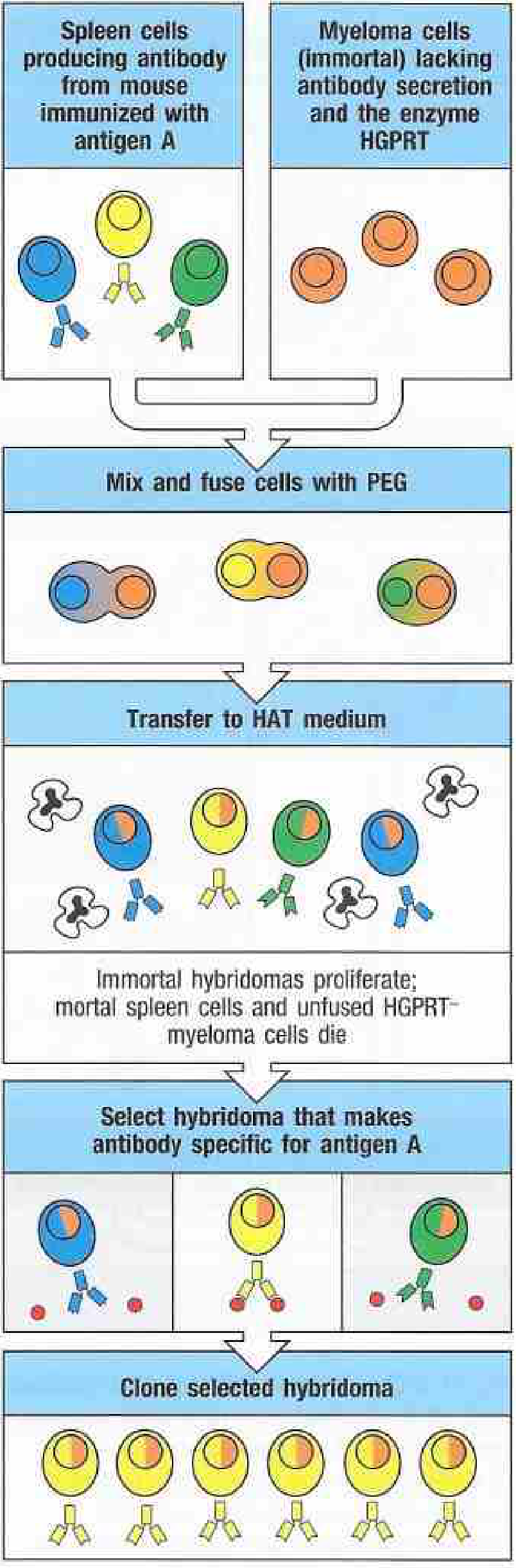
Fig. A.10 - Janeway's Immunobiology: production of monoclonal antibodies via the hybridoma technique
1. Immunization
A mouse is injected with the target antigen. An intravenous booster is given 3 days before sacrifice to maximize the population of spleen cells actively secreting specific antibody.
2. Spleen Cell Harvest
The spleen is removed and dissociated to yield B lymphocytes, many of which are secreting antibody against the antigen. These cells are mortal - they die within days in culture.
3. Cell Fusion with Myeloma Cells
The B cells are mixed with myeloma cells (immortal plasmacytoma cells) and fused using polyethylene glycol (PEG), which destabilizes cell membranes and promotes fusion. The myeloma cells used are specially selected to:
- Not secrete their own antibody
- Lack the enzyme HGPRT (hypoxanthine-guanine phosphoribosyltransferase)
4. HAT Medium Selection
The mixed cell population is transferred to HAT medium (Hypoxanthine-Aminopterin-Thymidine):
- Unfused myeloma cells die - they lack HGPRT, so the aminopterin blocks their only alternative nucleotide synthesis pathway
- Unfused spleen cells die naturally within a few days
- Only hybridomas survive - they inherit HGPRT from the spleen cell and immortality from the myeloma cell
5. Screening and Cloning
Individual hybridomas are isolated by limiting dilution (one cell per well) and screened for production of antibody with the desired specificity. Positive clones are expanded.
6. Bulk Production
Selected hybridoma clones are grown in large culture to produce unlimited quantities of the mAb with perfectly consistent affinity and specificity.
- Janeway's Immunobiology 10e; Katzung's Basic & Clinical Pharmacology, 16th Ed; Harper's Illustrated Biochemistry, 32nd Ed
Mechanism of Action
Monoclonal antibodies work by several mechanisms depending on their target:
| Mechanism | Description |
|---|---|
| Antigen neutralization | Directly block a receptor, ligand, or pathogen (e.g., anti-TNF-α mAbs block TNF from binding its receptor) |
| Opsonization | Fc region of mAb bound to target cell is recognized by phagocytes - antibody-dependent cellular phagocytosis (ADCP) |
| ADCC | Antibody-Dependent Cellular Cytotoxicity - Fc region recruits NK cells to kill the target cell |
| Complement activation | IgG mAbs can fix complement via the classical pathway, leading to membrane attack complex formation |
| Receptor blockade | Block growth factor receptors (e.g., anti-HER2, anti-EGFR in oncology) |
| Signaling agonism | Some mAbs activate receptors, e.g., immune checkpoint activators |
From Mouse to Human: Engineering Strategies
Native mouse (murine) antibodies given to humans trigger a Human Anti-Mouse Antibody (HAMA) response, which clears the therapeutic antibody rapidly and can cause allergic reactions. To overcome this, molecular engineering is used:
| Type | Description | Naming convention |
|---|---|---|
| Murine | 100% mouse protein | -omab (e.g., muromonab) |
| Chimeric | Mouse variable regions + human constant regions (~65% human) | -ximab (e.g., infliximab, rituximab) |
| Humanized | Only mouse CDRs retained in human framework (~95% human) | -zumab (e.g., natalizumab, bevacizumab) |
| Fully human | 100% human sequence (phage display or transgenic mice) | -umab (e.g., adalimumab, denosumab) |
Humanizing antibodies markedly reduces immunogenicity and extends the half-life in the patient. - Katzung's Basic & Clinical Pharmacology, 16th Ed; Harper's Illustrated Biochemistry, 32nd Ed
Modern Production Methods (Beyond Hybridomas)
-
Phage display - Combinatorial libraries of antibody variable (V) genes are expressed on the surface of bacteriophages and screened against purified antigen. This eliminates the need for animal immunization and generates fully human antibodies. Greg Winter received the Nobel Prize in Chemistry (2018) for this concept. - Henry's Clinical Diagnosis
-
Transgenic mice - Mice engineered to carry human immunoglobulin gene loci produce fully human antibodies upon immunization.
-
ADLib system (DT40 cells) - An ex vivo method using gene conversion in DT40 chicken B cells treated with trichostatin A, allowing rapid generation of mAbs using antigen-conjugated magnetic beads for selection.
-
Recombinant expression - Once the V-gene sequences are known, antibody fragments (Fab, scFv) or full-length antibodies can be expressed in E. coli, yeast, or mammalian (CHO) cell lines at industrial scale.
Key Advantages of Monoclonal Antibodies
- Unlimited production of a homogeneous reagent with highly consistent affinity and specificity
- Can be produced against non-purified antigens - only the desired epitope-specific clone is expanded
- Enable analysis of molecules on an epitope-by-epitope basis
- Identify isoenzymes, subtypes, isotypes, and conformational changes in molecules
- Highly useful in immunoassays, diagnostic probes, and therapeutic agents
Limitations
- A single mAb recognizes only one epitope, so antigens with heterogeneous epitopes are harder to characterize with a single mAb
- Insufficient reactivity in precipitation/agglutination assays because network formation is weak with a single antibody
- Can be cross-reactive if different antigens share the same epitope (e.g., CA 19-9)
- Production remains expensive and technically demanding
- Henry's Clinical Diagnosis and Management by Laboratory Methods
maturation of the lymphocytes
Lymphocyte maturation involves B cells developing in bone marrow (through pro-B, pre-B, immature B, and mature B cell stages) and T cells maturing in the thymus (through positive and negative selection). Both processes involve V(D)J recombination to generate diverse antigen receptors, followed by tolerance checkpoints to eliminate self-reactive clones.
Searching sources...
Finding Sources
Finding Sources
Finding Sources
Reading File
Good, rich content. Let me now read the B cell section from Henry's and get key sections from Janeway's on T cell development and thymic selection.
Reading File
Reading File
Good detailed content. Let me now get more on T cell thymic selection (positive/negative) and the B cell stages from Henry's (continuation), plus fetch key diagrams.
Reading File
Finding Sources
I have the T cell maturation table. Let me get the full content of this table and the B cell stages, plus fetch key diagrams.
Reading File
Loading Image
Excellent. Now let me fetch the thymus structure diagram and get more on thymic selection from Janeway's.
Loading Image
Finding Sources
Reading File
Reading File
I now have comprehensive material from multiple authoritative textbooks. Let me compile the full answer.
Maturation of Lymphocytes
Lymphocytes are the cellular basis of adaptive immunity. They arise from a common lymphoid progenitor in the bone marrow and mature along two major pathways - B cells (bone marrow) and T cells (thymus). A third lineage, NK cells, shares a common precursor with T cells.
Part 1: B Cell Maturation
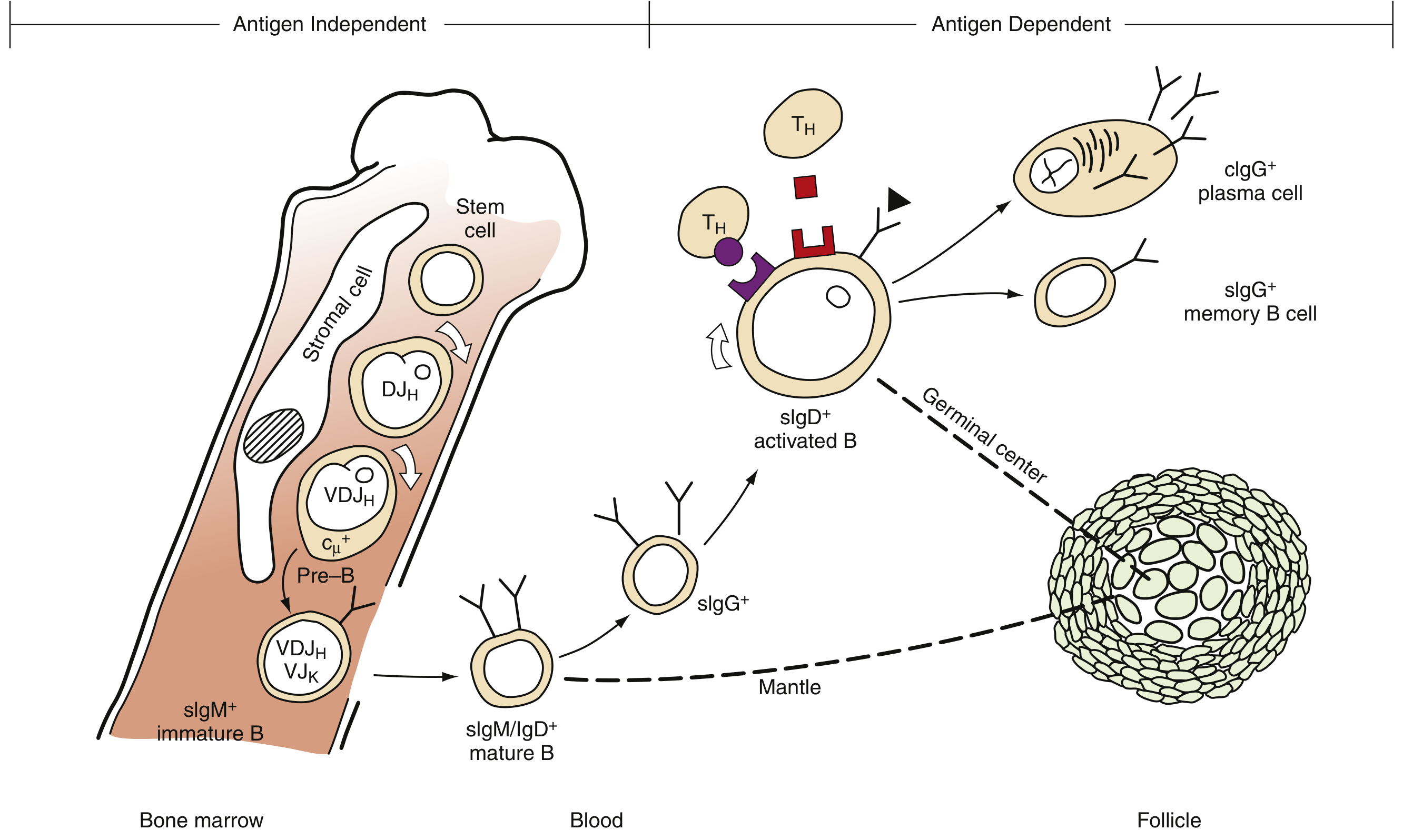
Fig. 32.27 - B cell differentiation: antigen-independent stages in bone marrow, antigen-dependent stages in peripheral lymphoid organs (Henry's Clinical Diagnosis)
B cell maturation has two broad phases:
Phase 1 - Antigen-Independent (Bone Marrow)
The bone marrow stroma provides essential cytokines and cell-contact signals that drive B cell development. IL-7 signals through IL-7R, activating JAK1/JAK3 and STAT5/STAT3, and is critical for progenitor survival and proliferation.
| Stage | Key Features | Surface Markers |
|---|---|---|
| Hematopoietic stem cell | Pluripotent; gives rise to all blood lineages | CD34+ |
| Pro-B cell | First committed B cell; RAG1/RAG2 and TdT activated; D-J rearrangement of heavy chain begins | CD19+, TdT+; no cytoplasmic or surface Ig |
| Pre-B cell (large) | V-DJ rearrangement completes functional μ heavy chain (V | HLA-DR+, CD19+, CD79a+; cμ+; no sIg |
| Immature B cell | VJ rearrangement of κ or λ light chain; complete IgM assembled and expressed on surface; central tolerance checkpoint - self-reactive cells deleted or undergo receptor editing | sIgM+ |
| Mature/naive B cell | Exits bone marrow; co-expresses sIgM and sIgD (via alternative RNA splicing); enters circulation as resting naive B cell | sIgM+, sIgD+ |
The key molecular events are V(D)J recombination - RAG1 and RAG2 cut the DNA at recombination signal sequences; TdT adds random nucleotides at V-D and D-J junctions, generating enormous diversity (the CDR3 region). - Henry's Clinical Diagnosis and Management by Laboratory Methods
Phase 2 - Antigen-Dependent (Peripheral Lymphoid Organs)
When a mature naive B cell encounters its cognate antigen in lymph nodes, spleen, or mucosal lymphoid tissue:
- Antigen binds the BCR (surface IgM/IgD) - activates the B cell
- T helper (TH) cell interaction via CD40-CD40L and cytokines (IL-4, IL-5, IL-6, etc.) drives proliferation in germinal centers
- Somatic hypermutation introduces point mutations in V-region genes, increasing affinity - a process called affinity maturation
- Class-switch recombination (isotype switching): intrachromosomal recombination between switch regions deletes intervening C-gene segments (e.g., Cμ→Cγ1 for IgG switch), allowing the same V-region to pair with a different constant region
- Activated B cells differentiate into:
- Plasma cells - terminally differentiated antibody secretion factories (secrete abundant cytoplasmic IgG)
- Memory B cells - long-lived cells with refined specificity, poised to deliver a faster, stronger anamnestic response on re-encounter with antigen
Part 2: T Cell Maturation
The Thymus
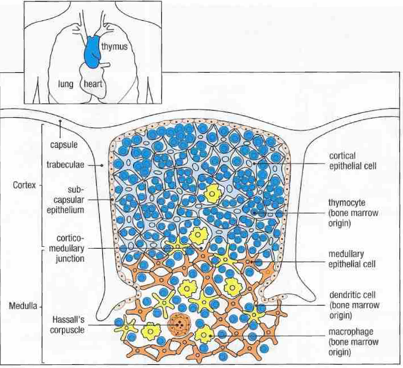
Fig. 8.16 - Thymus structure showing capsule, subcapsular epithelium, cortex (with dense thymocytes), corticomedullary junction, and medulla with Hassall's corpuscles (Janeway's Immunobiology 10e)
The thymus has two zones:
- Cortex - densely packed with immature thymocytes + cortical epithelial cells; site of positive selection
- Medulla - medullary epithelial cells, dendritic cells, macrophages; site of negative selection; contains Hassall's corpuscles
Without a thymus (DiGeorge syndrome - 22q11 deletion; nude mouse - Foxn1 defect), B cells develop normally but T cells are absent or severely reduced. - Janeway's Immunobiology 10e
Stages of T Cell Development
| Stage | Location | Maturation Events | Surface Markers |
|---|---|---|---|
| Pro-T cell | Bone marrow → subcapsular cortex | Migration from marrow; CD34 high | CD2, CD44; DN (CD4-CD8-) |
| Double-negative (DN) thymocyte | Subcapsular cortex | γ- and β-chain genes rearrange simultaneously; β-chain forms pre-TCR with surrogate α-chain | TdT+, CD1, CD2, CD7; CD4-CD8- |
| Double-positive (DP) thymocyte | Inner cortex | TCR α-chain gene rearranges; full αβ TCR expressed; positive selection occurs | CD1, CD2, CD3, CD4, CD5, CD7, CD8; TdT lost |
| Single-positive (SP) thymocyte | Medulla | CD4 or CD8 downregulated; negative selection occurs | CD4+ or CD8+; CD3+, CD5+, CD7+; mature TCR |
| Mature peripheral T cell | Blood/lymphoid tissue | Functional T cell; antigen-dependent activation pathway | CD4+ (helper) or CD8+ (cytotoxic) |
T lymphoblasts mature: common (cortical) thymocyte → medullary thymocyte → mature peripheral T cell of CD4 or CD8 phenotype - Henry's Clinical Diagnosis and Management by Laboratory Methods
The Two Critical Selection Events
Both occur through TCR engagement with self-MHC/peptide complexes - but with different outcomes depending on the avidity of binding. This is the avidity model:
Positive Selection (Cortex)
- Occurs on cortical epithelial cells
- Thymocytes whose TCR binds self-MHC with low-to-intermediate affinity receive a survival signal and continue maturing
- Thymocytes that fail to recognize self-MHC at all → death by neglect (~95% of thymocytes)
- Result: ensures all surviving T cells are MHC-restricted (can interact with antigen-presenting cells)
- CD4 or CD8 lineage commitment also determined here: cells whose TCR binds MHC class II keep CD4; those binding MHC class I keep CD8
Negative Selection (Medulla)
- Occurs primarily on medullary epithelial cells and dendritic cells
- Thymocytes whose TCR binds self-MHC + self-peptide with high affinity → clonal deletion (apoptosis)
- Prevents self-reactive T cells from escaping to the periphery - central to self-tolerance
- AIRE (autoimmune regulator) gene in medullary epithelial cells drives ectopic expression of peripheral self-antigens (e.g., insulin, thyroglobulin), ensuring T cells specific for these antigens are deleted. AIRE knockout → autoimmunity (type 1 diabetes model)
The same MHC-peptide signal has two outcomes: low avidity → positive selection; high avidity → negative selection. - Roitt's Essential Immunology
Mature Peripheral T Cell Subsets
After positive/negative selection, mature T cells leave the thymus and circulate. CD4+ T cells can further differentiate into functionally distinct subsets upon antigen stimulation:
| Subset | Cytokines Produced | Function |
|---|---|---|
| Th1 | IFN-γ, TNF-β, IL-2 | Defense against intracellular pathogens |
| Th2 | IL-4, IL-5 | Defense against extracellular parasites; allergy |
| Th17 | IL-17, IL-21, IL-22 | Defense against extracellular bacteria and fungi |
| iTreg | TGF-β, IL-10, IL-35 | Self-tolerance maintenance; immune regulation |
CD8+ cytotoxic T cells express αβ TCR and kill target cells presenting antigen on MHC class I.
γδ T cells (a minority) express γδ TCR, lack CD4/CD8, and function as cytotoxic cells in epithelial tissues.
Part 3: NK Cell Maturation
NK cells develop primarily in the bone marrow (and also in secondary lymphoid tissues). They share a common precursor with T cells - fetal NK cells express cytoplasmic CD3 proteins. Unlike T cells, NK cells do not require the thymus for development. They express CD56 and CD16 (FcγRIII), lack CD3 surface expression, and recognize targets via germline-encoded activating/inhibitory receptors rather than a rearranged antigen receptor. - Henry's Clinical Diagnosis and Management by Laboratory Methods
Summary Comparison
| Feature | B cells | T cells |
|---|---|---|
| Primary maturation site | Bone marrow | Thymus |
| Gene rearrangement | Ig heavy + light chain (V(D)J) | TCR α/β or γ/δ (V(D)J) |
| Enzyme driving diversity | RAG1/RAG2, TdT | RAG1/RAG2, TdT |
| Key tolerance mechanism | Receptor editing / clonal deletion (bone marrow) | Positive + negative selection (thymus) |
| Functional output | Plasma cells (antibodies) + memory B cells | CD4+ helper, CD8+ cytotoxic, memory T cells |
| Antigen recognition | Free antigen (no MHC needed) | Peptide-MHC complex (MHC-restricted) |
This is a shared conversation. Sign in to Orris to start your own chat.