
Lumber spine
Reading File
The query "Lumbar spine" is broad - it could cover anatomy, pathology, clinical conditions, imaging, or management. Let me clarify before spending research budget.
Asking for Preferences
What aspect of the lumbar spine are you interested in?
Searching sources...
Finding Sources
Finding Sources
Reading File
Reading File
Reading File
Finding Sources
Reading File
Loading Image
Finding Sources
Loading Image
Loading Image
Finding Sources
Finding Sources
Reading File
Reading File
Reading File
Loading Image
Finding Sources
Reading File
Loading Image
I now have comprehensive information from multiple authoritative textbooks. Let me compile the full anatomy overview.
Lumbar Spine - Anatomy & Structure
Overview
The lumbar spine consists of five vertebrae (L1-L5), forming the lowest movable segment of the vertebral column before the sacrum. It bears the greatest mechanical load of any spinal region and is characterized by having the largest vertebral bodies of any spinal level.
1. Bony Anatomy of Lumbar Vertebrae
Each lumbar vertebra has features that distinguish it from cervical and thoracic vertebrae:
Vertebral Body
- Massive, cylindrical body - largest of any vertebral region
- Bodies progressively increase in size from L1 to L5
- L5 is the largest individual lumbar vertebra
- Bear the greatest compressive loads in the body
- Covered by cartilaginous endplates superiorly and inferiorly
Vertebral Arch (Neural Arch)
- Pedicles: short and stocky; project posterolaterally from the body
- Laminae: flat plates that meet in the midline
- Together they form the triangular vertebral (spinal) foramen, which is larger than in the thoracic region
Processes
- Spinous processes: large, stubby, project straight posteriorly (horizontal orientation) - easily palpable
- Transverse processes (costal processes): long and slender - homologs of ribs, hence the term "costal process" in some atlases
- LV transverse processes are massive and cone-shaped, providing attachment for the iliolumbar ligaments
- Superior and inferior articular processes: large; oriented medially/laterally (coronal/sagittal plane), which promotes flexion-extension and limits rotation in the lumbar region
- Mammillary processes: small projections on the superior articular processes
- Accessory processes: small projections near the root of the transverse process
Superior view - Second lumbar vertebra (L2)
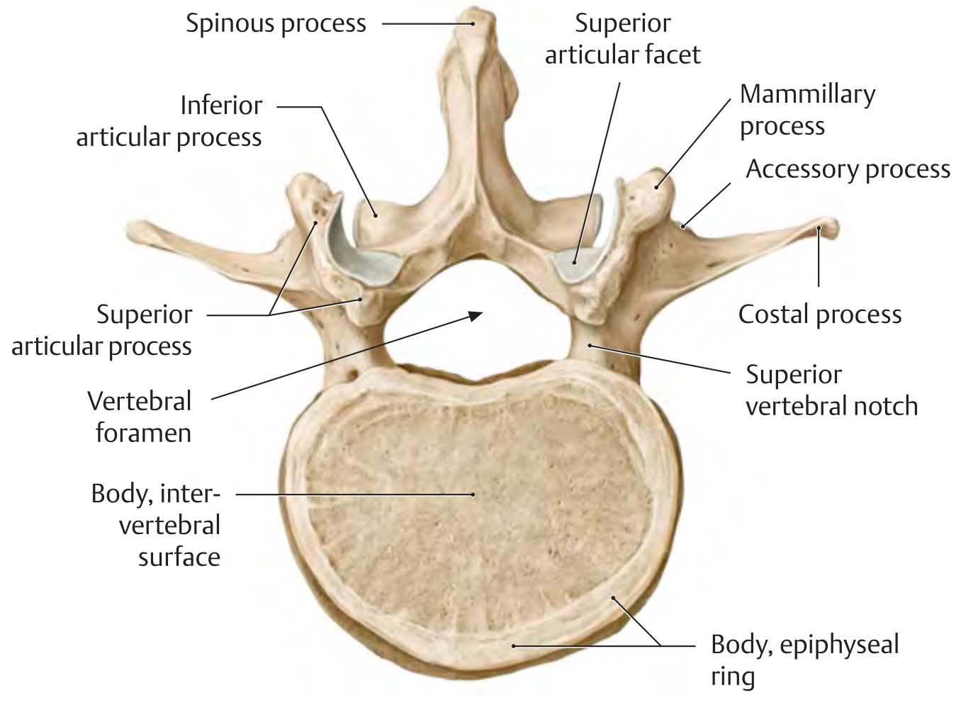
THIEME Atlas of Anatomy - Second lumbar vertebra, superior view
Lateral view - Fourth lumbar vertebra (L4)
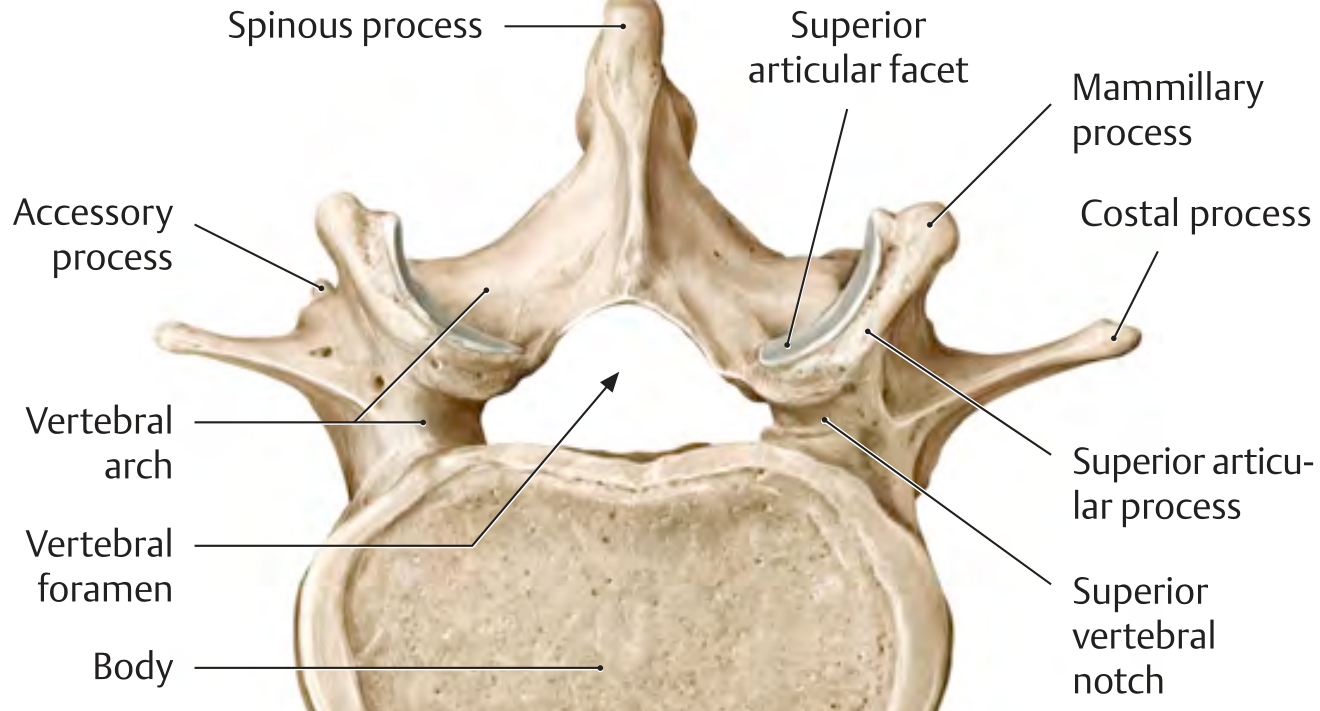
THIEME Atlas of Anatomy - Fourth lumbar vertebra, lateral view
2. Intervertebral Discs
Between each pair of adjacent lumbar vertebrae lies an intervertebral disc, which:
- Contributes to the lordotic curve of the lumbar region
- Provides shock absorption and allows movement
- Consists of two components:
- Nucleus pulposus (inner): gel-like, high water content (from glycosaminoglycans), provides compressive shock absorption
- Annulus fibrosus (outer): concentric rings of fibrocartilage that contain the nucleus and resist tensile forces
The disc at L5-S1 is the most commonly injured disc. With aging, the nucleus pulposus loses water content and the annulus frays, predisposing to disc herniation.
3. Ligaments
The lumbar spine is reinforced by several ligaments that provide stability through the full range of motion:
| Ligament | Location | Function |
|---|---|---|
| Anterior longitudinal ligament (ALL) | Runs along the anterior surface of the vertebral bodies | Resists extension and hyperextension |
| Posterior longitudinal ligament (PLL) | Runs along the posterior surface of the vertebral bodies (inside the canal) | Resists flexion; narrow in lumbar region |
| Ligamentum flavum | Connects adjacent laminae; yellow elastic tissue | Resists flexion; maintains upright posture; can hypertrophy causing stenosis |
| Interspinous ligaments | Connect adjacent spinous processes | Resist flexion |
| Supraspinous ligament | Connects tips of spinous processes | Resists flexion |
| Iliolumbar ligaments | Connect LV transverse processes to iliac crests | Stabilize the lumbosacral junction |
Sagittal section showing ligaments and disc
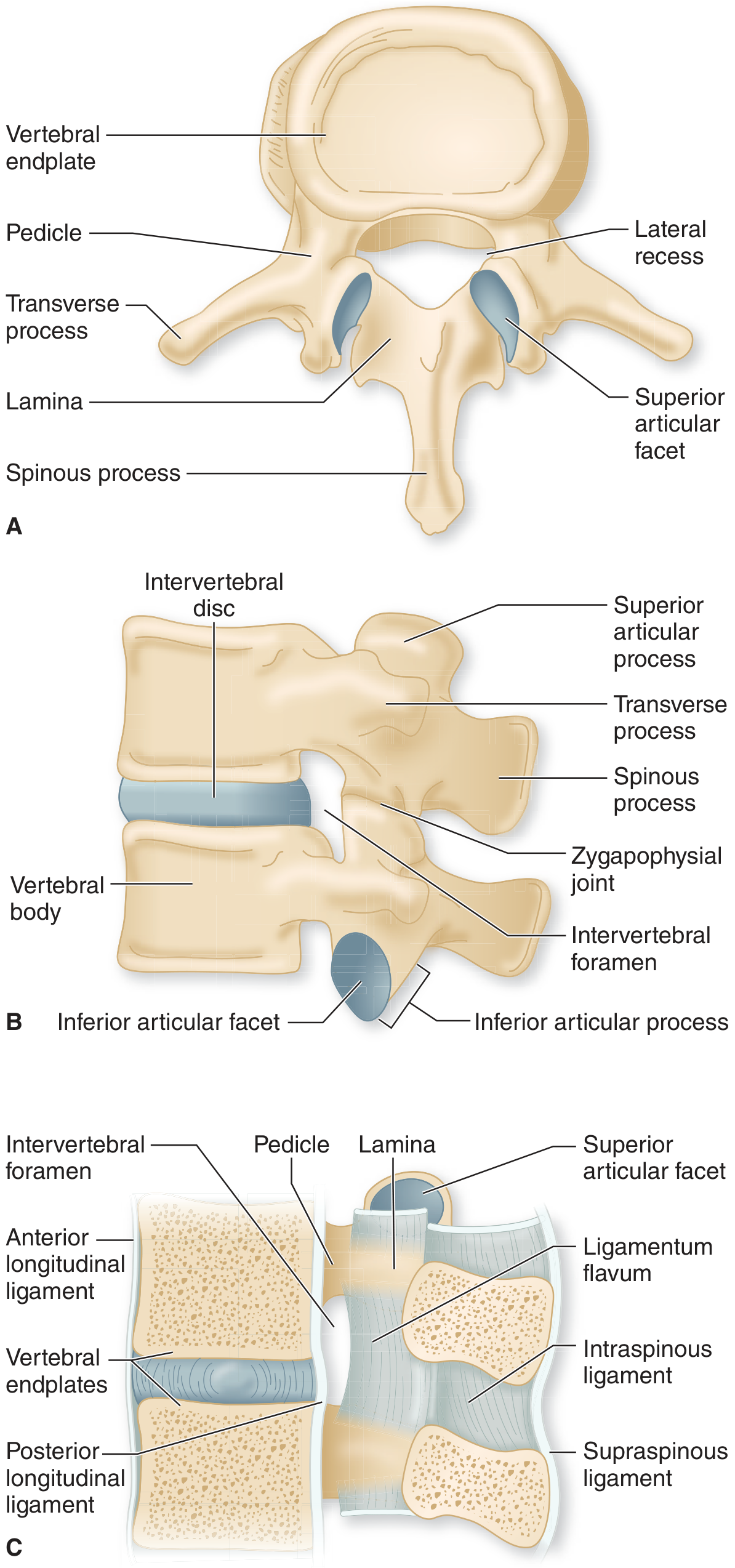
Adams and Victor's Principles of Neurology - Lumbar vertebra (A) superior view; (B) lateral view; (C) mid-sagittal section showing all major ligaments
4. Joints
Zygapophysial (Facet) Joints
- Synovial joints formed between superior articular process of the lower vertebra and inferior articular process of the upper vertebra
- In the lumbar region: oriented in the sagittal plane (medially/laterally), allowing flexion/extension but limiting rotation
- Each is surrounded by a fibrous joint capsule
- A common pain generator in lumbar back pain
Intervertebral (Cartilaginous) Joints
- Secondary cartilaginous joints between the disc and adjacent vertebral bodies
- Allow flexion, extension, lateral flexion, and limited rotation
Lumbosacral Joint (L5-S1)
- The most stressed joint in the entire spine
- Reinforced by the massive iliolumbar ligaments
5. Vertebral/Spinal Canal
- The lumbar vertebral foramen is triangular in shape and relatively large
- The lumbar spinal cord ends at L1-L2 (conus medullaris), tapering into the cauda equina
- Below L2, the spinal canal contains only nerve roots (cauda equina = "horse's tail")
- The lateral recess is a narrow groove along the inner surface of the pedicle where lumbar nerve roots lie before entering the foramen - a common site of root entrapment
6. Intervertebral Foramina
Each intervertebral foramen is bordered:
- Anteriorly: by the intervertebral disc and adjacent vertebral bodies
- Posteriorly: by the zygapophysial joint
- Superiorly: by the inferior vertebral notch of the upper pedicle
- Inferiorly: by the superior vertebral notch of the lower pedicle
The lumbar spinal roots in the cauda equina angulate sharply to exit horizontally through the foramina. Prior to entry, each root lies in the lateral recess - a common site of compression by disc fragments or osteophytes.
7. Regional Relationships and Curvature
The lumbar spine has a secondary (lordotic) curvature - a forward convexity - which develops when walking begins in childhood. This lumbar lordosis:
- Transfers body weight anteriorly to sit over the pelvis
- The five lumbar vertebral bodies project into the posterior abdominal cavity, forming the midline boundary of the posterior abdominal wall
The lumbar spine articulates:
- Superiorly with T12 (thoracolumbar junction)
- Inferiorly with the sacrum at the lumbosacral junction (L5-S1)
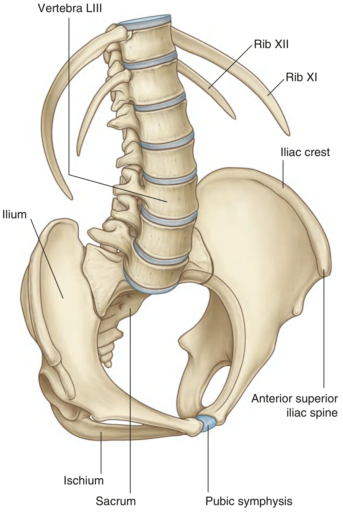
Gray's Anatomy for Students - Lumbar spine in relation to the posterior abdominal wall and pelvis
8. Muscles Acting on the Lumbar Spine
Posterior (extensor) muscles:
- Erector spinae (iliocostalis, longissimus, spinalis) - major extensors
- Multifidus - segmental stabilizer, important for proprioception and segmental control
- Quadratus lumborum - lateral flexion and extension
Anterior muscles:
- Psoas major - originates from lumbar vertebral bodies and transverse processes; flexes the hip and laterally flexes the spine
- Psoas minor (when present)
Abdominal muscles (indirect stabilizers via increased intra-abdominal pressure):
- Transversus abdominis, internal/external obliques, rectus abdominis
Summary Table
| Feature | Lumbar Specifics |
|---|---|
| Vertebrae | L1-L5 (5 total) |
| Vertebral body | Largest of any region; cylindrical |
| Vertebral foramen | Triangular; larger than thoracic |
| Spinous process | Large, stubby, horizontal |
| Transverse process | Long, slender (costal processes); L5 massive |
| Articular facets | Sagittal/medial-lateral orientation; allow flexion-extension, limit rotation |
| Discs | 5 (L1/2 to L5/S1); largest nucleus pulposus |
| Curvature | Lordotic (secondary, forward convexity) |
| Spinal cord end | Conus at L1-L2; cauda equina below |
| Key landmark | L4-L5 level = Tuffier's line (intercristal plane) |
Sources: Gray's Anatomy for Students, THIEME Atlas of General Anatomy and Musculoskeletal System, Adams and Victor's Principles of Neurology (12th Ed), Grainger & Allison's Diagnostic Radiology
This is a shared conversation. Sign in to Orris to start your own chat.